. 2021 Nov;73(11):2052-2058.
doi: 10.1002/art.41787. Epub 2021 Sep 29.

Estrogen-Induced hsa-miR-10b-5p Is Elevated in T Cells From Patients With Systemic Lupus Erythematosus and Down-Regulates Serine/Arginine-Rich Splicing Factor 1

Affiliations

Estrogen-Induced hsa-miR-10b-5p Is Elevated in T Cells From Patients With Systemic Lupus Erythematosus and Down-Regulates Serine/Arginine-Rich Splicing Factor 1

Suruchi A Ramanujan et al. Arthritis Rheumatol. 2021 Nov.

Abstract

Objective: Autoimmune diseases affect women disproportionately more than men. Estrogen is implicated in immune cell dysfunction, yet its precise molecular roles are not fully known. We recently identified new roles for serine/arginine-rich splicing factor 1 (SRSF1) in T cell function and autoimmunity. SRSF1 levels are decreased in T cells from patients with systemic lupus erythematosus (SLE) and are associated with active disease and comorbidity. However, the molecular mechanisms that control SRSF1 expression are unknown. Srsf1 messenger RNA (mRNA) has a long 3'-untranslated region (3'-UTR), suggesting posttranscriptional control. This study was undertaken to investigate the role of estrogen and posttranscriptional mechanisms of SRSF1 regulation in T cells and SLE.

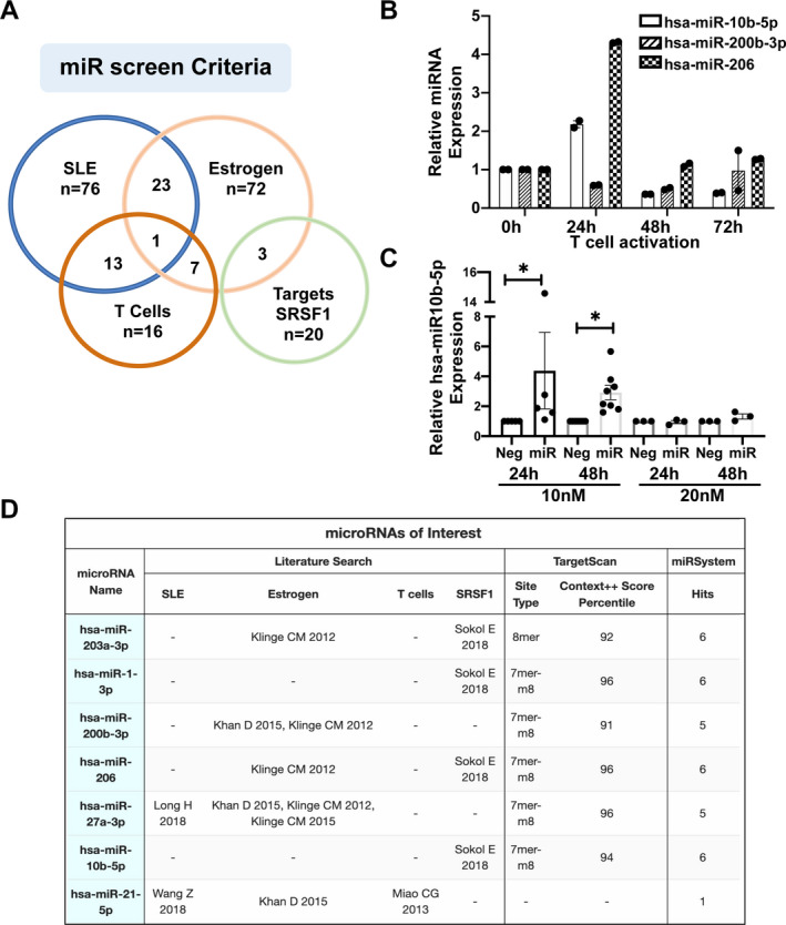
Methods: In silico bioinformatics analysis of Srsf1-3'-UTR revealed multiple microRNA (miRNA; miR)-binding sites. Additional screening and literature searches narrowed down hsa-miR-10b-5p for further study. Peripheral blood T cells from healthy individuals and SLE patients were evaluated for mRNA and miRNA expression by quantitative reverse transcription-polymerase chain reaction, and SRSF1 protein levels were assessed by immunoblotting. T cells were cultured with β-estradiol, and transient transfections were used to overexpress miRNAs. Luciferase assays were used to measure 3'-UTR activity.

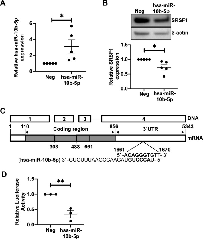
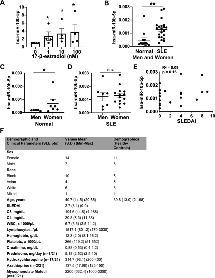
Results: We demonstrated that estrogen increased hsa-miR-10b-5p expression in human T cells, and hsa-miR-10b-5p down-regulated SRSF1 protein expression. Mechanistically, hsa-mir-10b-5p regulated SRSF1 posttranscriptionally via control of its 3'-UTR activity. Importantly, hsa-miR-10b-5p expression levels were elevated in T cells from healthy women compared to healthy men and also elevated in T cells from SLE patients.

Conclusion: We identified a previously unrecognized molecular link between estrogen and gene regulation in immune cells, with potential relevance to systemic autoimmune disease.


Figures

Figure 1
Figure 1
In silico screening and selection of microRNA (miRNA; miR) targeting Srsf1. A, Web‐based bioinformatics prediction tools (TargetScan and miRSystem) were used to screen for microRNAs targeting Srsf1, combined with a literature search to curate miRNAs that are up‐regulated in systemic lupus erythematosus (SLE), up‐regulated by estrogen, up‐regulated in T cells, and/or shown to target Srsf1. B, Quantitative reverse transcription–polymerase chain reaction was performed for 3 selected miRNAs in resting T cells (0 hours) and in activated T cells (24 hours, 48 hours, and 72 hours). Each symbol represents an individual subject; bars show the mean ± SEM relative miRNA expression from duplicates. C, Negative control (Neg) or hsa‐miR‐10b‐5p mimics were transiently transfected into HEK 293T cells in 10 nM or 20 nM concentrations, and cells were collected at 24 hours or 48 hours posttransfection. Expression levels of miRNA were measured by reverse transcription–polymerase chain reaction. Each symbol represents an individual subject; bars show the mean ± SEM relative miRNA expression from at least 3 independent experiments. * = P < 0.05. D, The chart shows the miRNAs narrowed for further study based on the above‐mentioned criteria. SRSF = serine/arginine‐rich splicing factor 1.
Figure 2
Figure 2
Overexpression of hsa‐miR‐10b‐5p leads to down‐regulation of SRSF1 via its 3′‐untranslated region (3′‐UTR). A, Negative control miR mimics or hsa‐miR‐10b‐5p mimics were transiently transfected into HEK 293T cells. Quantitative reverse transcription–polymerase chain reaction was performed to assess the levels of hsa‐miR‐10b‐5p. Each symbol represents an individual subject; bars show the mean ± SEM from 4 experiments. * = P < 0.05. B, Levels of SRSF1 total protein in transfected cells were assessed by Western immunoblotting. Representative blots are shown (top). Densitometric results from Western blotting were quantified as the expression of SRSF1 protein normalized to β‐actin (bottom). Each symbol represents an individual subject; bars show the mean ± SEM from 4 experiments. * = P < 0.05. C, Schematic shows DNA and mRNA sequence of Srsf1. The hsa‐miR‐10b‐5p sequence and its complementary binding site within the 3′‐UTR are shown. D, HEK 293T cells were cotransfected with the Srsf1 3ʹ‐UTR–luciferase plasmid and 10 nM of hsa‐miR‐10b‐5p mimic or negative control mimic. Cells were collected and 3ʹ‐UTR activity was measured by luciferase assay. Each symbol represents an individual subject; bars show the mean ± SEM from 3 experiments. ** = P < 0.01. See Figure 1 for other definitions.
Figure 3
Figure 3
Induction of hsa‐miR‐10b‐5p by estrogen and elevated levels of hsa‐miR‐10b‐5p in T cells from healthy women and systemic lupus erythematosus (SLE) patients. A, T cells from healthy women were treated with increasing concentrations of β‐estradiol, and hsa‐miR‐10b‐5p levels were assessed by real‐time quantitative reverse transcription–polymerase chain reaction. Results are from 4 experiments. * = P < 0.05 versus no β‐estradiol. B, Levels of hsa‐miR‐10b‐5p were measured by quantitative reverse transcription–polymerase chain reaction in T cells from SLE patients (n = 21) and age‐, race‐, and sex‐matched healthy controls (n = 12). ** = P < 0.01. C, Levels of hsa‐miR‐10b‐5p in healthy men and healthy women are shown. * = P < 0.05. D, Levels of hsa‐miR‐10b‐5p in men and women with SLE are shown. In A‐D, each symbol represents an individual subject; bars show the mean ± SEM. E, Correlations between Systemic Lupus Erythematosus Disease Activity Index (SLEDAI) scores and hsa‐miR‐10b‐5p levels in patients with SLE are shown. F, Demographic and clinical features of the SLE patients and healthy controls are shown. NS = not significant; WBC = white blood cells.

References

    1. Moulton VR. Sex hormones in acquired immunity and autoimmune disease. Front Immunol 2018;9:2279. - PMC - PubMed
    1. Yen EY, Singh RR. Lupus—an unrecognized leading cause of death in young females: a population‐based study using nationwide death certificates, 2000–2015. Arthritis Rheumatol 2018;70:1251–5. - PMC - PubMed
    1. Corradetti C, Jog NR, Cesaroni M, Madaio M, Caricchio R. Estrogen receptor α signaling exacerbates immune‐mediated nephropathies through alteration of metabolic activity. J Immunol 2018;200:512–22. - PMC - PubMed
    1. Klinge CM. miRNAs regulated by estrogens, tamoxifen, and endocrine disruptors and their downstream gene targets [review]. Mol Cell Endocrinol 2015;418:273–97. - PMC - PubMed
    1. Rider V, Abdou NI, Kimler BF, Lu N, Brown S, Fridley BL. Gender bias in human systemic lupus erythematosus: a problem of steroid receptor action? Front Immunol 2018;9:611. - PMC - PubMed

Publication types

MeSH terms