. 2021 Dec;73(12):2249-2260.
doi: 10.1002/art.41794. Epub 2021 Oct 28.

Amelioration of Posttraumatic Osteoarthritis in Mice Using Intraarticular Silencing of Periostin via Nanoparticle-Based Small Interfering RNA

Affiliations

Amelioration of Posttraumatic Osteoarthritis in Mice Using Intraarticular Silencing of Periostin via Nanoparticle-Based Small Interfering RNA

Xin Duan et al. Arthritis Rheumatol. 2021 Dec.

Abstract

Objective: Recent evidence delineates an emerging role of periostin in osteoarthritis (OA), since its expression after knee injury is detrimental to the articular cartilage. We undertook this study to examine whether intraarticular (IA) knockdown of periostin would ameliorate posttraumatic OA in a murine model.

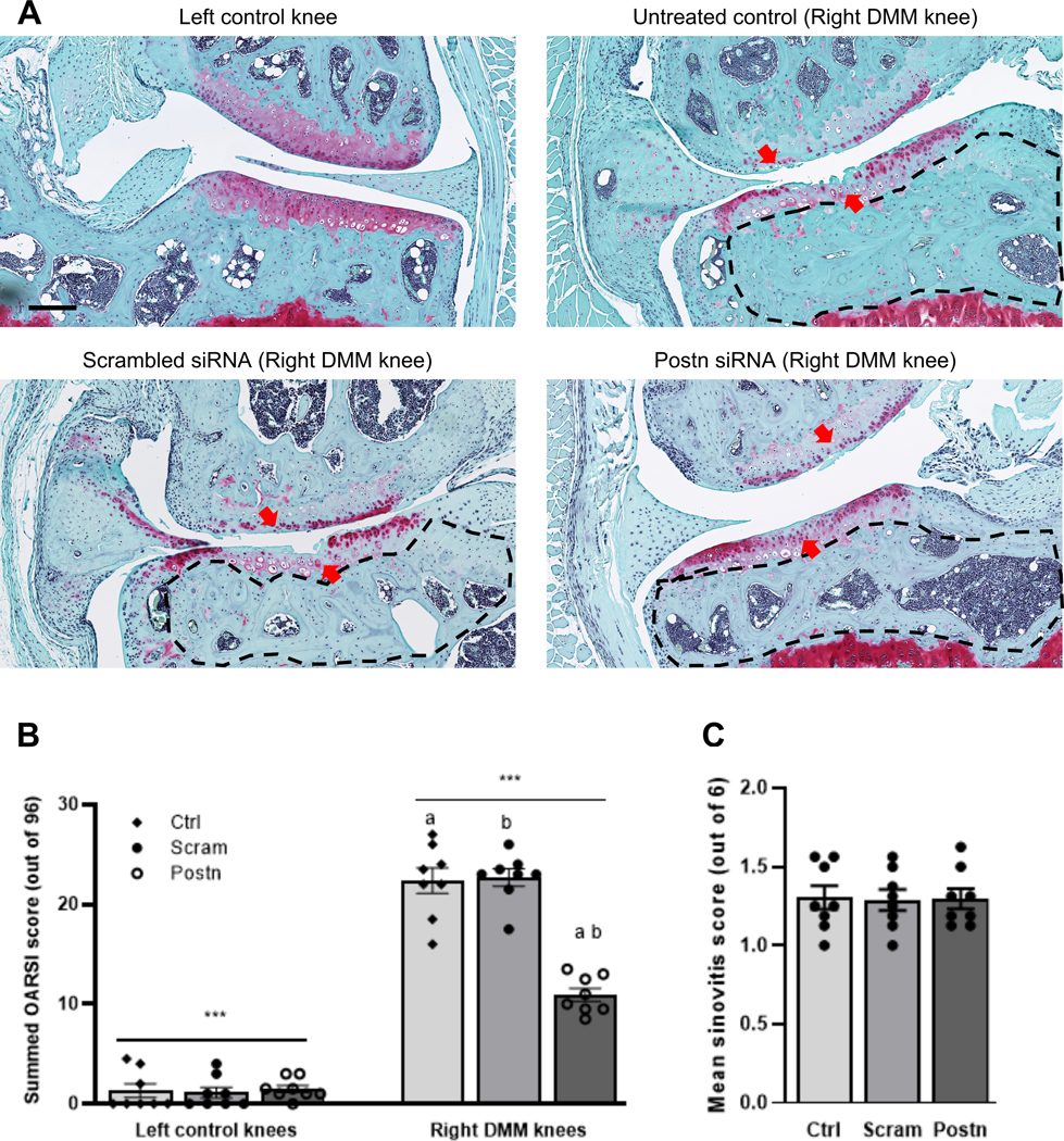
Methods: Posttraumatic OA was induced in 10-week-old male C57BL/6J mice (n = 24) by destabilization of the medial meniscus (DMM), and mice were analyzed 8 weeks after surgery. Periostin expression was inhibited by small interfering RNA (siRNA) delivered IA using a novel peptide-nucleotide polyplex. Following histologic assessment of the mouse knee cartilage, the extent of cartilage degeneration was determined using Osteoarthritis Research Society International (OARSI) cartilage damage score, and severity of synovitis was also assessed. Bone changes were measured using micro-computed tomography. The effect and mechanism of periostin silencing were investigated in human chondrocytes that had been stimulated with interleukin-1β (IL-1β) with or without the IκB kinase 2 inhibitor SC-514.

Results: Periostin expression in mice with posttraumatic OA was significantly abolished using IA delivery of a peptide-siRNA nanoplatform. OARSI cartilage damage scores were significantly lower in mice receiving periostin siRNA (mean ± SEM 10.94 ± 0.66) compared to untreated mice (22.38 ± 1.30) and mice treated with scrambled siRNA (22.69 ± 0.87) (each P = 0.002). No differences in the severity of synovitis were observed. Subchondral bone sclerosis, bone volume/total volume, volumetric bone mineral density, and heterotopic ossification were significantly lower in mice that had received periostin siRNA treatment. Immunostaining of cartilage revealed that periostin knockdown reduced the intensity of DMM-induced matrix metalloproteinase 13 (MMP-13) expression and also diminished the phosphorylation of p65 and immunoreactivity of the aggrecan neoepitope DIPEN. Periostin knockdown also suppressed IL-1β-induced MMP-13 and ADAMTS-4 expression in chondrocytes. Mechanistically, periostin-induced MMP-13 expression was abrogated by SC-514, demonstrating a link between periostin and NF-κB.

Conclusion: IA delivery of the periostin-siRNA nanocomplex represents a promising clinical approach to mitigate the severity of joint degeneration in OA. Our findings may thus provide an unequivocal scientific rationale for longitudinal studies of this approach. Utilizing a cartilage-specific gene-knockout strategy will further illuminate the functional role of periostin in OA.


Conflict of interest statement

Conflict of interest statement

All authors have declared that no conflict of interest exists in conjunction with this study.

Figures

Fig. 1:
Fig. 1:. Peptide Postn siRNA treatment suppressed Postn upregulation following DMM surgery.
(A) Experimental design: DMM surgery was performed in 10-week-old male mice (n=8 each group). HBSS (untreated control), scrambled siRNA, or Postn siRNA was injected intra-articularly at the following time points: immediately after DMM surgery, at week 1, 2, 4 and 6 post-surgery (5 injections in total). Knee joints were harvested and analyzed at 8-weeks after surgery. (B) Postn immunostaining revealed that Postn was upregulated (green) in the cartilage of knees subjected to DMM surgery compared to the uninjured control knees (see untreated control vs. left control knee). Intra-articular administration of Postn siRNA nanoparticles suppressed the expression of DMM-induced Postn compared with untreated control or scrambled siRNA treatment groups. The dotted lines indicate Col 2 positive cartilage area based on the images in the upper panel. DAPI (blue) was used to stain nuclei. Scale bar=50 μm. (C) Quantification of immunofluorescence intensity showed significant differences in Postn expression between scrambled siRNA and Postn siRNA treatment groups (Kruskal-Wallis test with Dunn’s multiple comparisons, P=0.033). Normalized mean Postn fluorescence intensity per chondrocyte was measured from Z-stack confocal images. Similar lowercase letters indicate statistically significant difference.
Fig. 2:
Fig. 2:. Peptide Postn siRNA treatment attenuated cartilage degeneration in a DMM-induced post-traumatic OA mouse model but did not affect synovitis.
(A) Cartilage sections stained with Safranin-O/Fast green displayed significantly less cartilage degeneration in the left control knees compared with DMM-operated right knees. Treatment with Postn siRNA resulted in reduced cartilage degeneration compared to treatment with untreated control or scrambled siRNA (red arrows). The area within dotted black lines in each image indicates the subchondral bone area. We observed pronounced sclerosis in the untreated control (HBSS) or scrambled siRNA than in the Postn siRNA group. (B) The summed OARSI score (based on the four compartments of each knee; highest possible score = 96) was significantly lower in the left control knee than right DMM knee (***2-way ANOVA with Tukey’s adjusted P<0.001). The summed OARSI score in the Postn siRNA treatment group was significantly lower than in the untreated control (P=0.002) or scrambled siRNA (P=0.002) groups (Kruskal-Wallis test with Dunn’s multiple comparisons). Similar lowercase letters indicate statistically significant differences. Scale bar=200 μm. (C) Histological assessment of synovial pathology (synovitis) displayed no significant differences across treatment groups (Kruskal-Wallis test with Dunn’s multiple comparisons).
Fig. 3:
Fig. 3:. Peptide Postn siRNA treatment blocked DMM-induced subchondral bone changes and heterotopic ossification.
(A) 3D reconstruction of μCT images showed that Postn siRNA treatment resulted in reduced subchondral bone sclerosis (red circles) of the medial tibial compartment in DMM-operated right knees compared to untreated control (HBSS) or scrambled siRNA. L, lateral; M, medial; F, femur; T, tibia. Scale bar=1 mm. (B) BV/TV was significantly lower in the Postn siRNA treatment group than untreated control (P=0.027) or scrambled siRNA (P=0.004) groups. Note: BV/TV of the right DMM-operated limb was normalized to the BV/TV of the left control limb of the same animal to eliminate intraindividual variations. (Kruskal-Wallis test with Dunn’s multiple comparisons). (C) Relative vBMD was significantly lower in the Postn siRNA group compared to the untreated control (P=0.010) or scrambled siRNA (P=0.022) groups. (Kruskal-Wallis test with Dunn’s multiple comparisons). (D) 3D reconstruction of micro-CT images to assess heterotopic ossification (white arrows) in DMM-induced OA knees. (E) Quantification of the number of ossified nodules indicated that Postn siRNA treatment reduced the number of nodules compared the scrambled siRNA group (P=0.018). (Kruskal-Wallis test with Dunn’s multiple comparisons). Similar lowercase letters indicate statistically significant differences.
Fig. 4:
Fig. 4:. Peptide Postn siRNA treatment abrogated MMP-13 upregulation and canonical NF-κB signaling following DMM surgery.
(A) Immunofluorescent analysis revealed that MMP-13 levels (green) were higher in DMM-operated knees than in uninjured control knees. Postn siRNA treatment reduced MMP-13 around the injury site. Yellow vertical lines mark the injury site in the cartilage, while white arrows mark MMP-13 in superficial zone chondrocytes. Col 2 (red) staining was used for cartilage. DAPI (blue) = nuclei. Scale bars=100 μm. (B) Quantification of immunofluorescence intensity showed significant differences in MMP-13 expression between scrambled siRNA and Postn siRNA treatment groups (Mann-Whitney test, P=0.029). (C) Upper and lower panels show 2D and 3D views, respectively, of immunofluorescence analysis of phosphorylated p65 subunit (green) in cartilage adjacent to the injury site. Less co-localization of activated/phosphorylated p65 (P-p65, green) was observed in the Postn siRNA treatment group than in the untreated control and scrambled siRNA groups. DAPI (blue) = nuclei. Scale bars=50 μm. Untreated control=DMM knees receiving HBSS. (D) Quantification of immunofluorescence intensity showed significant differences in P-p65 expression between scrambled siRNA and Postn siRNA treatment groups (Mann-Whitney test, P=0.029).
Fig. 5:
Fig. 5:. Peptide Postn siRNA nanoparticle treatment attenuated the catabolic effects of IL-1β in human primary chondrocytes.
(A) Human primary chondrocytes treated with p5RHH peptide, Cy5.5-labeled-siRNA, or peptide-Cy5.5-labled siRNA are shown. Our data show that the Cy5.5 labeled siRNAs delivered with nanoparticles were effectively taken up by the human primary chondrocytes (red color, indicated by arrows) and remained in the cells after 72h in culture. Scale bars=100 μm. (B) Real-time qPCR analysis showed that human chondrocytes treated with IL-1β and transfected with peptide Postn siRNA exhibited significantly lower Postn (P=0.016), MMP-13 (P=0.016), IL-1β (P=0.016), ADAMTS-4 (P=0.016), ACAN (P=0.016) and COL2A1 (P=0.016), mRNA levels. There was no effect of treatment on COL1A1 expression (P=0.688). (Wilcoxon matched-pairs signed rank test). (C) Western blot analysis confirmed that IL-1β treatment induced MMP-13 expression and activated p65 (phosphorylated, P-p65). Cells treated with peptide Postn siRNA displayed significantly lower levels of Postn (P=0.025, P=0.025) MMP-13 (P=0.079, P=0.030) and reduced phosphorylation of p65 (P=0.010, P=0.046) than the control and scrambled siRNA groups respectively. (Kruskal-Wallis test with Dunn’s multiple comparisons). Similar lowercase letters indicate statistically significant differences.
Fig. 6:
Fig. 6:. Mechanism and Pathway summary:
(A) An increased expression of MMP-13 in cells treated with IL-1β was noted, which was abrogated by SC-514 treatment (Wilcoxon matched-pairs signed rank test). (B) Exogenous recombinant Postn instigated the expression of MMP-13, which was mitigated by SC-514 treatment. (Kruskal-Wallis test with Dunn’s multiple comparisons). (C) Pathway summary: DMM surgery or IL-1β treatment activates NF-κB signaling pathway in chondrocytes, as evidenced by increased p65 phosphorylation, leading to its nuclear translocation, and the expression of catabolic mediators (e.g., MMP-13) that promote extracellular matrix degradation. Administration of p5RHH Postn siRNA reduces the activation of p65, blocks the downstream catabolic effects of NF-κB signaling directly or indirectly, and prevents the progressive cartilage characteristic of post-traumatic OA. i.a.=intra-articular. Similar lowercase letters indicate statistically significant differences.

References

    1. Hootman JM, Helmick CG, Barbour KE, Theis KA, Boring MA. Updated Projected Prevalence of Self-Reported Doctor-Diagnosed Arthritis and Arthritis-Attributable Activity Limitation Among US Adults, 2015–2040. Arthritis Rheumatol. 2016;68(7):1582–7. - PMC - PubMed
    1. Rai MF, Pham CT. Intra-articular drug delivery systems for joint diseases. Curr Opin Pharmacol. 2018;40:67–73. - PMC - PubMed
    1. Brown TD, Johnston RC, Saltzman CL, Marsh JL, Buckwalter JA. Posttraumatic osteoarthritis: a first estimate of incidence, prevalence, and burden of disease. J Orthop Trauma. 2006;20(10):739–44. - PubMed
    1. Rai MF, Brophy RH, Sandell LJ. Osteoarthritis following meniscus and ligament injury: insights from translational studies and animal models. Curr Opin Rheumatol. 2019;31(1):70–9. - PubMed
    1. Roos H, Adalberth T, Dahlberg L, Lohmander LS. Osteoarthritis of the knee after injury to the anterior cruciate ligament or meniscus: the influence of time and age. Osteoarthritis Cartilage. 1995;3(4):261–7. - PubMed

Publication types

MeSH terms

LinkOut - more resources