. 2021 May 26;13(20):23438-23451.
doi: 10.1021/acsami.1c03537. Epub 2021 May 13.

High Aspect Ratio and Light-Sensitive Micropillars Based on a Semiconducting Polymer Optically Regulate Neuronal Growth

Affiliations

High Aspect Ratio and Light-Sensitive Micropillars Based on a Semiconducting Polymer Optically Regulate Neuronal Growth

Frano Milos et al. ACS Appl Mater Interfaces. .

Abstract

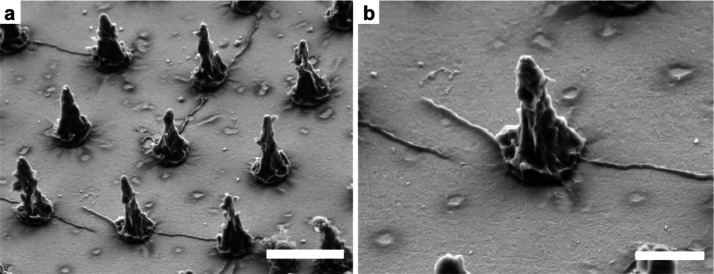
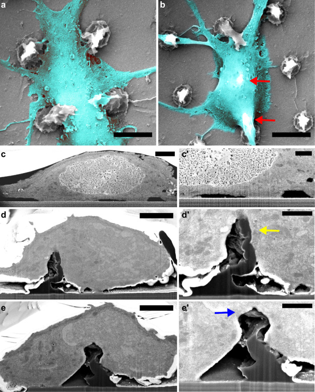
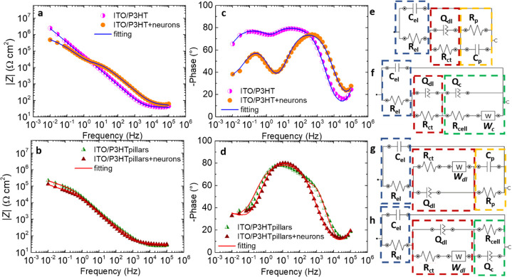
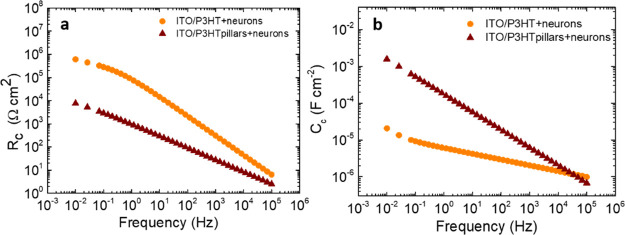
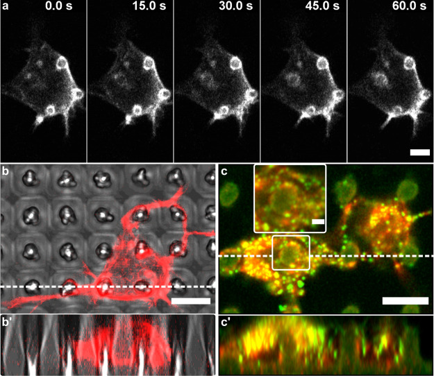
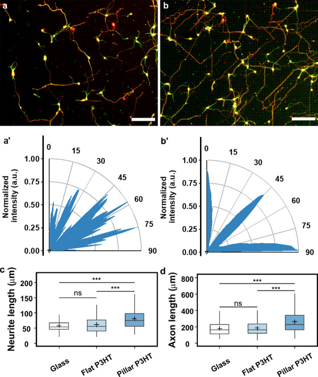
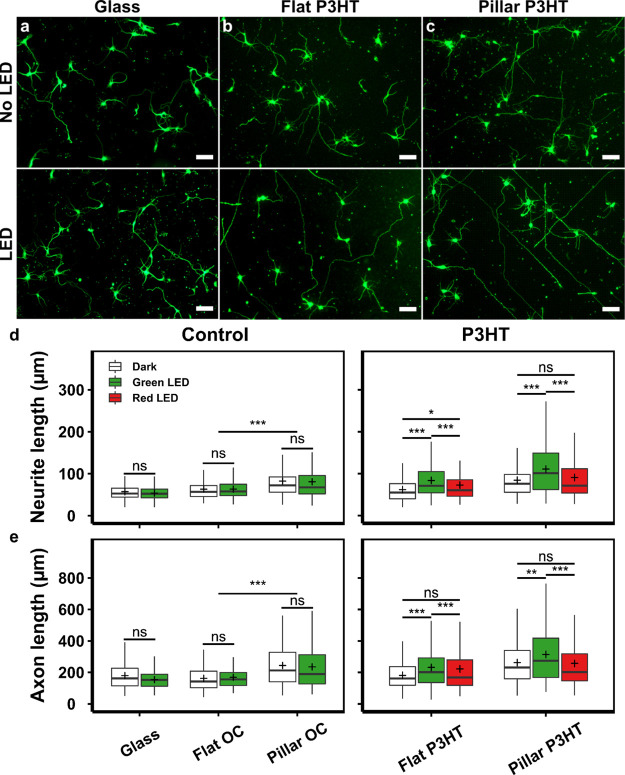
Many nano- and microstructured devices capable of promoting neuronal growth and network formation have been previously investigated. In certain cases, topographical cues have been successfully complemented with external bias, by employing electrically conducting scaffolds. However, the use of optical stimulation with topographical cues was rarely addressed in this context, and the development of light-addressable platforms for modulating and guiding cellular growth and proliferation remains almost completely unexplored. Here, we develop high aspect ratio micropillars based on a prototype semiconducting polymer, regioregular poly(3-hexylthiophene-2,5-diyl) (P3HT), as an optically active, three-dimensional platform for embryonic cortical neurons. P3HT micropillars provide a mechanically compliant environment and allow a close contact with neuronal cells. The combined action of nano/microtopography and visible light excitation leads to effective optical modulation of neuronal growth and orientation. Embryonic neurons cultured on polymer pillars show a clear polarization effect and, upon exposure to optical excitation, a significant increase in both neurite and axon length. The biocompatible, microstructured, and light-sensitive platform developed here opens up the opportunity to optically regulate neuronal growth in a wireless, repeatable, and spatio-temporally controlled manner without genetic modification. This approach may be extended to other cell models, thus uncovering interesting applications of photonic devices in regenerative medicine.

Keywords: cell optical excitation; cell−substrate interface; conjugated polymers; embryonic cortical neurons; microstructured cell interfaces; tissue engineering; topography.


Conflict of interest statement

The authors declare no competing financial interest.

Figures

Figure 1
Figure 1
P3HT micropillar array. Representative SEM images of a micropillar array (a) and an individual pillar (b). Micropillars had a conical shape with a high degree of nanoscale roughness on their sidewalls. Images were acquired with a 45° tilt angle. Scale bars: (a) 5 and (b) 2 μm.
Figure 2
Figure 2
FIB/SEM characterization of the cell–micropillar adhesion. (a) Neuronal soma positioned between the pillars and (b) suspended over two pillars (red arrows). Scale bars: 5 μm. (c,c′) FIB/SEM cross-sections of the neuronal soma positioned on the flat surface. (d,e′) FIB/SEM cross-sections of the neuronal soma positioned on a micropillar at the soma periphery (d,d′) and at the soma center (e,e′). Arrows in (d′,e′) indicate the pillar being pulled toward the center (yellow arrow) and pushed down (blue arrow), respectively. Scale bars: (c,d,e) 2 and (c′,d′,e′) 1 μm.
Figure 3
Figure 3
EIS characterization. Impedance modulus |Z| and phase angle recorded during EIS experiments for ITO/P3HT (a,c) and ITO/P3HT pillars (b,d) with and without cortical neurons. |Z| values were normalized to the geometrical and to the estimated effective area of the sample, in the flat and pillar cases, respectively. Equivalent circuits employed to model the experimental data related to the ITO/P3HT (e), ITO/P3HT + neurons (f), ITO/P3HT pillars (g), and ITO/P3HT pillars + neurons (h) systems. Numerical values of the circuital components are reported in Table S1. χ2 values: 0.08 and 0.05 for ITO/P3HT samples, with and without cells, respectively; 0.06 for cell-covered and uncovered ITO/P3HT pillars.
Figure 4
Figure 4
Cell resistance and capacitance. Resistance (Rc) (a) and capacitance (Cc) (b) values, obtained by the combination of circuital parameters (Table S1) of the cell equivalent circuital loops depicted in Figure 3, as a function of the frequency. Rc and Cc values were normalized to the geometrical and to the estimated effective area of the sample, in the flat and pillar cases, respectively.
Figure 5
Figure 5
Actin rings and paxillin adhesions on P3HT micropillars. (a) Time-lapse sequence of stable F-actin structures around the pillars. Additionally, the formation of a fourth ring can be observed. (b,b′) F-actin ring-like accumulations formed around the micropillars indicate membrane wrapping. (c,c′) These structures often overlapped with paxillin-rich adhesions (zoomed-in inset; green puncta). Images in (b′,c′) are Z-stack orthogonal projections of 30 slices (400 and 200 nm thickness, respectively), along the dashed lines in images (b,c). Scale bars: (a–c′) 5 μm; inset 2 μm. Cells in (a,b) were transfected with a fluorescent F-actin marker (Lifeact-RFP). Cells in image (c) were stained with TRITC-phalloidin (actin, red) and anti-paxillin antibodies (green).
Figure 6
Figure 6
Neuronal growth on P3HT micropillars. Cortical neurons cultured on (a) flat P3HT and (b) P3HT micropillars after 3 DIV. Neurons were fixed and fluorescently labeled for β-III-tubulin (green) and Tau-1 (red). Scale bar: 100 μm. Lower panels (a′,b′) represent the FFT-generated angle distribution of neurite alignment on flat and micropillar arrays, respectively. (c) Average neurite length. (d) Axon length. Number of neurons analyzed: glass = 257, flat P3HT = 225, and pillar P3HT = 229. Data were compared using the nonparametric Mann–Whitney U-test with Bonferroni–Holm multiple comparison correction (0.05 significance level). ***p < 0.001, ns—not significant.
Figure 7
Figure 7
Photostimulation of neuronal growth on P3HT substrates. Representative images of primary neurons (DIV 3) grown with/without photostimulation on (a) glass, (b) flat P3HT, and (c) P3HT micropillars. Neurons were fixed and fluorescently labeled for: β-III-tubulin (green). Scale bar: 50 μm. (d) Average neurite length. (e) Axon length. More than 200 neurons from three independent experiments were analyzed for each substrate and condition. Data in (d,e) were compared using the non-parametric Mann–Whitney U-test with Bonferroni–Holm multiple comparison correction (0.05 significance level). *p < 0.05, **p < 0.01, ***p < 0.001, ns—not significant.

Similar articles

Cited by

References

    1. Franze K.; Guck J. The Biophysics of Neuronal Growth. Rep. Prog. Phys. 2010, 73, 094601.10.1088/0034-4885/73/9/094601. - DOI
    1. Fu L.; Xie J.; Carlson M. A.; Reilly D. A. Three-Dimensional Nanofiber Scaffolds with Arrayed Holes for Engineering Skin Tissue Constructs. MRS Commun. 2017, 7, 361–366. 10.1557/mrc.2017.49. - DOI
    1. Ayala-Caminero R.; Pinzón-Herrera L.; Rivera Martinez C. A.; Almodovar J. Polymeric Scaffolds for Three-Dimensional Culture of Nerve Cells: A Model of Peripheral Nerve Regeneration. MRS Commun. 2017, 7, 391–415. 10.1557/mrc.2017.90. - DOI - PMC - PubMed
    1. Spira M. E.; Hai A. Multi-Electrode Array Technologies for Neuroscience and Cardiology. Nat. Nanotechnol. 2013, 8, 83–94. 10.1038/nnano.2012.265. - DOI - PubMed
    1. Hoffman-Kim D.; Mitchel J. A.; Bellamkonda R. V. Topography, Cell Response, and Nerve Regeneration. Annu. Rev. Biomed. Eng. 2010, 12, 203–231. 10.1146/annurev-bioeng-070909-105351. - DOI - PMC - PubMed

LinkOut - more resources