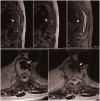
Case report of a rare giant bone island in a vertebral body combined with hemangioma
- PMID: 33983052
- PMCID: PMC8127800
- DOI: 10.1177/03000605211010699
Case report of a rare giant bone island in a vertebral body combined with hemangioma
Abstract
This case report describes a rare giant bone island combined with hemangioma diagnosed in a patient with osteolytic vertebral metastases. The bone island's greatest diameter was 3.15 cm, and bone islands of this size are rare in the literature. This article aims to provide clinicians with information about the diagnosis and relevant literature of bone islands.
Keywords: Bone island; case report; hemangioma; osteoblastic metastases; radiographic images; spine.
Conflict of interest statement
Figures


References
-
- Park HS, Kim JR, Lee SY, et al.. Symptomatic giant (10-cm) bone island of the tibia. Skeletal Radiol 2005; 34: 347–350. - PubMed
-
- Onitsuka H. Roentgenologic aspects of bone islands. Radiology 1977; 123: 607–612. - PubMed
-
- Smith J. Giant bone islands. Radiology 1973; 107: 35–36. - PubMed
-
- Gold RH, Mirra JM, Remotti F, et al.. Case report 527: giant bone island of tibia. Skeletal Radiol 1989; 18: 129–132. - PubMed
-
- Hwang YJ. Follow-up CT and MR findings of osteoblastic spinal metastatic lesions after stereotactic radiotherapy. Jpn J Radiol 2012; 30: 492–498. - PubMed
Publication types
MeSH terms
LinkOut - more resources
Full Text Sources
Other Literature Sources
Medical

