. 2021 May 14;128(10):1533-1553.
doi: 10.1161/CIRCRESAHA.121.318346. Epub 2021 May 13.

Molecular Genetic Basis of Hypertrophic Cardiomyopathy

Affiliations

Molecular Genetic Basis of Hypertrophic Cardiomyopathy

A J Marian. Circ Res. .

Abstract

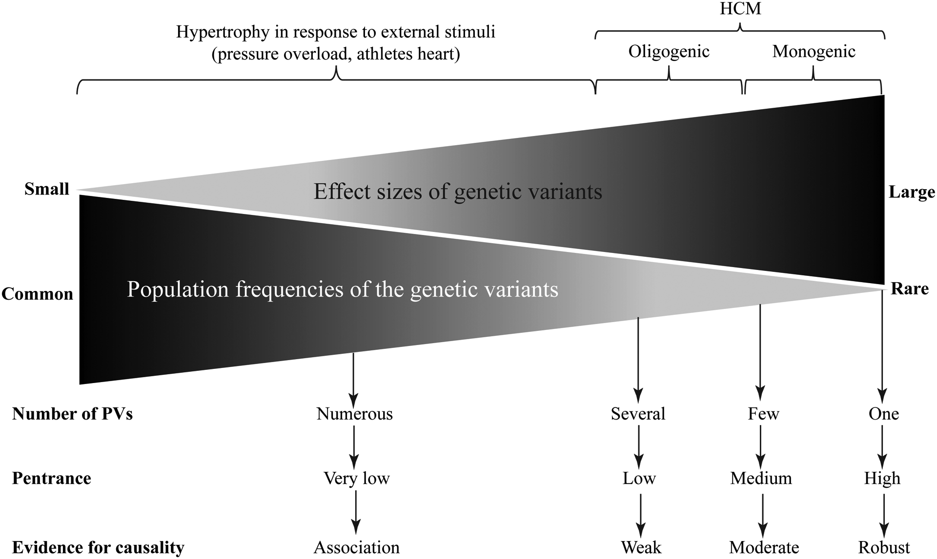
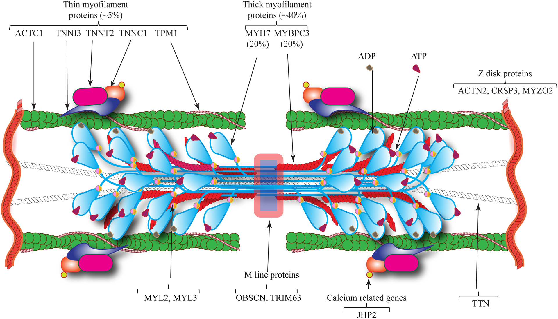
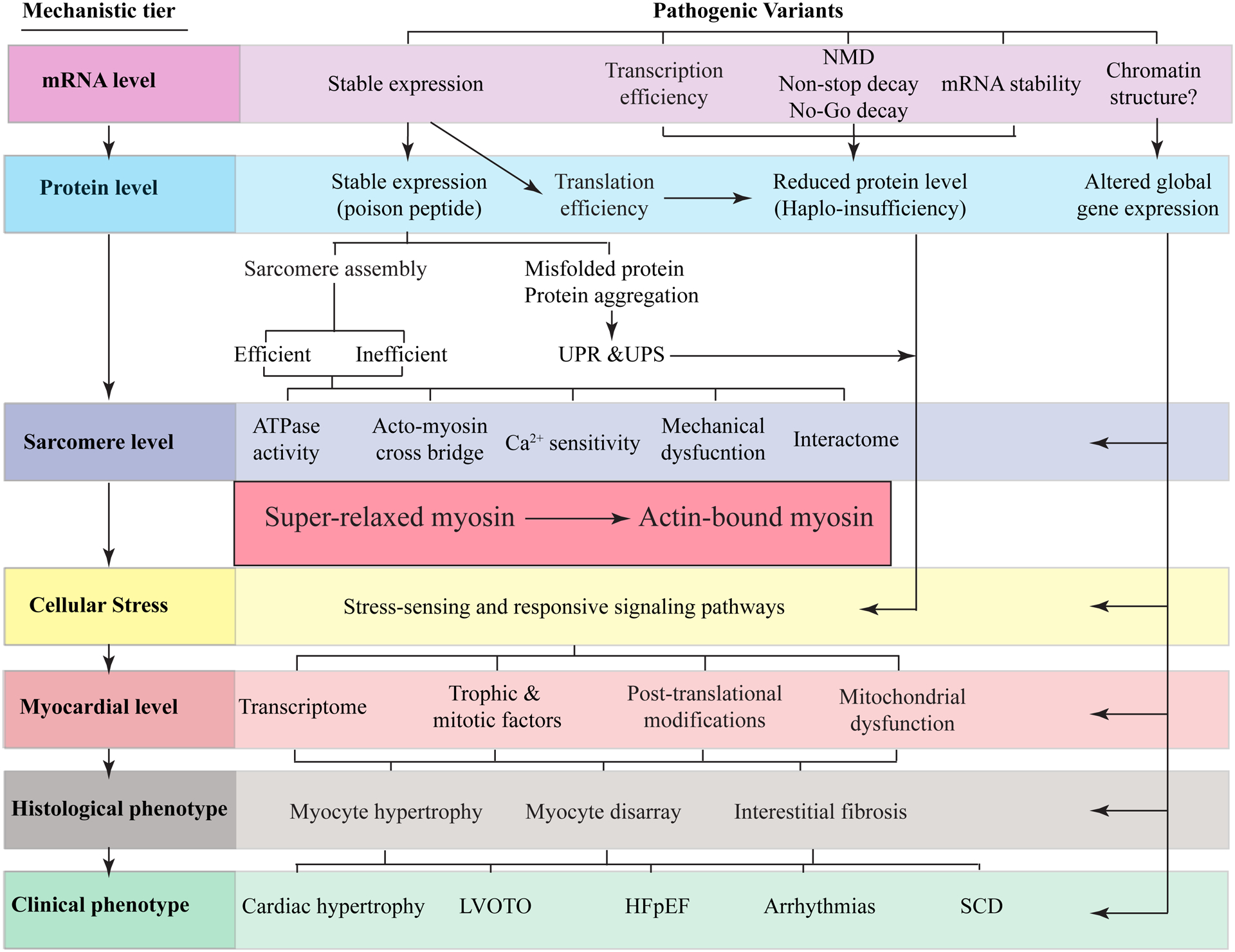
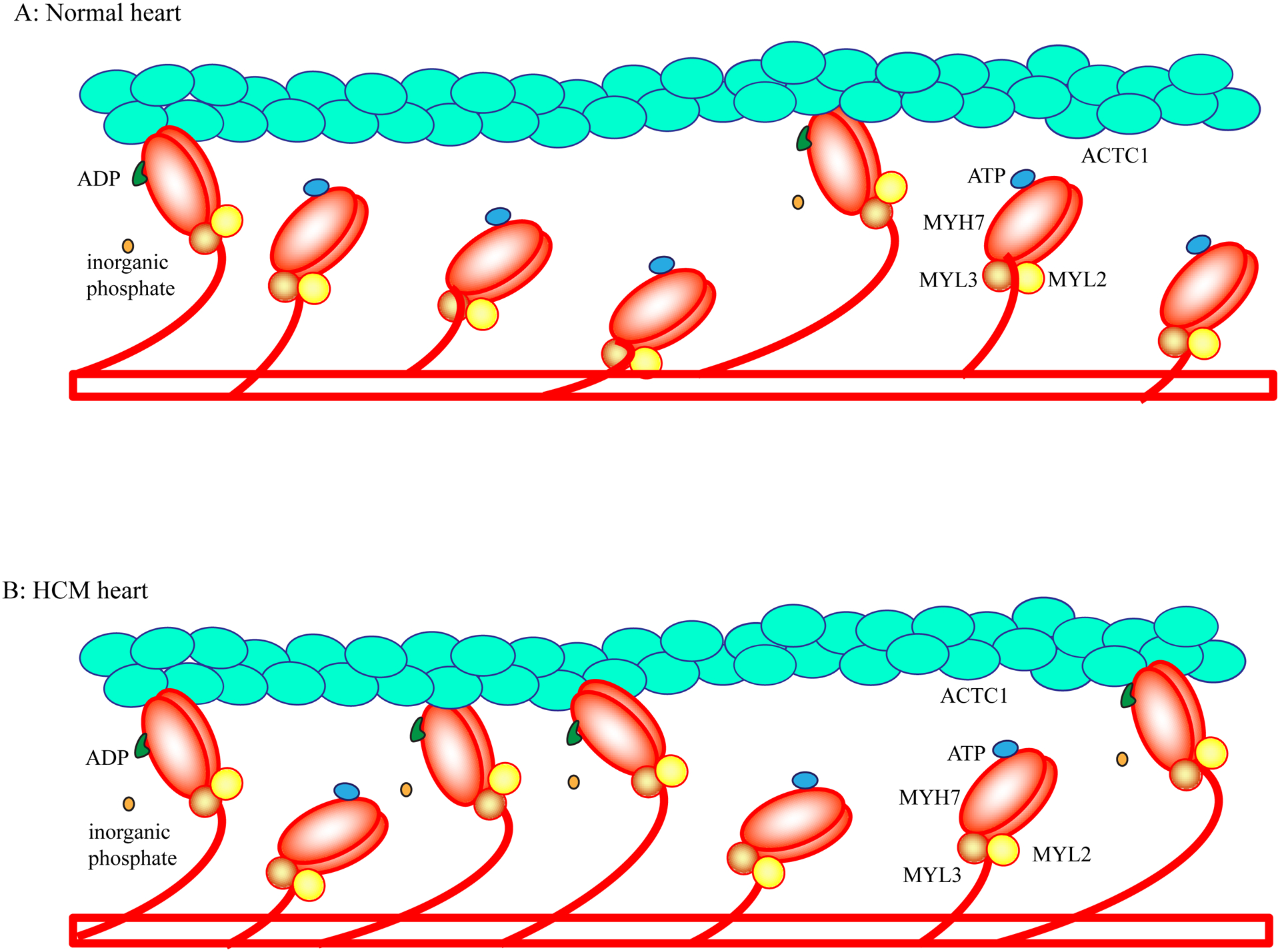
Hypertrophic cardiomyopathy (HCM) is a genetic disease of the myocardium characterized by a hypertrophic left ventricle with a preserved or increased ejection fraction. Cardiac hypertrophy is often asymmetrical, which is associated with left ventricular outflow tract obstruction. Myocyte hypertrophy, disarray, and myocardial fibrosis constitute the histological features of HCM. HCM is a relatively benign disease but an important cause of sudden cardiac death in the young and heart failure in the elderly. Pathogenic variants (PVs) in genes encoding protein constituents of the sarcomeres are the main causes of HCM. PVs exhibit a gradient of effect sizes, as reflected in their penetrance and variable phenotypic expression of HCM. MYH7 and MYBPC3, encoding β-myosin heavy chain and myosin binding protein C, respectively, are the two most common causal genes and responsible for ≈40% of all HCM cases but a higher percentage of HCM in large families. PVs in genes encoding protein components of the thin filaments are responsible for ≈5% of the HCM cases. Whereas pathogenicity of the genetic variants in large families has been firmly established, ascertainment causality of the PVs in small families and sporadic cases is challenging. In the latter category, PVs are best considered as probabilistic determinants of HCM. Deciphering the genetic basis of HCM has enabled routine genetic testing and has partially elucidated the underpinning mechanism of HCM as increased number of the myosin molecules that are strongly bound to actin. The discoveries have led to the development of mavacamten that targets binding of the myosin molecule to actin filaments and imparts beneficial clinical effects. In the coming years, the yield of the genetic testing is expected to be improved and the so-called missing causal gene be identified. The advances are also expected to enable development of additional specific therapies and editing of the mutations in HCM.

Keywords: death, sudden, cardiac; genetics; heart failure; hypertrophy; mutation.


Conflict of interest statement

DISCLOSURE/CONFLICT OF INTEREST

None

Figures

Figure 1.
Figure 1.. Gradient of effect sizes of the pathogenic variants (PVs) and their population frequencies in the context of familial and sporadic HCM
A schematic presentation of the effect sizes of the PVs that vary from small to large. The effect size typically inversely correlates with the population frequency of the variant. Rare variants with large effect sizes are more common in familial HCM and those with moderate effect sizes are often found in the sporadic cases and in small families with HCM.
Figure 2.
Figure 2.. Schematic illustration of sarcomere proteins involved in HCM.
HCM is mainly a disease of sarcomere proteins, which are comprised of thick and thin filaments. Thick filaments are mainly composed of myosin heavy chain 7 (MYH7) and myosin binding protein C3 (MYBPC3), which are the most commonly affected proteins by mutations causing HCM. Thin filaments are comprised of cardiac α actin 1 (ACTC1), troponin complex (TNNT2, TNNI3, and TNNC1) and α tropomyosin (TPM1), which are affected by mutations in about 5% of the HCM cases. Examples of the Z and M line proteins implicated in HCM are also illustrated, which are rare causes of HCM.
Figure 3.
Figure 3.. Pathogenesis of HCM.
The primary defect in HCM is the mutation in the sarcomere proteins, composed of thick and thin filaments, M line and the Z disks (or lines). The effects of the mutations on mRNA, protein, sarcomere, myocyte, myocardium (molecular level), histology, and clinical phenotypes are depicted as sequential layers. A change in the amino acid sequence in the sarcomere protein (dominant-negative effect) or the deficiency of a sarcomere protein (haplo-insufficiency) instigates a series of proximal functional defects in cardiac sarcomeres, such as altered calcium sensitivity and ATPase activity. A consequence of these initial defects is a shift in the number of myosin molecules in the super-relaxed state with a low ATPase activity toward the myosin molecules that are in strong bound state with the actin molecule and have a high ATPase activity. The changes activate expression of a stress-sensing (mechanical, biochemical, and energetics) intermediary molecular phenotypes, such as altered transcriptomics and expression of trophic and mitotic factors. The latter set of the molecular changes induce histological and morphological changes in the myocardium, such as myocyte hypertrophy and fibrosis, which collectively lead to the clinical phenotypes, such as cardiac arrhythmias and heart failure.
Figure 4.
Figure 4.. Impaired acto-myosin interaction in the pathogenesis of HCM
Binding of ATP to the globular head of myosin results in its dissociation from the thin filament actin, whereas hydrolysis of ATP to ADP and inorganic phosphate leads to association of the MYH7 and ACTC1 molecules. Release of the inorganic phosphate induces conformational changes in the MYH7 and flexion of the myosin globular head at lever arm over ACTC1 in the thin filament. The flexion displaces the thin filament by about 10 nm. In the normal heart, at any given moment during diastole, only a small fraction of the MYH7 molecules (estimated to be about 10%) is bound to the actin (ACTC1) molecule. Approximately 50–60% of the myosin molecules are totally dissociated from the actin molecule and are in the super-relaxed state. The remaining myosin molecules are weakly associated with the actin filaments (Panel A). In HCM caused by mutations in the MYH7 and MYBPC3 genes, there is a shift from myosin molecules in the super-relax state to the myosin molecules in the strong bound state with the actin molecule (Panel B). The increased number of acto-myosin complex (strong bound state) is responsible for increased contractility and impaired relaxation.

References

    1. Marian AJ, Braunwald E. Hypertrophic cardiomyopathy: Genetics, pathogenesis, clinical manifestations, diagnosis, and therapy. Circ Res. 2017;121:749–770 - PMC - PubMed
    1. Braunwald E Hypertrophic cardiomyopathy: The first century 1869–1969. Glob Cardiol Sci Pract. 2012;2012:5. - PMC - PubMed
    1. Dahm R Friedrich miescher and the discovery of DNA. Dev Biol. 2005;278:274–288 - PubMed
    1. Evans W Familial cardiomegaly. Br Heart J. 1949;11:68–82 - PMC - PubMed
    1. Teare D Asymmetrical hypertrophy of the heart in young adults. Br Heart J. 1958;20:1–8 - PMC - PubMed

Publication types

MeSH terms