Review
. 2021 May 14;128(10):1451-1467.
doi: 10.1161/CIRCRESAHA.121.318159. Epub 2021 May 13.

From Systemic Inflammation to Myocardial Fibrosis: The Heart Failure With Preserved Ejection Fraction Paradigm Revisited

Affiliations
Review

From Systemic Inflammation to Myocardial Fibrosis: The Heart Failure With Preserved Ejection Fraction Paradigm Revisited

Walter J Paulus et al. Circ Res. .

Abstract

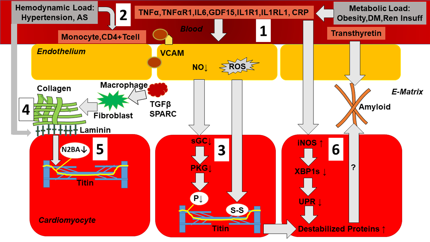
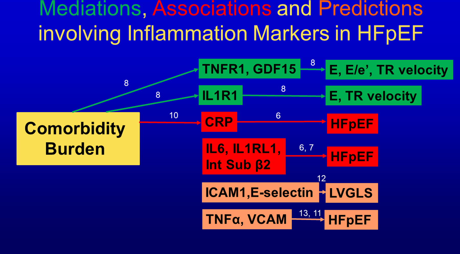
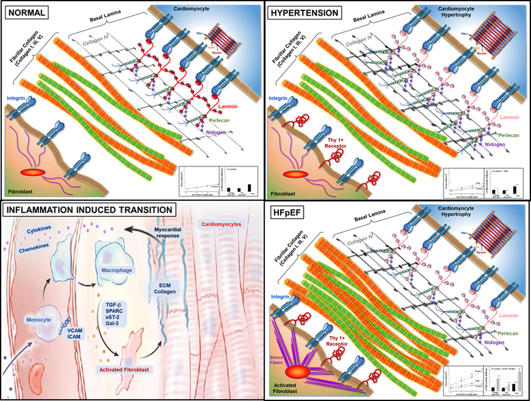
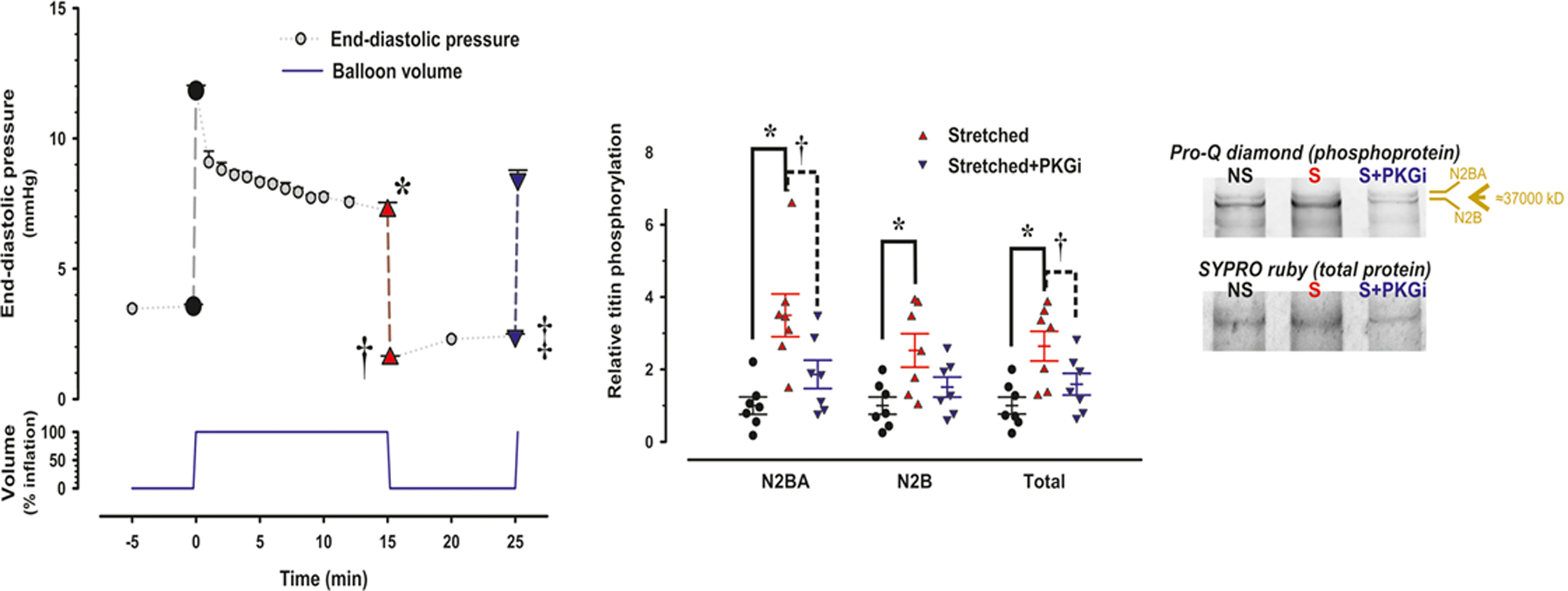
In accordance with the comorbidity-inflammation paradigm, comorbidities and especially metabolic comorbidities are presumed to drive development and severity of heart failure with preserved ejection fraction through a cascade of events ranging from systemic inflammation to myocardial fibrosis. Recently, novel experimental and clinical evidence emerged, which strengthens the validity of the inflammatory/profibrotic paradigm. This evidence consists among others of (1) myocardial infiltration by immunocompetent cells not only because of an obesity-induced metabolic load but also because of an arterial hypertension-induced hemodynamic load. The latter is sensed by components of the extracellular matrix like basal laminin, which also interact with cardiomyocyte titin; (2) expression in cardiomyocytes of inducible nitric oxide synthase because of circulating proinflammatory cytokines. This results in myocardial accumulation of degraded proteins because of a failing unfolded protein response; (3) definition by machine learning algorithms of phenogroups of patients with heart failure with preserved ejection fraction with a distinct inflammatory/profibrotic signature; (4) direct coupling in mediation analysis between comorbidities, inflammatory biomarkers, and deranged myocardial structure/function with endothelial expression of adhesion molecules already apparent in early preclinical heart failure with preserved ejection fraction (HF stage A, B). This new evidence paves the road for future heart failure with preserved ejection fraction treatments such as biologicals directed against inflammatory cytokines, stimulation of protein ubiquitylation with phosphodiesterase 1 inhibitors, correction of titin stiffness through natriuretic peptide-particulate guanylyl cyclase-PDE9 (phosphodiesterase 9) signaling and molecular/cellular regulatory mechanisms that control myocardial fibrosis.

Keywords: biomarker; fibrosis; heart failure; inflammation; obesity.


Figures

Figure 1
Figure 1. Pathophysiological mechanisms linking systemic inflammation to myocardial stiffness.
1) Metabolic load induces proinflammatory signaling: Metabolic load related to obesity, diabetes mellitus (DM) and renal insufficiency (Ren Insuff) triggers systemic inflammation evident from raised plasma levels of TNFα (Tumor Necrosis Factor α), TNFαR1 (Tumor Necrosis Factor α Receptor 1), IL6 (Interleukin 6), GDF15 (Growth Differentiation Factor 15), IL1R1 (Interleukin 1 Receptor 1), IL1RL1 (Interleukin 1 Receptor Like 1) and CRP (C-Reactive Protein). Systemic inflammation triggers endothelial expression of adhesion molecules (VCAM: Vascular Cell Adhesion Molecule), which attracts monocytes, lowers endothelial production of nitric oxide (NO) and raises endothelial production of Reactive Oxygen Species (ROS); 2) Hemodynamic load as occurs in arterial hypertension and aortic stenosis induces proinflammatory and fibrotic signaling evident from myocardial infiltration of monocytes and CD4+ T cells; 3) Low NO reduces activity of soluble Guanylyl Cyclase (sGC) and Protein Kinase G (PKG). This leads to hypophosphorylation of titin (P↓). ROS cause formation of disulfide bonds within titin. Both these titin modifications raise cardiomyocyte stiffness; 4) Myocardial collagen homeostasis: Infiltrating monocytes become macrophages with production of Transforming Growth Factor β (TGFβ) and secreted protein acidic and rich in cysteine (SPARC) which stimulate collagen production by fibroblasts; 5) Crosstalk between hemodynamic load, extracellular matrix basal laminin and cardiomyocyte titin results in changed titin isoform expression with less N2BA isoform (N2BA↓); 6) Myocardial accumulation of degraded proteins: Expression in cardiomyocytes of inducible Nitric Oxide Synthase (iNOS) lowers inositol-requiring enzyme1α (IRE1α), spliced X-box binding protein1 (XBP1s) and the Unfolded Protein Response (UPR). The latter leads to build-up of destabilized proteins which could potentially also accumulate in the extracellular matrix as occurs in transthyretin amyloidosis. (Illustration credit: Ben Smith)
Figure 2
Figure 2. Mediations, associations and predictions involving inflammation markers in HFpEF.
Mediations (Green) between comorbidity burden, inflammatory biomarkers and echocardiographic cardiac function (TNFR1: Tumor Necrosis Factor Receptor 1; GDF15: Growth Differentiation Factor 15; IL1R1: Interleukin 1 Receptor 1; E: Early diastolic mitral flow velocity; E/e’: ratio of Early diastolic mitral flow velocity over Early diastolic long axis lengthening velocity; TR: Tricuspid Regurgitation velocity). Associations (Red) between inflammatory biomarkers and presence of HFpEF (CRP: C-Reactive Protein; IL6: Interleukin 6; IL1RL1: Interleukin 1 Receptor Like 1; Int Sub β 2: Integrin Subunit Beta 2). Biomarkers predicting (Orange) myocardial function or HFpEF (ICAM1: Intercellular Adhesion Molecule 1; TNFα: Tumor Necrosis Factor α; VCAM: Vascular Cell Adhesion Molecule; LVGLS: Left Ventricular Global Longitudinal Strain). Numbers indicate corresponding reference.
Figure 3
Figure 3. Hemodynamic load-induced myocardial inflammatory/fibrotic signaling.
Sequential changes in myocardial cardiomyocytes, fibroblasts, fibrillar collagen, and basal lamina structures result from the imposition of increased hemodynamic load that lead to the development of HFpEF. (Illustration credit: Ben Smith). Compared to Panel A, showing normal myocardium, Panel B depicts changes that result from increased hemodynamic load, such as that which occurs in systemic arterial hypertension. The change in load is sensed by cardiomyocytes, fibroblasts and resident macrophages and leads to alterations in the basal lamina structures. Basal lamina changes include changes in laminin isoform to a more compliant form, with increases in perlican, nitogen and collagen IV which may compliment and compensate for these changes in laminin. Cardiomyocytes undergo parallel addition of sarcomeres and increased cross-sectional area; these cellular changes lead to concentric LV hypertrophy. However, these changes in myocardial structure does not result in increased myocardial diastolic stiffness (inset). Panel C depicts the inflammation induced transition from hypertensive heart disease to HFpEF. This transition is led by proinflammatory signaling of increased cytokines and chemokines causing cell recruitment to the myocardium of macrophages and T/B cells. These cytokines and chemokines are secreted by the myocardium and enter the circulation. Circulating monocytes, both from the bone marrow and the spleen, migrate to myocardial endothelial cell surfaces, with attachment and extravasation into the interstitial space facilitated by vascular cell adhesion proteins and become activated macrophages. These macrophages both secrete matricellular proteins that facilitate procollagen processing and collagen fiber assembly and may further lead to fibroblasts activation. Panel D shows the results of this transition in HFpEF and the aggregate profibrotic changes in interstitial ECM and their resultant increase in myocardial diastolic stiffness (inset).
Figure 4.
Figure 4.. Stretch-induced titin phosporylation.
In a Langendorff preparation an acute volume load causes a brisk rise in LV end-diastolic pressure (LVEDP), which gradually falls over a 15 minute period. Subsequent removal of the volume load causes LVEDP to drop below baseline without full recovery over the next 10 minutes period (Left hand panel; *,†,‡: p<0.001). In stretched muscle strips, titin phosphorylation (total and isoform specific) was signficantly higher than in non-stretched muscle strips (Middle panel) as also evident from Pro-Q diamond staining (Right hand panel). Stretch induced effects disappeared following inhibition of PKG (PKGi) and were absent in hypertrophied myocardium.
Figure 5.
Figure 5.. Molecular and cellular processes that control collagen homeostasis.
Panel A: Myocardial fibroblasts synthesize and secrete procollagen into the extracellular matrix (ECM) stimulated by sST-2, galectin-3 (gal-3), aldosterone (Aldo) and other proinflammatory and profibrotic factors (green dashed box). Panel B: Procollagen is processed in the ECM chaperoned by matricellular proteins (such as SPARC, periostin and thrombospondin) through sequential steps that remove c-terminal by bone morphometric peptide 1 (BMP-1) and n-terminal propeptides by ADAM-TS2. Plasma/serum concentrations of the resulting propeptides (PICP, PIIICP, PINP,PIIINP) reflect collagen synthesis rate. Panel C: Collagen cross-linking finalizes processing into mature insoluble collagen under the influence of lysyl oxidase (LOX), advanced glycation end products (AGE) and transglutaminase (TG). Panel D: Collagen degradation by matrix metalloproteinases (MMPs) into telopeptides (CITP) occurs under regulation of the endogenous tissue inhibitors of MMPs (TIMPs). Each of the biomarkers within the green and purple dashed boxes can be measured in the circulation. (Illustration credit: Ben Smith).
Figure 6.
Figure 6.. Prognostic value of biomarker data from PARAGON-HF Study.
Biomarkers that reflect mechanisms that increase procollagen synthesis, such as soluble ST2, are increased in HFpEF patients; biomarkers that reflect mechanisms that decrease collagen degradation, such as increased tissue inhibitor of matrix metalloproteinase-1 (TIMP-1) are increased in HFpEF patients. Both baseline and change from baseline levels of these profibrotic biomarkers provide prognostic value. Continuous relationships of TIMP-1 and sST2 baseline values and 16-week change from baseline values with incidence of subsequent heart failure (HF) hospitalization and cardiovascular (CV) death are plotted. (A, B) The x-axis and histogram represent plasma biomarker level at baseline. The solid line represents estimated incidence rate of the primary endpoint, total HF hospitalizations and CV death. The dashed lines represent 95% confidence intervals for the estimated incidence rate. Incidence rate is displayed on the primary (left-sided) y-axis. (C, D) The x-axis and histogram represent change in biomarker level between pre-run-in baseline visit and the week 16 visit. The solid line represents estimated incidence rate of the primary endpoint, total HF hospitalizations and CV death, that occurred after 16 weeks, relative to patients with no change in biomarker level, adjusted for log-transformed baseline value. The dashed lines represent 95% confidence intervals for the estimated incidence rate. Incidence rate ratio is displayed on the primary (left-sided) y-axis. The higher the baseline value of TIMP-1 and sST2, the higher the rate of HF hospitalization and CV mortality. Over 16 weeks follow-up if TIMP-1 or sST2 decreased, the primary endpoints decreased.
Figure 7.
Figure 7.. Compartimentalization and counterregulation of cardiomyocyte cGMP stimulation
Cyclic guanosine monophosphate (cGMP) produced by soluble guanylyl cyclase (sGC) from guanosine triphosphate (GTP) is mainly localized in sarcomeres around Z discs and preferably degraded to guanosine monophosphate (GMP) by phosphodiesterase 5 (PDE5). PDE5 activity rises when cGMP is elevated, thus providing a counterregulatory feedback. cGMP produced by particulate guanylyl cyclase (pGC) is mainly localized around myofilamentary proteins like titin and preferably degraded by PDE9. PDE9 lacks a cGMP sensitive regulatory site and is therefore not subject to counterregulation by cGMP.

References

    1. Shah SJ, Borlaug BA, Kitzman DW, McCulloch AD, Blaxall BC, Agarwal R, Chirinos JA, Collins S, Deo RC, Gladwin MT et al. Research Priorities for Heart Failure With Preserved Ejection Fraction: National Heart, Lung, and Blood Institute Working Group Summary. Circulation. 2020;141:1001–1026. - PMC - PubMed
    1. Vasan RS, Xanthakis V, Lyass A, Andersson C, Tsao C, Cheng S, Aragam J, Benjamin EJ, Larson MG. Epidemiology of Left Ventricular Systolic Dysfunction and Heart Failure in the Framingham Study: An Echocardiographic Study Over 3 Decades. JACC Cardiovasc Imaging. 2018;11:1–11. - PMC - PubMed
    1. Shah SJ, Kitzman DW, Borlaug BA, van Heerebeek L, Zile MR, Kass DA, Paulus WJ. Phenotype-Specific Treatment of Heart Failure With Preserved Ejection Fraction: A Multiorgan Roadmap. Circulation. 2016;134:73–90. - PMC - PubMed
    1. Borlaug BA. Evaluation and management of heart failure with preserved ejection fraction. Nat Rev Cardiol. 2020;17:559–573. - PubMed
    1. Santhanakrishnan R, Chong JP, Ng TP, Ling LH, Sim D, Leong KT, Yeo PS, Ong HY, Jaufeerally F, Wong R et al. Growth differentiation factor 15, ST2, high-sensitivity troponin T, and N-terminal pro brain natriuretic peptide in heart failure with preserved vs. reduced ejection fraction. Eur J Heart Fail 2012;14:1338–1347. - PubMed

Publication types

MeSH terms