Review
. 2021 May 14;128(10):1576-1593.
doi: 10.1161/CIRCRESAHA.121.318223. Epub 2021 May 13.

Heart Failure With Targeted Cancer Therapies: Mechanisms and Cardioprotection

Affiliations
Review

Heart Failure With Targeted Cancer Therapies: Mechanisms and Cardioprotection

Virginia S Hahn et al. Circ Res. .

Abstract

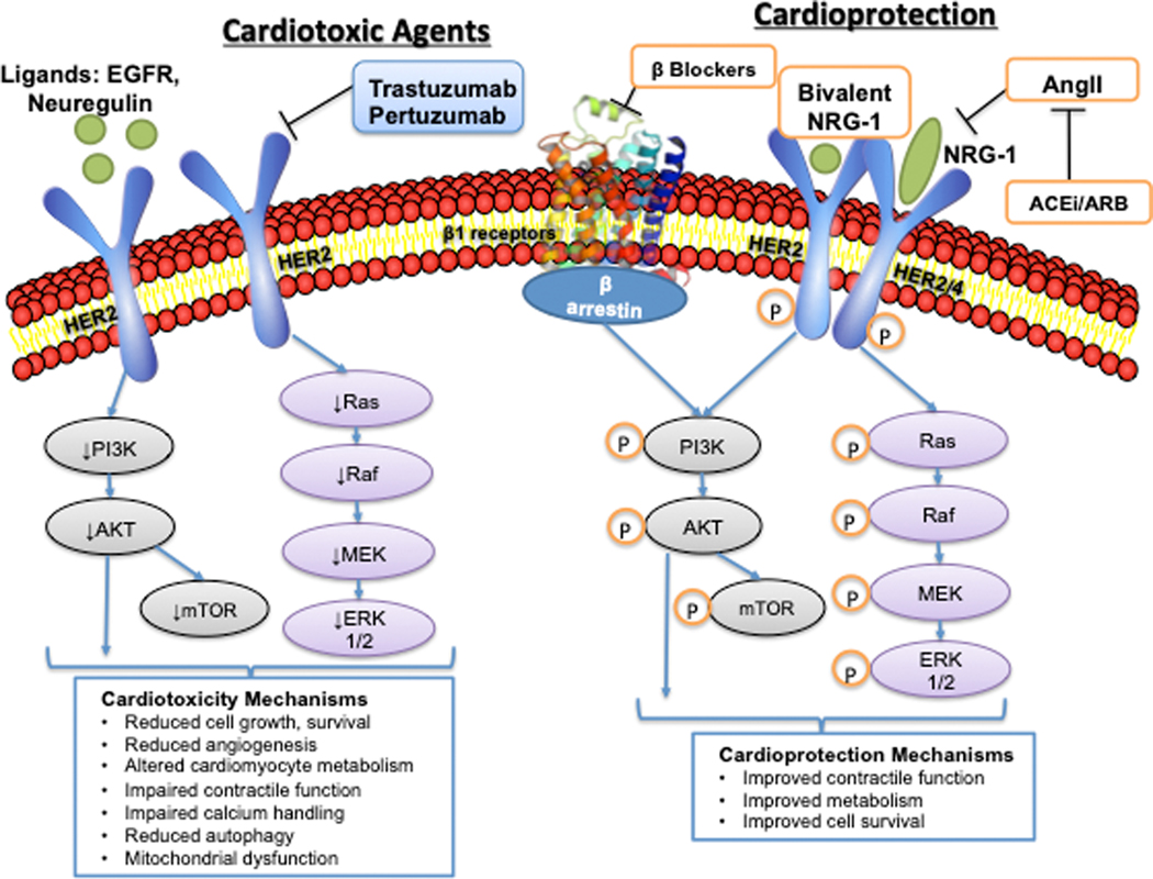
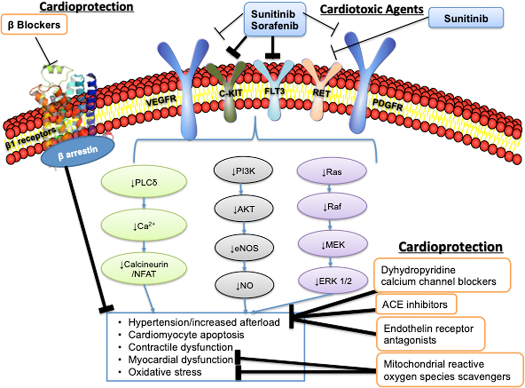
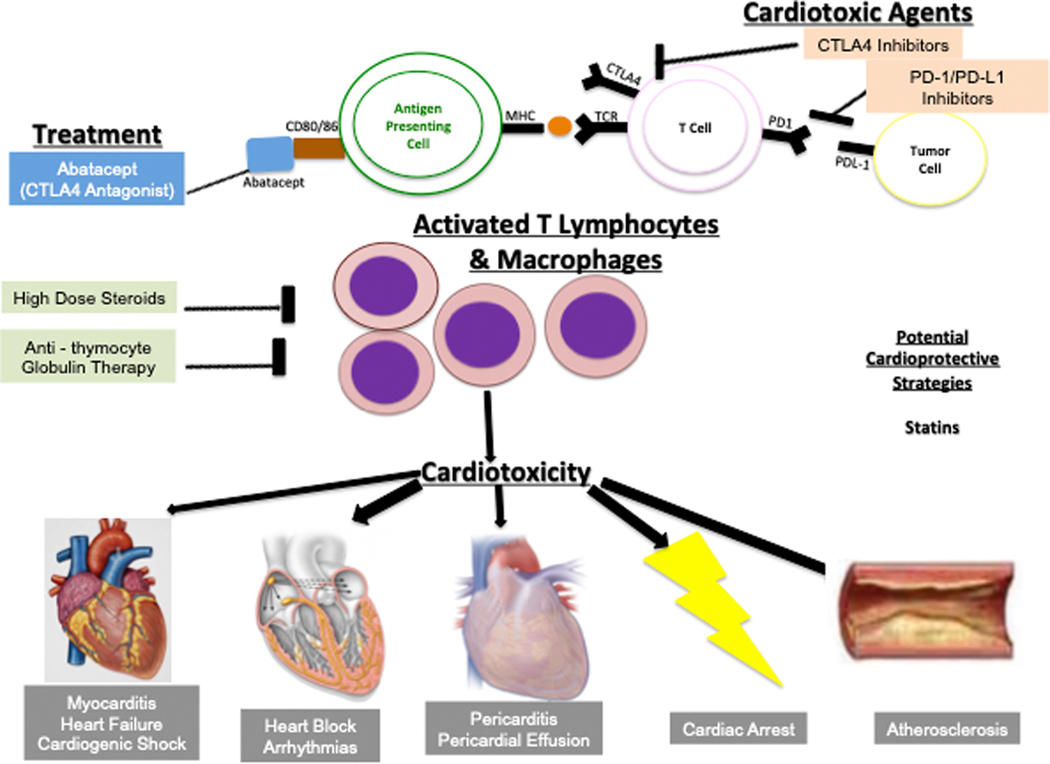
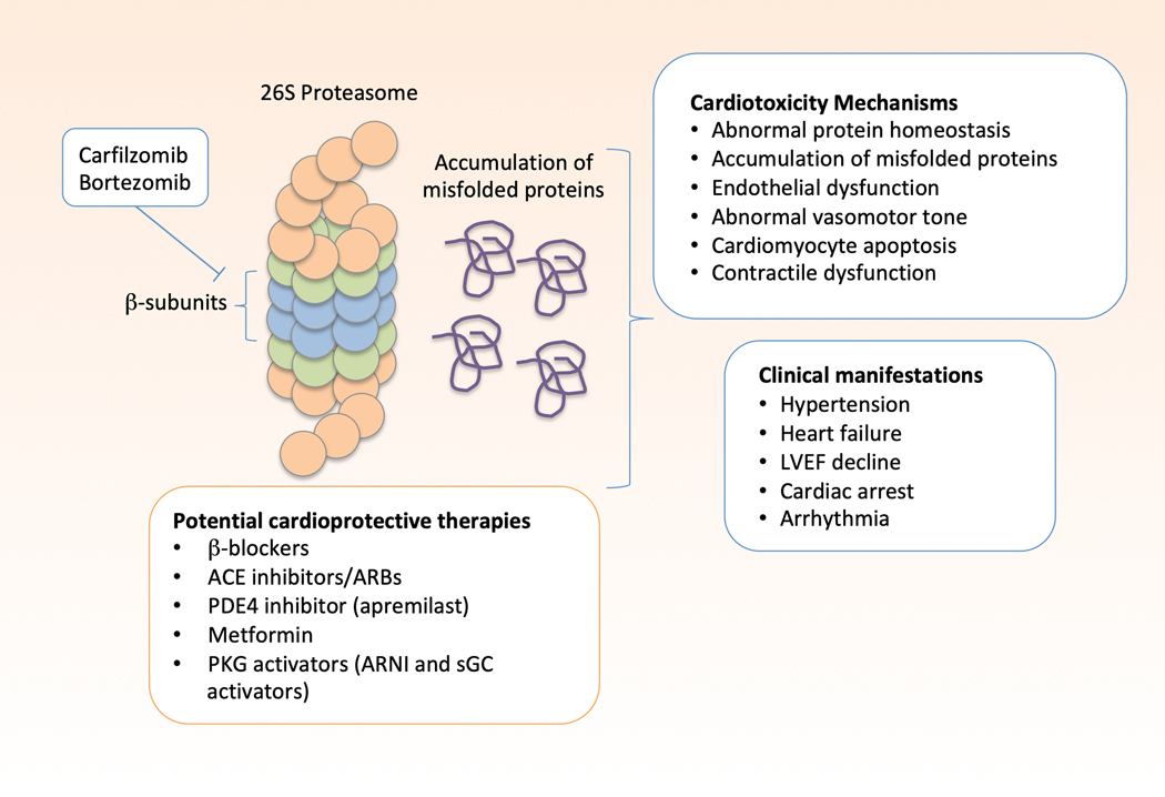
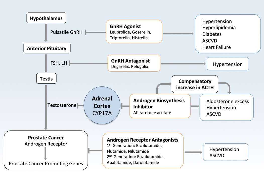
Oncology has seen growing use of newly developed targeted therapies. Although this has resulted in dramatic improvements in progression-free and overall survival, challenges in the management of toxicities related to longer-term treatment of these therapies have also become evident. Although a targeted approach often exploits the differences between cancer cells and noncancer cells, overlap in signaling pathways necessary for the maintenance of function and survival in multiple cell types has resulted in systemic toxicities. In particular, cardiovascular toxicities are of important concern. In this review, we highlight several targeted therapies commonly used across a variety of cancer types, including HER2 (human epidermal growth factor receptor 2)+ targeted therapies, tyrosine kinase inhibitors, immune checkpoint inhibitors, proteasome inhibitors, androgen deprivation therapies, and MEK (mitogen-activated protein kinase kinase)/BRAF (v-raf murine sarcoma viral oncogene homolog B) inhibitors. We present the oncological indications, heart failure incidence, hypothesized mechanisms of cardiotoxicity, and potential mechanistic rationale for specific cardioprotective strategies.

Keywords: cardiotoxicity; heart failure; incidence; survival; tyrosine.


Figures

Figure 1:
Figure 1:
Human Epidermal Growth Factor Receptor Targeted Therapies: Mechanisms of Cardiotoxicity and Potential Cardioprotective Mechanisms. Left side: Human epidermal growth factor receptor 2 (HER2) is a transmembrane tyrosine kinase that dimerizes with other members of the epidermal growth factor receptor family to activate downstream signaling that promotes cell survival and proliferation. Ligands of HER2 include epidermal growth factor (EGF) and neuregulin-1 (NRG-1). Trastuzumab and pertuzumab inhibit HER2 signaling, leading to downstream downregulation of PI3K/AKT, mTOR, and Ras/RAF/MEK/ERK signaling. In the cardiovascular system this leads to reduced growth and survival of cardiomyocytes, altered metabolism, reduced angiogenesis, altered calcium handling, reduced autophagy, and mitochondrial dysfunction. Right side: Potential cardioprotective mechanisms include beta-blockers that transactivate β-arrestin, promoting PI3K/Akt signaling in the cardiomyocyte. Additional cardioprotective mechanisms include ACE inhibitors/ARBs that reduce angiotensin-II mediated downregulation of NRG-1, and bivalent NRG-1. HER2- human epidermal growth factor receptor 2; EGF- epidermal growth factor; NRG-1- Neuregulin-1; PI3K- phosphoinositide 3-kinase; Akt- Ak transforming factor; mTOR- mammalian target of rapamycin; RAS- Ras GTPase; RAF- Raf-1; MEK- Ras/Raf/mitogen-activated-protein kinase kinase; ERK- extracellular signal-regulated kinase; ACEi- angiotensin converting enzyme inhibitor; ARB- angiotensin II receptor blocker (Illustration credit: Ben Smith).
Figure 2:
Figure 2:
Vascular Endothelial Growth Factor Signaling Pathway Inhibitors: Mechanisms of Cardiotoxicity and Potential Cardioprotective Mechanisms. Sunitinib and sorafenib are the most commonly used targeted VEGF pathway signaling inhibitors. They both target multiple tyrosine kinase receptors including the VEGFR, PDGFR, cKit, FLT3, and sunitinib targets RET. Inhibition of these receptors leads to downregulation of pro-survival signaling and angiogenesis, with additional effects on nitric oxide (NO) and calcineurin/NFAT. This leads to hypertension, cardiomyocyte apoptosis, contractile dysfunction, mitochondrial dysfunction, and oxidative stress. Potential cardioprotective mechanisms include dihydropyridine calcium channel blockers, ACE inhibitors, endothelin receptor antagonists, and scavengers of mitochondrial ROS. VEGFR- vascular endothelial growth factor receptor receptor; PDGFR- platelet derived growth factor receptor; FLT3- fms like tyrosine kinase 3; PLCgamma- phospholipase C gamma; NFAT- nuclear factor of activated T cells; PI3K- phosphoinositide 3-kinase; Akt- Ak transforming factor; eNOS- endothelial nitric oxide synthase; NO- nitric oxide; RAS- Ras GTPase; RAF- Raf-1; MEK- Ras/Raf/mitogen-activated-protein kinase kinase; ERK- extracellular signal-regulated kinase; ACEi- angiotensin converting enzyme inhibitor. (Illustration credit: Ben Smith).
Figure 3:
Figure 3:
Immune Checkpoint Inhibitors: Mechanisms of Cardiotoxicity and Potential Treatments. Inhibitors of CTLA-4 and PD-1/PD-L1 are designed to activate anti-tumor T lymphocytes to harness the immune system to attack cancer cells. This leads to several autoimmune off-target effects including cardiotoxicity affecting the myocardium, pericardium, and conduction system. Immune checkpoint inhibitors can cause myocarditis, heart failure, cardiogenic shock, heart block, arrhythmias, pericarditis, pericardial effusion, cardiac arrest, and accelerated atherosclerosis. Potential treatment options include high dose steroids, abatacept (a CTLA-4 agonist), and anti-thymocyte globulin, while statins may be cardioprotective via pleiotropic effects. CTLA-4- cytotoxic T lymphocyte associated antigen 4; PD-1- programmed death 1; PD-L1- programmed death 1 ligand; TCR- T cell receptor; MHC- major histocompatibility complex. (Illustration credit: Ben Smith)
Figure 4:
Figure 4:
Proteasome Inhibitors: Mechanisms of Cardiotoxicity and Potential Treatments. Carfilzomib and bortezomib inhibit the catalytically active beta-subunits of the 26S proteasome, impairing its ability to break down and recycle misfolded proteins. This leads to the accumulation of misfolded proteins and cellular proteotoxicity. This leads to abnormal protein homeostasis, accumulation of misfolded proteins, endothelial dysfunction, abnormal vasomotor tone, cardiomyocyte apoptosis, and contractile dysfunction. Clinical manifestations of proteasome inhibitor cardiotoxicity include hypertension, heart failure with preserved or reduced ejection fraction, left ventricular ejection fraction decline, arrhythmias, and cardiac arrest. Activators of protein kinase G (PKG) may counter these effects by activating the proteasome. Other potentially cardioprotective therapies include beta-blockers, ACE inhibitors, ARBs, apremilast, and metformin. PKG-protein kinase G; ARNI- angiotensin receptor-neprilysin inhibitor; sGC-soluble guanylate cyclase; ACEi- angiotensin converting enzyme inhibitor; ARB- angiotensin II receptor blocker; PDE4-phosphodiesterase 4; LVEF, left ventricular ejection fraction
Figure 5:
Figure 5:
Androgen Deprivation Therapy and Androgen Receptor Signaling Inhibitors. GnRH agonist and GnRH agonists lead to increased risk of cardiometabolic traits of diabetes, hypertension, hyperlipidemia, and ultimately atherosclerotic cardio- and cerebrovascular disease. Abiraterone inhibits the CYP17A enzyme that leads to conversion of pregnenolone to 17-hydroxyl-pregnenalone, and then to dehydroepiandrosterone (DHEA), ultimately leading to reduced estrogen and testosterone levels. This also leads to accumulation of pregnenolone, which is ultimately converted to aldosterone, contributing to the mineralocorticoid excess seen with abiraterone. Direct androgen receptor antagonists such as enzalutamide, do not have this mineralocorticoid excess, but can still lead to hypertension. Androgen receptor signaling inhibitors also increase the risk of atherosclerotic cardio- and cerebrovascular events. GnRH-gonadotropin-releasing hormone; FSH- follicle-stimulating hormone; LH- luteinizing hormone; Gen- generation

References

    1. Cardinale D, Colombo A, Bacchiani G, Tedeschi I, Meroni CA, Veglia F, Civelli M, Lamantia G, Colombo N, Curigliano G, Fiorentini C and Cipolla CM. Early detection of anthracycline cardiotoxicity and improvement with heart failure therapy. Circulation. 2015;131:1981–8. - PubMed
    1. Narayan HK, Finkelman B, French B, Plappert T, Hyman D, Smith AM, Margulies KB and Ky B. Detailed Echocardiographic Phenotyping in Breast Cancer Patients: Associations With Ejection Fraction Decline, Recovery, and Heart Failure Symptoms Over 3 Years of Follow-Up. Circulation. 2017;135:1397–1412. - PMC - PubMed
    1. Hahn VS, Lenihan DJ and Ky B. Cancer therapy-induced cardiotoxicity: basic mechanisms and potential cardioprotective therapies. J Am Heart Assoc. 2014;3:e000665. - PMC - PubMed
    1. Bhatia S. Genetics of Anthracycline Cardiomyopathy in Cancer Survivors. JACC: CardioOncology. 2020;2:539–552. - PMC - PubMed
    1. Macedo AVS, Hajjar LA, Lyon AR, Nascimento BR, Putzu A, Rossi L, Costa RB, Landoni G, Nogueira-Rodrigues A and Ribeiro ALP. Efficacy of Dexrazoxane in Preventing Anthracycline Cardiotoxicity in Breast Cancer. JACC: CardioOncology. 2019;1:68–79. - PMC - PubMed

Publication types

MeSH terms