Review
. 2021 May 14;128(10):1487-1513.
doi: 10.1161/CIRCRESAHA.121.318241. Epub 2021 May 13.

Cardiac Energy Metabolism in Heart Failure

Affiliations
Review

Cardiac Energy Metabolism in Heart Failure

Gary D Lopaschuk et al. Circ Res. .

Abstract

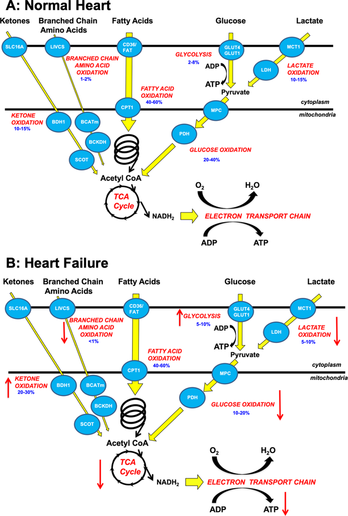
Alterations in cardiac energy metabolism contribute to the severity of heart failure. However, the energy metabolic changes that occur in heart failure are complex and are dependent not only on the severity and type of heart failure present but also on the co-existence of common comorbidities such as obesity and type 2 diabetes. The failing heart faces an energy deficit, primarily because of a decrease in mitochondrial oxidative capacity. This is partly compensated for by an increase in ATP production from glycolysis. The relative contribution of the different fuels for mitochondrial ATP production also changes, including a decrease in glucose and amino acid oxidation, and an increase in ketone oxidation. The oxidation of fatty acids by the heart increases or decreases, depending on the type of heart failure. For instance, in heart failure associated with diabetes and obesity, myocardial fatty acid oxidation increases, while in heart failure associated with hypertension or ischemia, myocardial fatty acid oxidation decreases. Combined, these energy metabolic changes result in the failing heart becoming less efficient (ie, a decrease in cardiac work/O2 consumed). The alterations in both glycolysis and mitochondrial oxidative metabolism in the failing heart are due to both transcriptional changes in key enzymes involved in these metabolic pathways, as well as alterations in NAD redox state (NAD+ and nicotinamide adenine dinucleotide levels) and metabolite signaling that contribute to posttranslational epigenetic changes in the control of expression of genes encoding energy metabolic enzymes. Alterations in the fate of glucose, beyond flux through glycolysis or glucose oxidation, also contribute to the pathology of heart failure. Of importance, pharmacological targeting of the energy metabolic pathways has emerged as a novel therapeutic approach to improving cardiac efficiency, decreasing the energy deficit and improving cardiac function in the failing heart.

Keywords: acetylation; diabetic cardiomyopathies; insulin resistance; ketones; mitochondria.


Conflict of interest statement

Conflict of interest: None declared

Figures

Figure 1:
Figure 1:. Overview of energy metabolism in the normal heart and failing heart
A: Glucose is transported into the cell via glucose transporter 1 or 4 (GLUT1, GLUT4), it then undergoes glycolysis to produce pyruvate. Lactate is taken up by the cardiomyocytes via monocarboxylic acid transporters (MCT) and converted to pyruvate by lactate dehydrogenase (LDH). The pyruvate from glucose and lactate is transported into the mitochondria via the mitochondrial pyruvate carrier (MPC) and is converted to acetyl CoA by pyruvate dehydrogenase (PDH). Fatty acids are transported into the cardiomyocyte, partly via CD36 and FA transport protein-1 (FATP-1), where they are esterified to fatty acyl-CoA. The acyl group are transferred to carnitine by carnitine palmitoyl transferase (CPT-1) and transported into the mitochondria where CPT-2 converts it back to fatty acyl CoA which can then undergo β-oxidation producing acetyl CoA. Ketones (i.e., ß-hydroxybutyrate) are transported into the cell via SLC16A1 where βOHB dehydrogenase 1 (BDH1) catalyses the oxidation of βOHB to acetoacetate (AcAc). AcAc is then activated by succinyl-CoA:3 oxoacid-CoA transferase (SCOT) to acetoacetyl-CoA (AcAc-CoA) which undergoes a thiolysis reaction producing acetyl-CoA. Branched chain amino acids (BCAAs) are transported into the cell by the branched chain amino acid:cation symporter family (LIVCS). In the mitochondria, BCAAs are converted to ketoacids by branched chain aminotransferase (BCATm). Acetyl CoA and succinyl CoA are subsequently formed from branch chain acid alpha-keto acid dehydrogenase (BCKDH). The acetyl CoA generated by fatty acid β-oxidation, glucose oxidation, ketone oxidation and BCAA oxidation enter the tricarboxylic acid (TCA) cycle, generating flavin adenine dinucleotide (FADH2) and nicotinamide adenine dinucleotide (NADH) which then enters the electron transport chain, consuming oxygen (O2) to generate adenosine triphosphate (ATP). Numbers in blue represent the contribution of the individual pathways to overall ATP production. B: Alterations in ketone oxidation, amino acid oxidation, fatty acid oxidation, glycolysis, glucose oxidation, and lactate oxidation in the failing heart. An arrow facing up indicates an increase and down indicates a decrease. Numbers in blue represent the contribution of the individual pathways to overall ATP production. Abbreviations: Glucose transporter 1 and 4 (GLUT1, GLUT4), mitochondrial pyruvate carrier (MPC), pyruvate dehydrogenase (PDH), monocarboxylate transporter (MCT), lactate dehydrogenase (LDH), CD36/fatty acid transporter (CD36/FAT), carnitine palmitoyl transferase (CPT-1), nicotinamide adenine dinucleotide (NADH2), adenosine triphosphate (ATP), adenosine diphosphate (ADP), tricarboxylic acid (TCA), β-hydroxybutyrate (βOHβ), monocarboxylate transporter 1 (SLC16A1), β-hydroxybutyrate dehydrogenase 1 (BDH1), succinyl-CoA:3 oxoacid-CoA transferase (SCOT), branched chain amino acid cation symporter (LIVCS), mitochondrial branched chain amino-transaminase (BCATm), branched chain α-keto acid dehydrogenase (BCKDH).
Figure 2:
Figure 2:. NAD Metabolism and its biological role in mammalian cells.
NAD carries electrons generated from substrate catabolism, e.g. TCA cycle or glycolysis, to oxidative phosphorylation for ATP production. The NAD+/NADH ratio determines cellular redox and metabolic fluxes. NAD+ is consumed by multiple enzymes, e.g. Sirtuins and PARPs, for protein and nucleotide modification thus regulating signal transduction. These reactions generate nicotinamide (NAM), which, through the salvage pathway, is converted into nicotinamide mononucleotide (NMN) by nicotinamide phosphoribosyl transferase (NAMPT). The NMN is converted to NAD+ by nicotinamide mononucleotide adenyltransferase (NMNAT). Alternatively, nicotinamide riboside (NR) can be phosphorylated by nicotinamide riboside kinase (NRK) 1 or 2 to form NMN. NAD+ can also be synthesized from nicotinic acid (NA) in the Preiss-Handler pathway or from tryptophan in the de novo pathway. Both pathways generate nicotinic acid mononucleotide (NaMN). NMNAT then converts NaMN to nicotinic acid adenine dinucleotide (NaAD) which is further metabolized into NAD+ by NAD synthase (NADS).
Figure 3:
Figure 3:. Metabolic signaling to epigenetic transcriptional control in the failing heart.
Left: Mitochondrial metabolic flux of primary cardiac metabolites, glucose and fatty acids, directly alter NAD+/NADH ratios as well as the Acetyl-CoA pool that signal to the nucleus to impact the epigenetic environment. Additionally, glucose that does not enter oxidative metabolism in the mitochondria can signal through the HBP. Additional metabolic intermediates discussed in the text, βOHB, can also regulate activity of epigenetic regulating enzymes as can single carbon metabolism via folate and SAM. Middle: Metabolites and metabolic pathways regulate the activity and/or substrate availability of epigenetic modifiers of histones (e.g., HDAC, HAT, HDM, HMT) and DNA (e.g., DNMT and TET). Right: A) Histone modifications include inhibitory tri-methylation as well as activating acetylation. B) DNA modifications such as DNA methylation (5mC) at CGI in promoters are associated with gene silencing while CGI demethylation and gene body DNA hydroxymethylation (5hmC) modifications are associated with gene activation. C) Although metabolites are not known to be directly regulated by metabolic intermediates, a number of metabolic genes are regulated by miR and lncRNA expression. Please see references included in the main text for additional details. (Illustration credit: Ben Smith). Abbreviations: α-ketoglutarate (αKG), β-hydroxybutyrate (βOHB), CpG island (CGI), DNA methyltransferase (DNMT), fatty acid oxidation (FAO), hexosamine biosynthetic pathway (HBP), histones (H2A, H2B, H3, H4), histone acetyltransferase (HAT), histone deacetylase (HDAC), histone demethylase (HDM), histone methyltransferase (HMT), long noncoding RNA (lncRNA), microRNA (miR), oxidative phosphorylation (OXPHOS), pyruvate dehydrogenase (PDH) complex, S-adenosylmethionine (SAM), ten-eleven translocation (TET), and tricarboxylic acid (TCA) cycle.
Figure 4:
Figure 4:. Pathways of non-oxidative glucose metabolism whose by-products contribute to cardiac remodeling
Schematic summary of glucose uptake via GLUT1 and GLUT4 transporters and entry of glucose into the glycolytic pathway (purple arrows) leading to the generation of pyruvate. In heart failure, impaired entry of pyruvate into mitochondria or decreased mitochondrial pyruvate metabolism leads to accumulation of glycolytic intermediates and increased flux into accessory pathways (blue arrows) such as the polyol pathway, pentose phosphate pathway, hexosamine biosynthetic pathway, mannose and galactosamine synthetic pathways and one carbon metabolism pathways, products of which have been linked to the activation of signaling pathways that may contribute to left ventricular modelling. Regulatory steps in glycolysis that have been implicated in heart failure include phosphofructokinase (PFK), pyruvate kinase (PKM1), the mitochondrial pyruvate carrier (MPC) and pyruvate dehydrogenase (PDH). (Illustration credit: Ben Smith).
Figure 5:
Figure 5:. Targeting cardiac metabolism to protect the failing heart.
MCD, malonyl CoA decarboxylase; DCA, dichloroacetate; IRS, insulin receptor substrate; PI3K, phosphatidylinositol 3-kinase; PDK1, 3-phosphoinositide-dependent protein kinase-1; Akt, protein kinase B; PDH, pyruvate dehydrogenase; BCAA, branched chain amino acids; BT2, branched chain keto acid dehydrogenase kinase inhibitor; SGLT2i, sodium/glucose cotransporter-2 inhibitors. (Illustration credit: Ben Smith).

References

    1. Lopaschuk GD, Ussher JR, Folmes CD, Jaswal JS, Stanley WC. Myocardial fatty acid metabolism in health and disease. Physiol. Rev. 2010;90:207–258 - PubMed
    1. Karwi QG, Uddin GM, Ho KL, Lopaschuk GD. Loss of metabolic flexibility in the failing heart. Front Cardiovasc Med. 2018;5:68. - PMC - PubMed
    1. Neubauer S The failing heart — an engine out of fuel. N. Engl. J. Med 2007;356:1140–1151 - PubMed
    1. Kochanek KD, Xu J, Murphy SL, Minino AM, Kung HC. Deaths: Final data for 2009. Natl. Vital Stat. Rep 2011;60:1–116 - PubMed
    1. Mozaffarian D, Benjamin EJ, Go AS, Arnett DK, Blaha MJ, Cushman M, Das SR, de Ferranti S, Despres JP, Fullerton HJ, et al. Heart disease and stroke statistics-2016 update: A report from the american heart association. Circ. 2016;133:e38–360 - PubMed

Publication types