Finite element analysis of cutting balloon expansion in a calcified artery model of circular angle 180°: Effects of balloon-to-diameter ratio and number of blades facing calcification on potential calcification fracturing and perforation reduction
- PMID: 33984003
- PMCID: PMC8118280
- DOI: 10.1371/journal.pone.0251404
Finite element analysis of cutting balloon expansion in a calcified artery model of circular angle 180°: Effects of balloon-to-diameter ratio and number of blades facing calcification on potential calcification fracturing and perforation reduction
Abstract
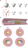
Calcified artery lesions cause stent under-expansion and increase the risk of in-stent restenosis and stent thrombosis. Cutting balloons facilitate the fracturing of calcification prior to stent implantation, although vessel dissection and perforation are potential issues. In clinical practice, calcifications having maximum calcium angles ≤ 180° are rarely fractured during conventional balloon angioplasty. We hypothesize that the lesion/device diameter ratio and the number of blades facing a non-circular calcified lesion may be crucial for fracturing the calcification while avoiding vessel injury. The geometries of the cutting balloons were constructed and their finite-element models were generated by folding and wrapping the balloon model. Numerical simulations were performed for balloons with five different diameters and two types of blade directions in a 180° calcification model. The calcification expansion ability was distinctly higher when two blades faced the calcification than when one blade did. Moreover, when two blades faced the calcification model, larger maximum principal stresses were generated in the calcification even when using undersized balloons with diameters reduced by 0.25 or 0.5 mm from the reference diameter, when compared with the case where one blade faced the calcified model and a balloon of diameter equal to the reference diameter was used. When two blades faced the calcification, smaller stresses were generated in the artery adjacent to the calcification; further, the maximum stress generated in the artery model adjacent to the calcification under the rated pressure of 12 atm when employing undersized balloons was smaller than that when only one blade faced the calcification and when lesion-identical balloon diameters were used under a nominal pressure of 6 atm. Our study suggested that undersized balloons of diameters 0.25 or 0.5 mm less than the reference diameter might be effective in not only expanding the calcified lesion but also reducing the risk of dissection.
Conflict of interest statement
The authors have declared that no competing interests exist.
Figures


References
-
- Vavuranakis M, Toutouzas K, Stefanadis C, Chrisohou C, Markou D, Toutouzas P. Stent deployment in calcified lesions: Can we overcome calcific restraint with high‐pressure balloon inflations? Catheter Cardiovasc Interv. 2001;52: 164–172. 10.1002/1522-726x(200102)52:2<164::aid-ccd1041>3.0.co;2-s - DOI - PubMed
Publication types
MeSH terms
LinkOut - more resources
Full Text Sources
Other Literature Sources
Medical
Research Materials
Miscellaneous

