. 2021 May 13;16(5):e0251454.
doi: 10.1371/journal.pone.0251454. eCollection 2021.

Effect of illumination level [18F]FDG-PET brain uptake in free moving mice

Affiliations

Effect of illumination level [18F]FDG-PET brain uptake in free moving mice

Alexandra de Francisco et al. PLoS One. .

Abstract

In both clinical and preclinical scenarios, 2-deoxy-2[18F]fluoro-D-glucose ([18F]FDG) is the radiotracer most widely used to study brain glucose metabolism with positron emission tomography (PET). In clinical practice, there is a worldwide standardized protocol for preparing patients for [18F]FDG-PET studies, which specifies the room lighting. However, this standard is typically not observed in the preclinical field, although it is well known that animal handling affects the biodistribution of [18F]FDG. The present study aimed to evaluate the effect of ambient lighting on brain [18F]FDG uptake in mice. Two [18F]FDG-PET studies were performed on each animal, one in light and one in dark conditions. Thermal video recordings were acquired to analyse animal motor activity in both conditions. [18F]FDG-PET images were analysed with the Statistical Parametric Mapping method. The results showed that [18F]FDG uptake is higher in darkness than in light condition in mouse nucleus accumbens, hippocampus, midbrain, hindbrain, and cerebellum. The SPM analysis also showed an interaction between the illumination condition and the sex of the animal. Mouse activity was significantly different (p = 0.01) between light conditions (632 ± 215 s of movement) and dark conditions (989 ± 200 s), without significant effect of sex (p = 0.416). We concluded that room illumination conditions during [18F]FDG uptake in mice affected the brain [18F]FDG biodistribution. Therefore, we highlight the importance to control this factor to ensure more reliable and reproducible mouse brain [18F]FDG-PET results.


Conflict of interest statement

The authors have declared that no competing interests exist.

Figures

Fig 1
Fig 1. Experimental design.
Awake animals were exposed to light or darkness in two different days during 33.6 ± 2.7 min before the PET/CT acquisition (n = 24) or video recording (n = 15). Before the illumination condition animals received an intravenous administration of [18F]FDG or saline.
Fig 2
Fig 2. SPM results overlapped to an MRI scan template.
The results are represented in axial (A), coronal (B) and sagittal (C) planes (p<0.05, t-test, k = 25 neighbor voxels). (D) 3D-render of the results. Red color represents the areas with higher [18F]FDG uptake in darkness while green color represents the interaction area between illumination conditions and gender. NAc: nucleus accumbes; HP: hippocampus; MB: midbrain; HB: hidbrain; CB: cerebellum.
Fig 3
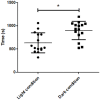
Fig 3. Animals activity.
Individual values of the activity recorded over 30 min under light (circles) and dark (squares) conditions. Statistically significant differences are found between these two groups (*p<0.05).

Similar articles

References

    1. Manook A, Yousefi BH, Willuweit A, Platzer S, Reder S, Voss A, et al.. Small-animal PET imaging of amyloid-beta plaques with [11 C] PiB and its multi-modal validation in an APP/PS1 mouse model of Alzheimer’s disease. PloS one. 2012;7(3):e31310. 10.1371/journal.pone.0031310 - DOI - PMC - PubMed
    1. Walker-Samuel S, Ramasawmy R, Torrealdea F, Rega M, Rajkumar V, Johnson SP, et al.. In vivo imaging of glucose uptake and metabolism in tumors. Nature medicine. 2013;19(8):1067–72. 10.1038/nm.3252 . - DOI - PMC - PubMed
    1. Cusso L, Reigadas E, Munoz P, Desco M, Bouza E. Evaluation of Clostridium difficile Infection with PET/CT Imaging in a Mouse Model. Molecular imaging and biology. 2019. 10.1007/s11307-019-01408-4 . - DOI - PubMed
    1. Cherry SR, Gambhir SS. Use of positron emission tomography in animal research. ILAR journal. 2001;42(3):219–32. 10.1093/ilar.42.3.219 . - DOI - PubMed
    1. Varrone A, Asenbaum S, Vander Borght T, Booij J, Nobili F, Nagren K, et al.. EANM procedure guidelines for PET brain imaging using [18F]FDG, version 2. European journal of nuclear medicine and molecular imaging. 2009;36(12):2103–10. 10.1007/s00259-009-1264-0 . - DOI - PubMed

Publication types

Substances

LinkOut - more resources