. 2021 Aug:294:114174.
doi: 10.1016/j.jviromet.2021.114174. Epub 2021 May 10.

Optimized protocol for a quantitative SARS-CoV-2 duplex RT-qPCR assay with internal human sample sufficiency control

Affiliations

Optimized protocol for a quantitative SARS-CoV-2 duplex RT-qPCR assay with internal human sample sufficiency control

Aileen G Rowan et al. J Virol Methods. 2021 Aug.

Abstract

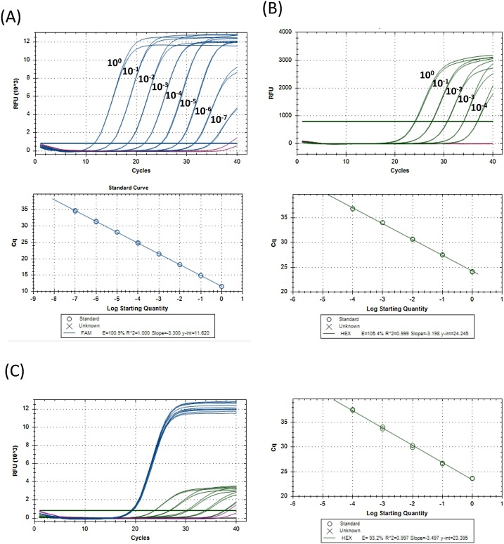
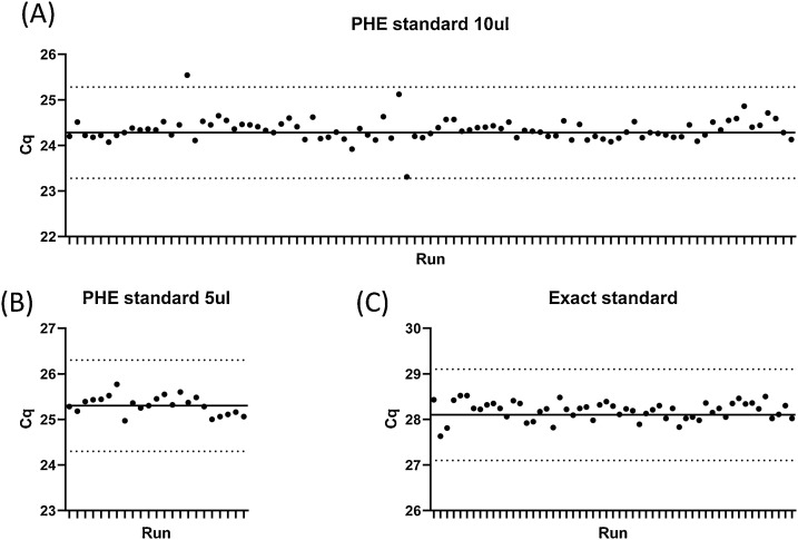
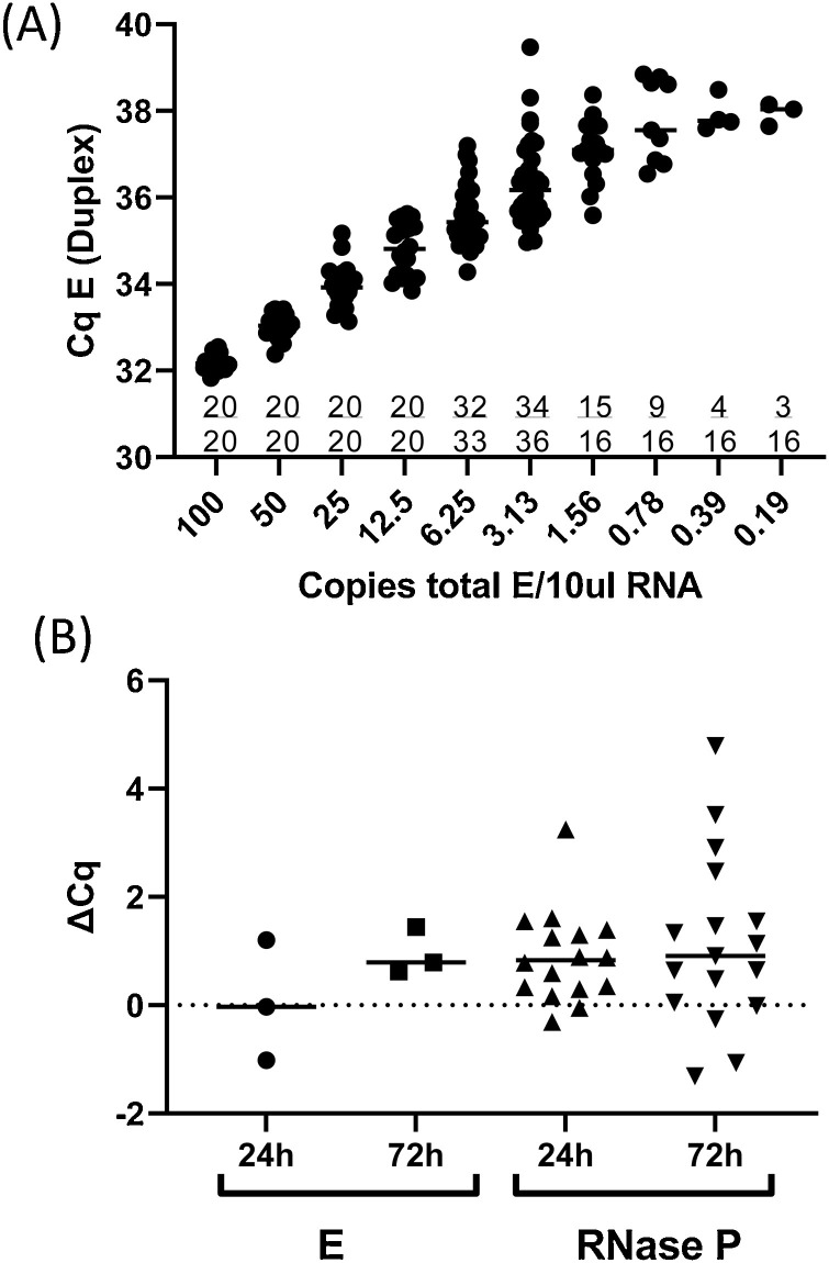
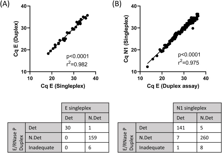
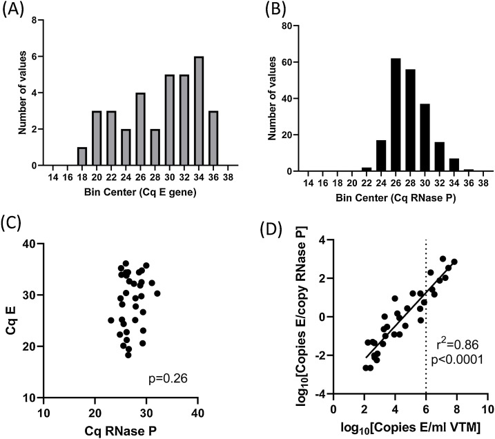
There is growing evidence that measurement of SARS-CoV-2 viral copy number can inform clinical and public health management of SARS-CoV-2 carriers and COVID-19 patients. Here we show that quantification of SARS-CoV-2 is feasible in a clinical setting, using a duplex RT-qPCR assay which targets both the E gene (Charité assay) and a human RNA transcript, RNase P (CDC assay) as an internal sample sufficiency control. Samples in which RNase P is not amplified indicate that sample degradation has occurred, PCR inhibitors are present, RNA extraction has failed or swabbing technique was insufficient. This important internal control reveals that 2.4 % of nasopharyngeal swabs (15/618 samples) are inadequate for SARS-CoV-2 testing which, if not identified, could result in false negative results. We show that our assay is linear across at least 7 logs and is highly reproducible, enabling the conversion of Cq values to viral copy numbers using a standard curve. Furthermore, the SARS-CoV-2 copy number was independent of the RNase P copy number indicating that the per-swab viral copy number is not dependent on sampling- further allowing comparisons between samples. The ability to quantify SARS-CoV-2 viral copy number will provide an important opportunity for viral burden-guided public health and clinical decision making.

Keywords: COVID-19; RNase P; RT-qPCR; SARS-CoV-2; Viral burden.


Conflict of interest statement

The authors report no declarations of interest.

Figures

Fig. 1
Fig. 1
Linear range and PCR efficiency for E and RNase P. RNA was extracted from SARS-CoV-2-infected Vero cells (A), serially diluted (10 fold) in nuclease-free water with carrier RNA and assayed in duplicate using the E/RNase P RT-qPCR assay. Relative fluorescence units (RFU) detected in the FAM channel for the E gene for samples containing viral RNA (blue) or carrier RNA alone (purple) are shown. (B) RNA was extracted from human peripheral blood mononuclear cells (PBMC), serially diluted (10 fold) in nuclease-free water with carrier RNA and assayed in duplicate using the E/RNase P RT-qPCR assay. RFU detected in the HEX channel for the RNase P for samples containing PBMC RNA (blue) or carrier RNA alone (purple) are shown. (C) RNA from SARS-CoV-2-infected Vero cells was spiked into serial dilutions of PBMC RNA. RFU from samples with PBMC and infected Vero cells RNA is shown in blue (FAM) and green (HEX), and carrier RNA alone is shown in purple (FAM) and black (HEX). (For interpretation of the references to colour in this figure legend, the reader is referred to the web version of this article).
Fig. 2
Fig. 2
Assay reproducibility. Aliquots of viral RNA (A, B) extracted from culture supernatant or a commercial synthetic RNA construct (C) were assayed using the E/RNase P duplex PCR. Each dot represents the E Cq of an independent run. The solid line represents the mean Cq of four runs (not included in the graph), and the dotted lines indicate the mean plus or minus 1 cycle.
Fig. 3
Fig. 3
Limit of detection and effect of sample storage. (A) The EXACT synthetic RNA standard was diluted in nuclease free water with 1 μg/μl PolyA carrier RNA (Qiagen) to a range of concentrations from 100 copies/10 μl- 0.19 copies/10 μl. Replicates of each concentration were assayed using the E/RNase P duplex assay. Cqs for E are shown for each replicate within each concentration, and the number of replicates in which a signal was detected is indicated on the graph as a fraction. (B) RNA was extracted from clinical samples immediately on receipt or after 24 h (n = 16) or 72 h (n = 17) storage at room temperature. The difference in Cq values (ΔCq) was calculated by subtracting the E or RNase P Cq observed in the sample processed and assayed on receipt from the E/ RNase P Cq observed in the sample processed after storage.
Fig. 4
Fig. 4
Comparison of E and N1 Singleplex and Duplex E/RNase P assays. (A) Clinical samples (n = 196) were assayed using the E primers and FAM-labelled probe alone (E singleplex) or in combination with RNase P primers and HEX-labelled probe (E/RNase P Duplex) (B) Clinical samples (n = 422) were assayed using the CDC N1 primers and FAM-labelled probe alone (N1 singleplex) or with a combination with E and RNase P primers and probes (E/RNase P Duplex).
Fig. 5
Fig. 5
Distribution of E and RNase P Cq in clinical samples. Clinical samples (n = 204) were assayed using the E primers and FAM-labelled probe in combination with RNase P primers and HEX-labelled probe (Duplex). N = 34 samples had detectable SARS-CoV-2, 6 were classed as inadequate due to lack of amplification of RNAse P, and the remainder were classified as ‘SARS-CoV-2 not detected’. Histograms of Cq values for E gene in samples with detectable SARS-CoV-2 (A) and Cq values for RNase P in all adequate samples (B). (C) E versus RNAse P Cq values in all samples with detectable SARS-CoV-2. (D) Normalised copy number of E versus per-swab copy number. Dotted line indicates 1,000,000 copies/mL (Cq E gene = 24 in the duplex assay).

References

    1. Arnaout R., Lee R.A., Lee G.R., Callahan C., Cheng A., Yen C.F., et al. The limit of detection matters: the case for benchmarking severe acute respiratory syndrome coronavirus 2 testing. Clin. Infect. Dis. 2021 2020.06.02.131144. - PMC - PubMed
    1. Bustin S.A., Benes V., Garson J.A., Hellemans J., Huggett J., Kubista M., et al. The MIQE guidelines: minimum information for publication of quantitative real-time PCR experiments. Clin. Chem. 2009;55:611–622. - PubMed
    1. Centre for Disease Control and Prevention . 2020. Novel Coronavirus (2019-nCoV) Real-time RT-PCR Primers and Probes.https://www.cdc.gov/coronavirus/2019-ncov/lab/rt-pcr-panel-primer-probes...
    1. Corman V.M., Landt O., Kaiser M., Molenkamp R., Meijer A., Chu D.K., et al. Detection of 2019 novel coronavirus (2019-nCoV) by real-time RT-PCR. Euro Surveill. 2020;25:1–8. - PMC - PubMed
    1. Crone M.A., Priestman M., Ciechonska M., Jensen K., Sharp D.J., Anand A., et al. A role for Biofoundries in rapid development and validation of automated SARS-CoV-2 clinical diagnostics. Nat. Commun. 2020;11:1–11. - PMC - PubMed

Publication types

LinkOut - more resources