

A dual autoencoder and singular value decomposition based feature optimization for the segmentation of brain tumor from MRI images
- PMID: 33985449
- PMCID: PMC8117624
- DOI: 10.1186/s12880-021-00614-3
A dual autoencoder and singular value decomposition based feature optimization for the segmentation of brain tumor from MRI images
Abstract
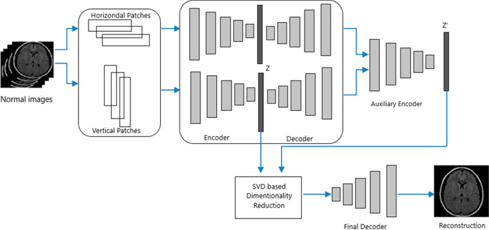
Background: The brain tumor is the growth of abnormal cells inside the brain. These cells can be grown into malignant or benign tumors. Segmentation of tumor from MRI images using image processing techniques started decades back. Image processing based brain tumor segmentation can be divided in to three categories conventional image processing methods, Machine Learning methods and Deep Learning methods. Conventional methods lacks the accuracy in segmentation due to complex spatial variation of tumor. Machine Learning methods stand as a good alternative to conventional methods. Methods like SVM, KNN, Fuzzy and a combination of either of these provide good accuracy with reasonable processing speed. The difficulty in processing the various feature extraction methods and maintain accuracy as per the medical standards still exist as a limitation for machine learning methods. In Deep Learning features are extracted automatically in various stages of the network and maintain accuracy as per the medical standards. Huge database requirement and high computational time is still poses a problem for deep learning. To overcome the limitations specified above we propose an unsupervised dual autoencoder with latent space optimization here. The model require only normal MRI images for its training thus reducing the huge tumor database requirement. With a set of normal class data, an autoencoder can reproduce the feature vector into an output layer. This trained autoencoder works well with normal data while it fails to reproduce an anomaly to the output layer. But a classical autoencoder suffer due to poor latent space optimization. The Latent space loss of classical autoencoder is reduced using an auxiliary encoder along with the feature optimization based on singular value decomposition (SVD). The patches used for training are not traditional square patches but we took both horizontal and vertical patches to keep both local and global appearance features on the training set. An Autoencoder is applied separately for learning both horizontal and vertical patches. While training a logistic sigmoid transfer function is used for both encoder and decoder parts. SGD optimizer is used for optimization with an initial learning rate of .001 and the maximum epochs used are 4000. The network is trained in MATLAB 2018a with a processor capacity of 3.7 GHz with NVIDIA GPU and 16 GB of RAM.
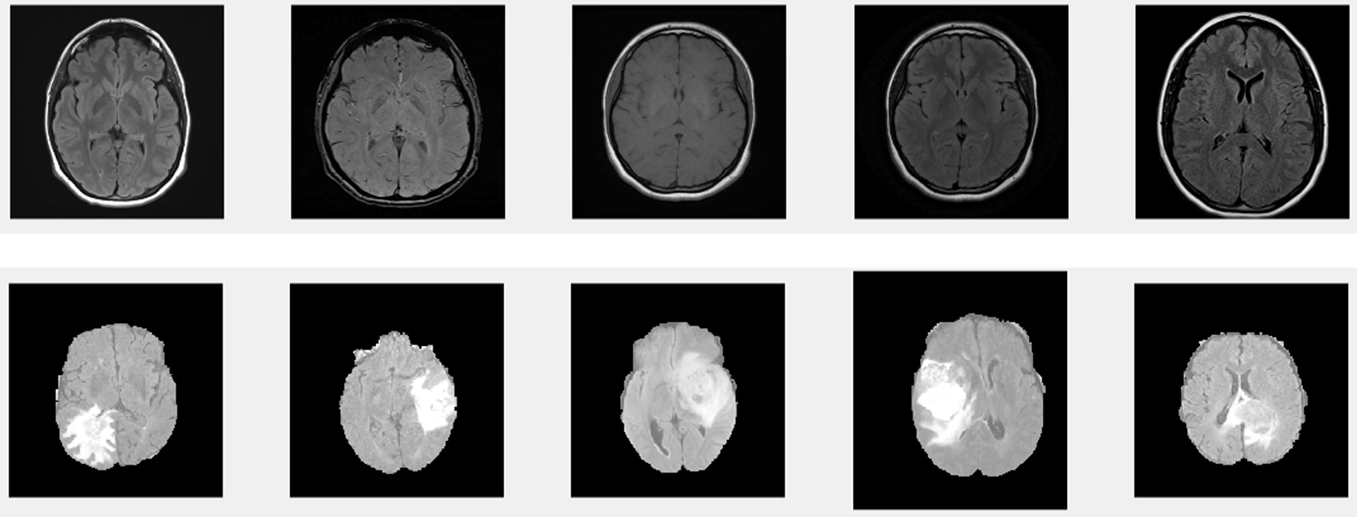
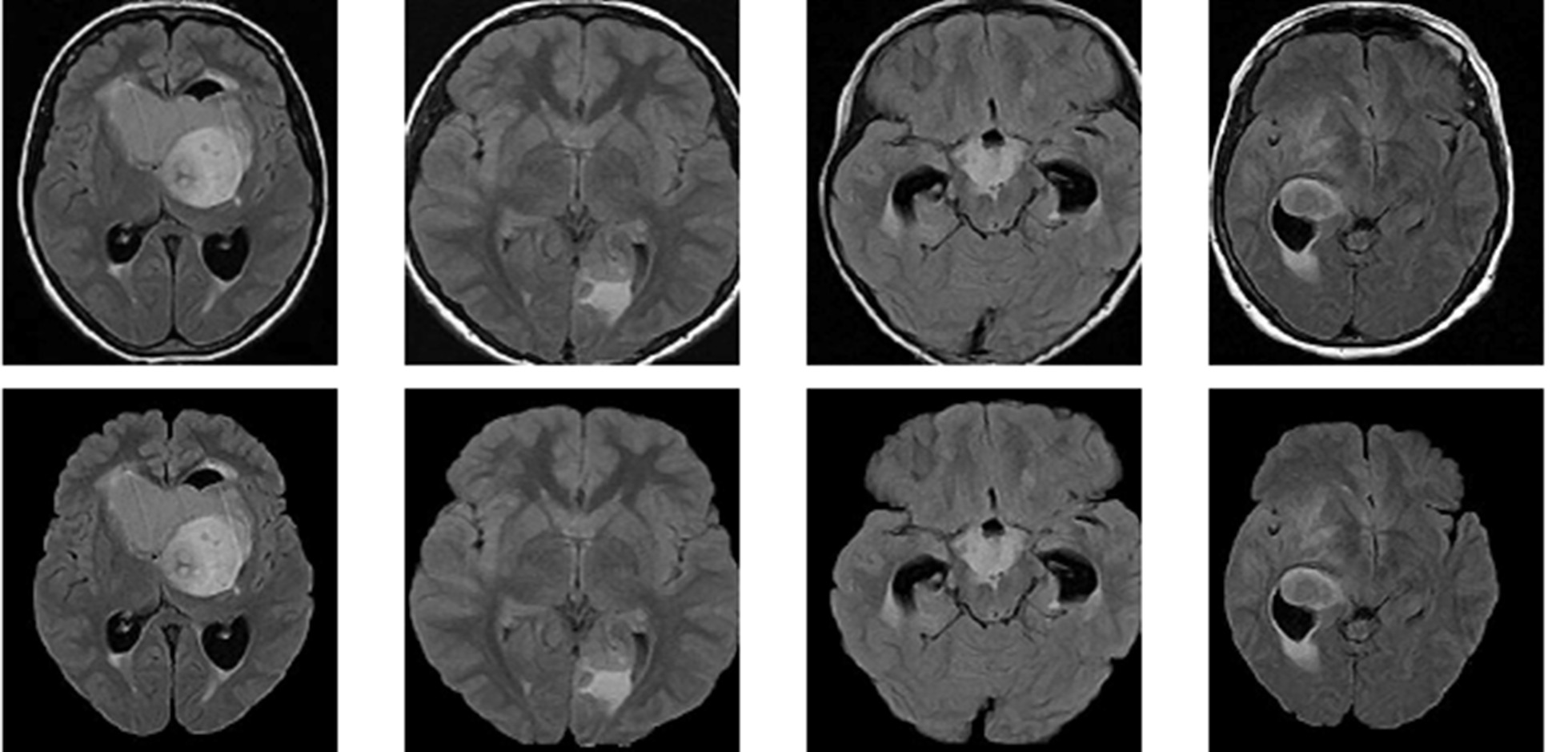
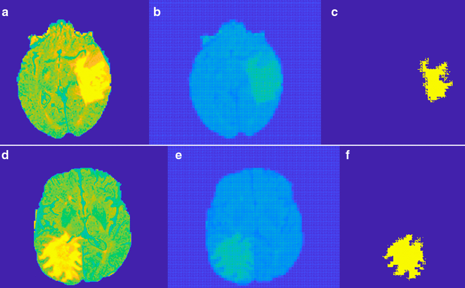
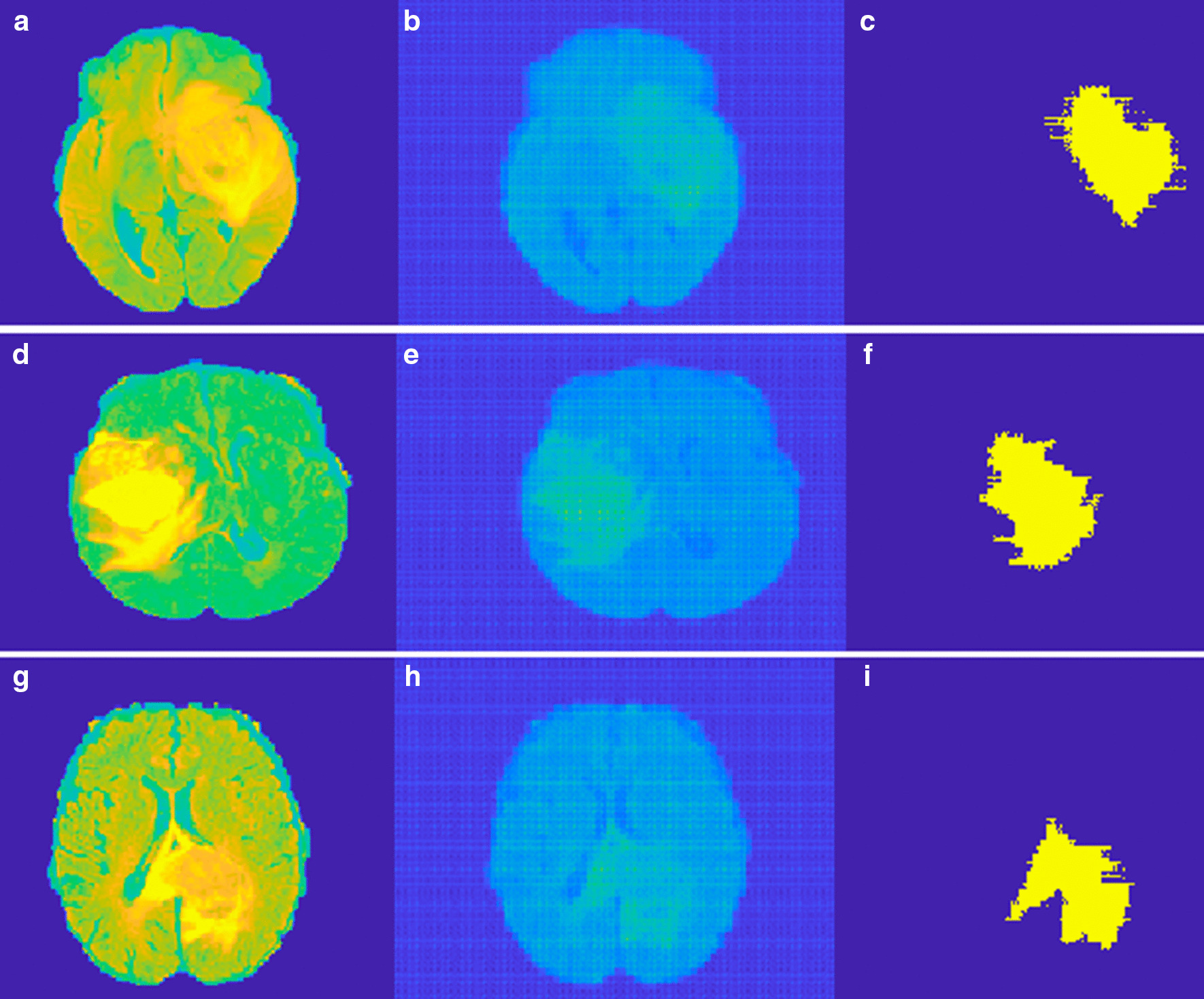
Results: The results are obtained using a patch size of 16 × 64, 64 × 16 for horizontal and vertical patches respectively. In Glioma images tumor is not grown from a point rather it spreads randomly. Region filling and connectivity operations are performed to get the final tumor segmentation. Overall the method segments Meningioma better than Gliomas. Three evaluation metrics are considered to measure the performance of the proposed system such as Dice Similarity Coefficient, Positive Predictive Value, and Sensitivity.
Conclusion: An unsupervised method for the segmentation of brain tumor from MRI images is proposed here. The proposed dual autoencoder with SVD based feature optimization reduce the latent space loss in the classical autoencoder. The proposed method have advantages in computational efficiency, no need of huge database requirement and better accuracy than machine learning methods. The method is compared Machine Learning methods Like SVM, KNN and supervised deep learning methods like CNN and commentable results are obtained.
Keywords: Anomaly prediction; Brain tumor; Computer vision; Deep learning; MRI.
Conflict of interest statement
The authors declare that they have no competing interests.
Figures
Similar articles
-
Performance of Convolutional Neural Network Models in Meningioma Segmentation in Magnetic Resonance Imaging: A Systematic Review and Meta-Analysis.Neuroinformatics. 2025 Jan;23(1):14. doi: 10.1007/s12021-024-09704-3. Epub 2024 Dec 28. Neuroinformatics. 2025. PMID: 39777602 Free PMC article.
-
Brain tumor segmentation and detection in MRI using convolutional neural networks and VGG16.Cancer Biomark. 2025 Mar;42(3):18758592241311184. doi: 10.1177/18758592241311184. Epub 2025 Apr 4. Cancer Biomark. 2025. PMID: 40183298
-
A Hybrid Approach Based on Deep CNN and Machine Learning Classifiers for the Tumor Segmentation and Classification in Brain MRI.Comput Math Methods Med. 2022 Aug 5;2022:6446680. doi: 10.1155/2022/6446680. eCollection 2022. Comput Math Methods Med. 2022. Retraction in: Comput Math Methods Med. 2023 Dec 13;2023:9848046. doi: 10.1155/2023/9848046. PMID: 36035291 Free PMC article. Retracted.
-
MRI-based brain tumor detection using convolutional deep learning methods and chosen machine learning techniques.BMC Med Inform Decis Mak. 2023 Jan 23;23(1):16. doi: 10.1186/s12911-023-02114-6. BMC Med Inform Decis Mak. 2023. PMID: 36691030 Free PMC article.
-
Machine learning and deep learning for brain tumor MRI image segmentation.Exp Biol Med (Maywood). 2023 Nov;248(21):1974-1992. doi: 10.1177/15353702231214259. Epub 2023 Dec 16. Exp Biol Med (Maywood). 2023. PMID: 38102956 Free PMC article. Review.
Cited by
-
A systematic review on deep learning-based automated cancer diagnosis models.J Cell Mol Med. 2024 Mar;28(6):e18144. doi: 10.1111/jcmm.18144. J Cell Mol Med. 2024. PMID: 38426930 Free PMC article.
-
Integrating artificial intelligence in healthcare: applications, challenges, and future directions.Future Sci OA. 2025 Dec;11(1):2527505. doi: 10.1080/20565623.2025.2527505. Epub 2025 Jul 4. Future Sci OA. 2025. PMID: 40616302 Free PMC article. Review.
-
Automatic quadriceps and patellae segmentation of MRI with cascaded U2 -Net and SASSNet deep learning model.Med Phys. 2022 Jan;49(1):443-460. doi: 10.1002/mp.15335. Epub 2021 Nov 22. Med Phys. 2022. PMID: 34755359 Free PMC article.
-
Performance of Convolutional Neural Network Models in Meningioma Segmentation in Magnetic Resonance Imaging: A Systematic Review and Meta-Analysis.Neuroinformatics. 2025 Jan;23(1):14. doi: 10.1007/s12021-024-09704-3. Epub 2024 Dec 28. Neuroinformatics. 2025. PMID: 39777602 Free PMC article.
References
-
- Umamaheswari K, Rajesh P, Srinivasa Rao S, Vinodh Babu P. Application of segmentation methodology for extracting MRI brain tumor duly mitigating the noise. In: 2015 international conference on computational intelligence and communication networks.
-
- Deepthi Murthy TS, Sadashivappa G. Brain tumor segmentation using thresholding, morphological operations and extraction of features of tumor. In: 2014 international conference on advances in electronics, computers and communications (ICAECC).
-
- Dawngliana M, Deb D, Handique M, Roy S. Automatic brain tumor segmentation in MRI: hybridized multilevel thresholding and level set. In: 2015 international symposium on advanced computing and communication (ISACC).
-
- Salwe S, Raut R, Hajare P. Brain tumor pixels detection using adaptive wavelet based histogram thresholding and fine windowing. In: 2016 international conference on information technology (InCITe)—the next generation IT summit.
-
- Parveen AS. Detection of brain tumor in MRI images, using combination of fuzzy C-means and SVM. In: 2015 2nd international conference on signal processing and integrated networks (SPIN).
MeSH terms
LinkOut - more resources
Full Text Sources
Other Literature Sources
Medical
