Review
. 2021 Aug;28(8):1058-1071.
doi: 10.1016/j.acra.2021.04.007. Epub 2021 May 1.

Lymphadenopathy Following COVID-19 Vaccination: Imaging Findings Review

Affiliations
Review

Lymphadenopathy Following COVID-19 Vaccination: Imaging Findings Review

Pedram Keshavarz et al. Acad Radiol. 2021 Aug.

Abstract

Rationale and objectives: Despite all the benefits and effectiveness of the coronavirus disease 2019 (COVID-19) vaccines mentioned in recent clinical trials, some post-vaccination side effects such as lymphadenopathy (LAP) were observed. The present study reviewed all studies with imaging findings presentation of LAP after COVID-19 vaccination.

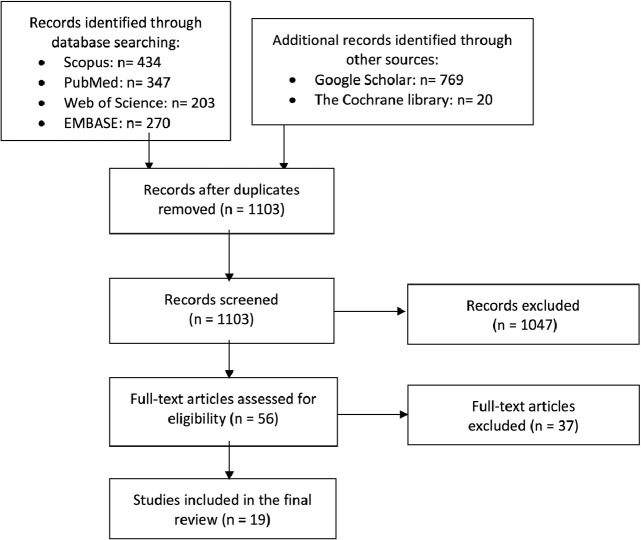
Materials and methods: We conducted a literature search in online databases, including Scopus, Medline (PubMed), Web of Science, Embase (Elsevier), Cochrane library, and Google Scholar.

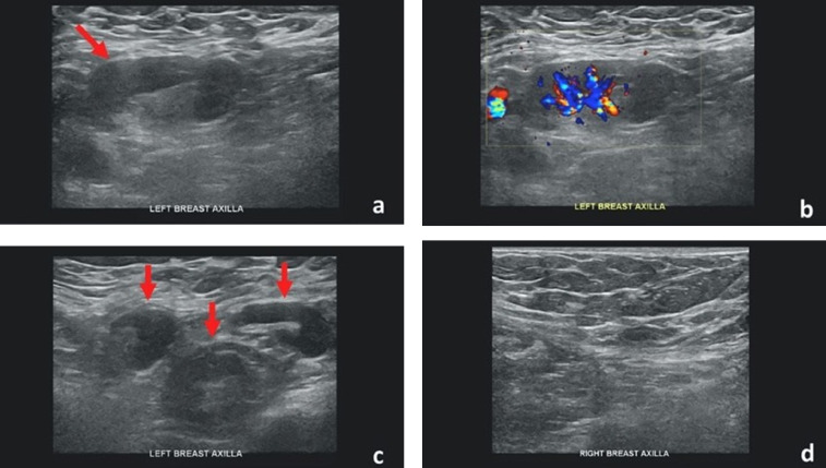
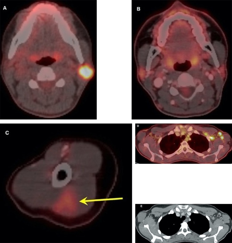
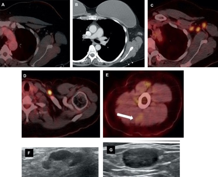
Results: A total of 19 studies (68 cases), including 60 (88.2%) females and eight (11.8%) males with a presentation of LAP after COVID-19 vaccination, were reviewed. LAP was identified after first or second dosages of three types of COVID-19 vaccines, including Pfizer-BioNTech (n = 30, 44.1%), Moderna (n = 17, 25%), and Oxford-AstraZeneca (n = 1, 1.5%). In 20 (29.4%) cases, vaccine type was not reported or only reported as mRNA COVID-19 vaccine. The median days of LAP presentation after the first and second dosages of COVID-19 vaccination, were 12 and 5 days, respectively. Most of the LAP imaging findings related to COVID-19 vaccination (n = 66, 97%) were seen from first day to 4 weeks after vaccination. However, LAP remained after 5 and 6 weeks of the first and second dosages of COVID-19 vaccination with decreased lymph nodes' size and residual cortical thickening in two cases.

Conclusion: This review study of cases with LAP-associated COVID-19 vaccination guides radiologists and physicians to rely on patient's clinical context and updated resources to prevent potential disease upstaging and change in therapy.

Keywords: Adenopathy; Coronavirus; Moderna; Oxford-AstraZeneca; Pfizer-BioNTech; Radiology; SARS-CoV-2; Vaccination.


Figures

Figure 1
Figure 1
Flow diagram of the study selection process.
Figure 2
Figure 2
Forty-two-year-old female with unilateral left axillary adenopathy noted 5 days after receiving the second dose of the Pfizer-BioNTech COVID-19 vaccine in her left upper extremity. (a) Gray-scale and (b) color Doppler images of an enlarged left axillary lymph node with cortical thickening (arrow). (c) Multiple additional morphologically abnormal left axillary lymph nodes were also present (arrows). Unremarkable right axilla was documented (d). Images obtained from Mehta et al., (38) Clinical Imaging, 2021, Vol. 75:12-15, and permission to use granted by Elsevier and Copyright Clearance Center. (Color version of figure is available online.)
Figure 3
Figure 3
Thirty-two-year-old female. A, Axial fused 18-FDG PET/CT showed hypermetabolic biopsy proven intraparotid lymph node with metastatic malignant melanoma. B, Three-month follow-up axial fused 18-FDG PET/CT shows complete resolution of the neck mass following chemotherapy, C, while left arm shows hypermetabolic triangular shaped inflammation (arrow) at the COVID vaccine injection site. D, Axial fused images at the axilla level shows multiple new hypermetabolic lymph nodes. E, Axial contrast enhanced CT demonstrates mild fat stranding surrounding the ovoid lymph nodes with preserved fatty hilum. Images obtained from Özütemiz et al., (39) Radiology, published online: February 24, 2021, and permission to use granted by LaShundra Carson, Coordinator, Journal Business Publications, Radiological Society of North America (RSNA). (Color version of figure is available online.)
Figure 4
Figure 4
Forty-six-year-old female with triple negative left breast cancer, disease free for three years. A, Axial fused 18-FDG PET/CT three years earlier with no concerning lymph node in the left axilla. B, Surveillance contrast enhanced axial chest CT showed new left axillary lymphadenopathy with fat stranding 15 days after the first Covid-19 vaccine. Further evaluation with PET/CT six days after the 2nd dose of vaccine, demonstrated, C, multiple enlarged hypermetabolic left axillary lymph nodes and, D, a hypermetabolic round shaped left supraclavicular lymph node in axial fused 18-FDG PET/CT images. E, A subtle wedge-shaped intramuscular hypermetabolism (white arrow) was also noted in this case, similar to first and third cases. Ultrasonography guided core needle biopsy was performed. F, On ultrasonography, axillary lymph nodes had thickened cortex while the supraclavicular lymph node demonstrated, G, thickened cortex with loss of normal fatty hilum. I, Hematoxylin and eosin staining under 40x magnification shows enlarged germinal center with interfollicular expansion by small lymphocytes. 100x magnification images show, J, prominent germinal center with tingible body macrophage and, K, reactive germinal center with expansion of interfollicular regions by small lymphocytes and focally prominent endothelial cells. Images obtained from Özütemiz et al. (39) Radiology, published online: February 24, 2021, and permission to use granted by LaShundra Carson, Coordinator, Journal Business Publications, Radiological Society of North America (RSNA). (Color version of figure is available online.)
Figure 4
Figure 4
Forty-six-year-old female with triple negative left breast cancer, disease free for three years. A, Axial fused 18-FDG PET/CT three years earlier with no concerning lymph node in the left axilla. B, Surveillance contrast enhanced axial chest CT showed new left axillary lymphadenopathy with fat stranding 15 days after the first Covid-19 vaccine. Further evaluation with PET/CT six days after the 2nd dose of vaccine, demonstrated, C, multiple enlarged hypermetabolic left axillary lymph nodes and, D, a hypermetabolic round shaped left supraclavicular lymph node in axial fused 18-FDG PET/CT images. E, A subtle wedge-shaped intramuscular hypermetabolism (white arrow) was also noted in this case, similar to first and third cases. Ultrasonography guided core needle biopsy was performed. F, On ultrasonography, axillary lymph nodes had thickened cortex while the supraclavicular lymph node demonstrated, G, thickened cortex with loss of normal fatty hilum. I, Hematoxylin and eosin staining under 40x magnification shows enlarged germinal center with interfollicular expansion by small lymphocytes. 100x magnification images show, J, prominent germinal center with tingible body macrophage and, K, reactive germinal center with expansion of interfollicular regions by small lymphocytes and focally prominent endothelial cells. Images obtained from Özütemiz et al. (39) Radiology, published online: February 24, 2021, and permission to use granted by LaShundra Carson, Coordinator, Journal Business Publications, Radiological Society of North America (RSNA). (Color version of figure is available online.)

References

    1. Hsiang S, Allen D, Annan-Phan S. The effect of large-scale anti-contagion policies on the COVID-19 pandemic. Nature. 2020;584(7820):262–267. doi: 10.1038/s41586-020-2404-8. - DOI - PubMed
    1. World Health Organization Website. WHO coronavirus (COVID-19) dashboard. Accessed March 8, 2021. Available from: https://covid19.who.int/?gclid=Cj0KCQiAvvKBBhCXARIsACTePW9nQttX871YsapnS....
    1. Yazdanpanah F, Hamblin MR, Rezaei N. The immune system and COVID-19: Friend or foe? Life Sci. 2020;256 doi: 10.1016/j.lfs.2020.117900. - DOI - PMC - PubMed
    1. Vabret N, Britton GJ, Gruber C. Immunology of COVID-19: current state of the science. Immunity. 2020;52(6):910–941. doi: 10.1016/j.immuni.2020.05.002. - DOI - PMC - PubMed
    1. Centers for Disease Control and Prevention. Different COVID-19 vaccines. Accessed March 3, 2021. Available from: https://www.cdc.gov/coronavirus/2019-ncov/vaccines/different-vaccines.html.

Substances