. 2021 Jul 1;27(13):3744-3756.
doi: 10.1158/1078-0432.CCR-21-0164. Epub 2021 May 13.

Combining AFM13, a Bispecific CD30/CD16 Antibody, with Cytokine-Activated Blood and Cord Blood-Derived NK Cells Facilitates CAR-like Responses Against CD30+ Malignancies

Affiliations

Combining AFM13, a Bispecific CD30/CD16 Antibody, with Cytokine-Activated Blood and Cord Blood-Derived NK Cells Facilitates CAR-like Responses Against CD30+ Malignancies

Lucila N Kerbauy et al. Clin Cancer Res. .

Abstract

Purpose: Natural killer (NK)-cell recognition and function against NK-resistant cancers remain substantial barriers to the broad application of NK-cell immunotherapy. Potential solutions include bispecific engagers that target NK-cell activity via an NK-activating receptor when simultaneously targeting a tumor-specific antigen, as well as enhancing functionality using IL12/15/18 cytokine pre-activation.

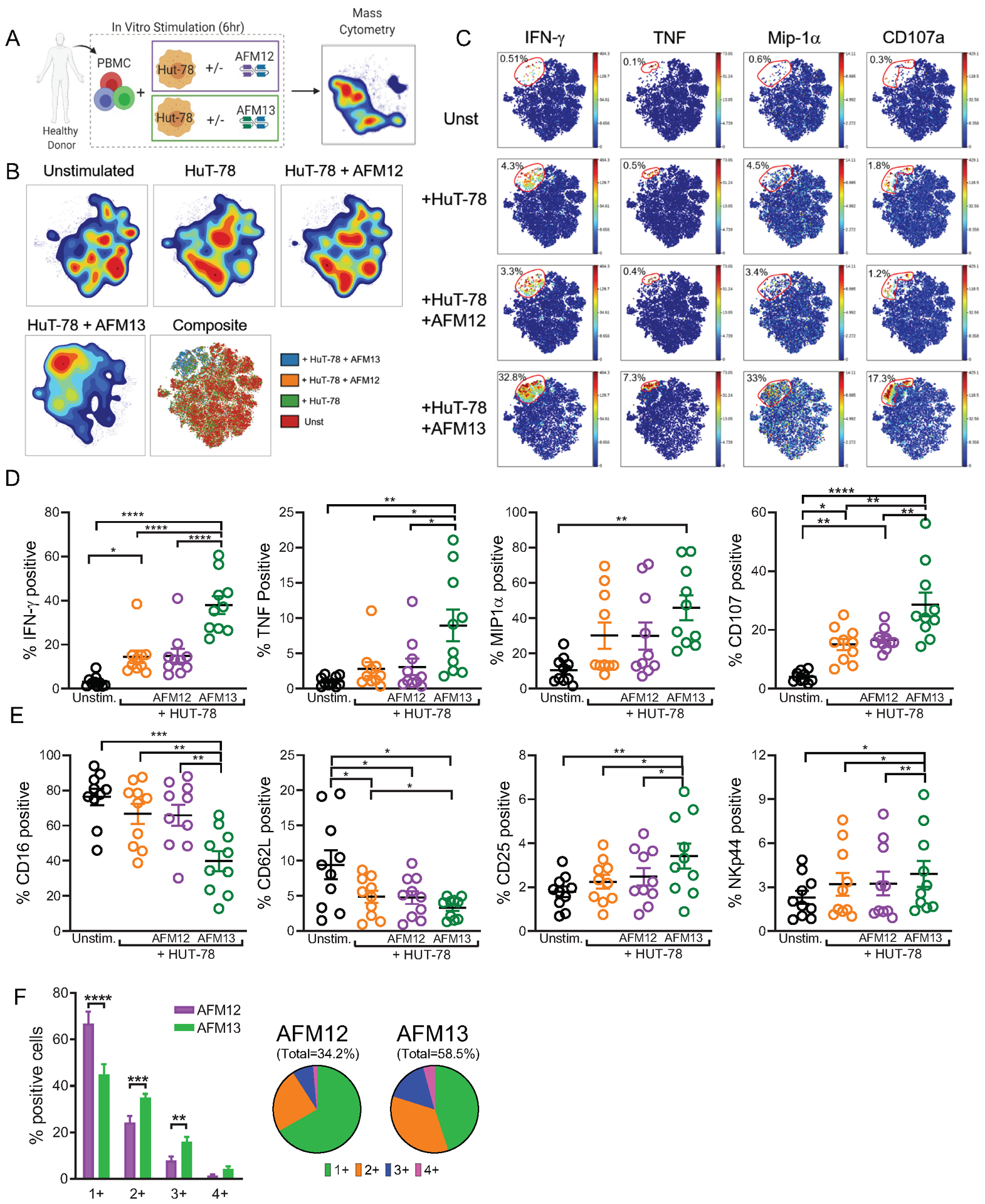
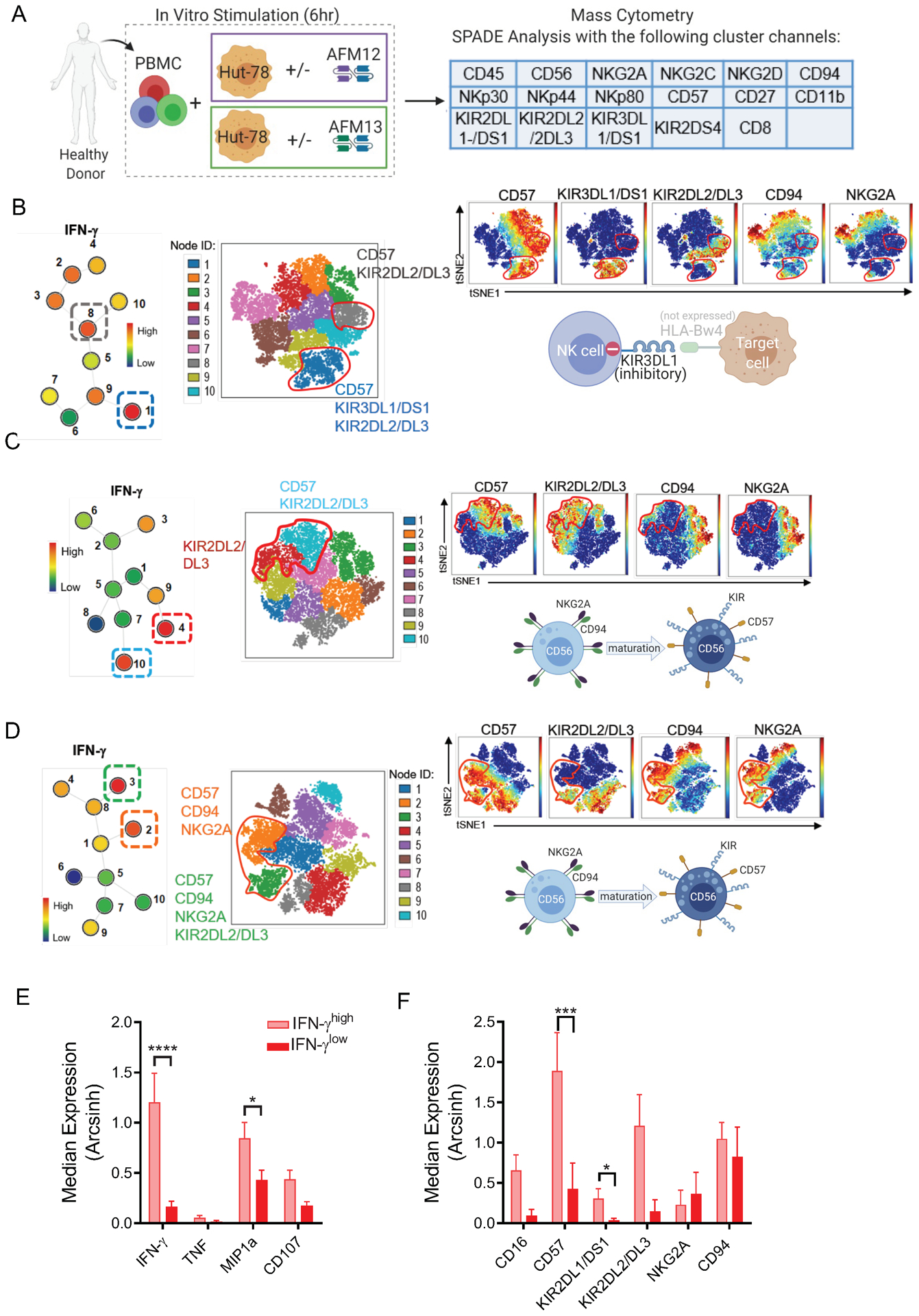
Experimental design: We assessed single-cell NK-cell responses stimulated by the tetravalent bispecific antibody AFM13 that binds CD30 on leukemia/lymphoma targets and CD16A on various types of NK cells using mass cytometry and cytotoxicity assays. The combination of AFM13 and IL12/15/18 pre-activation of blood and cord blood-derived NK cells was investigated in vitro and in vivo.

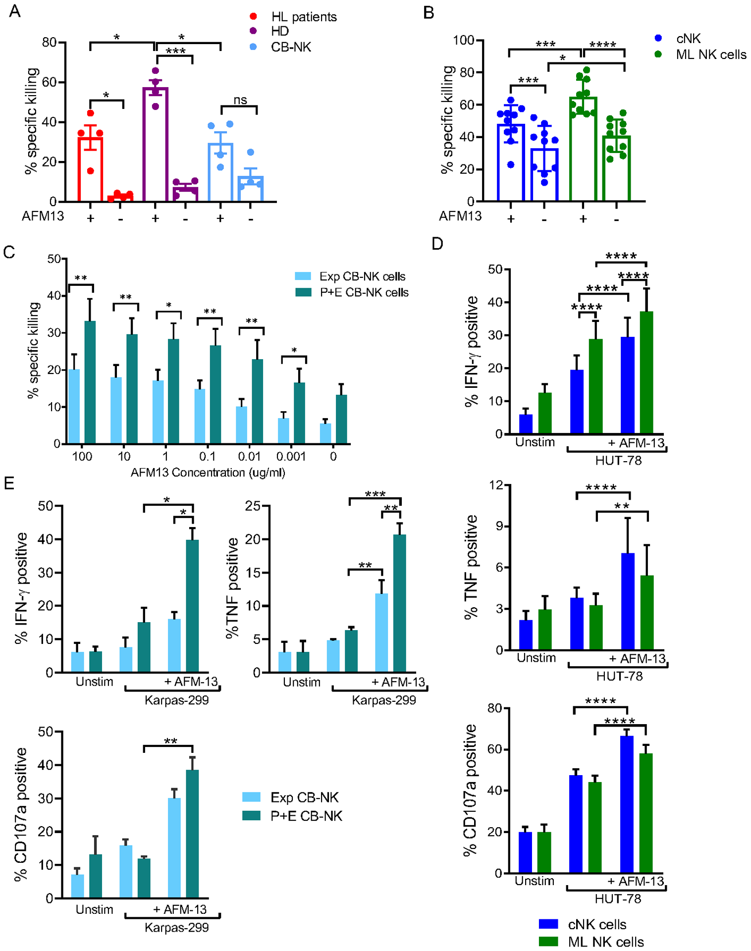
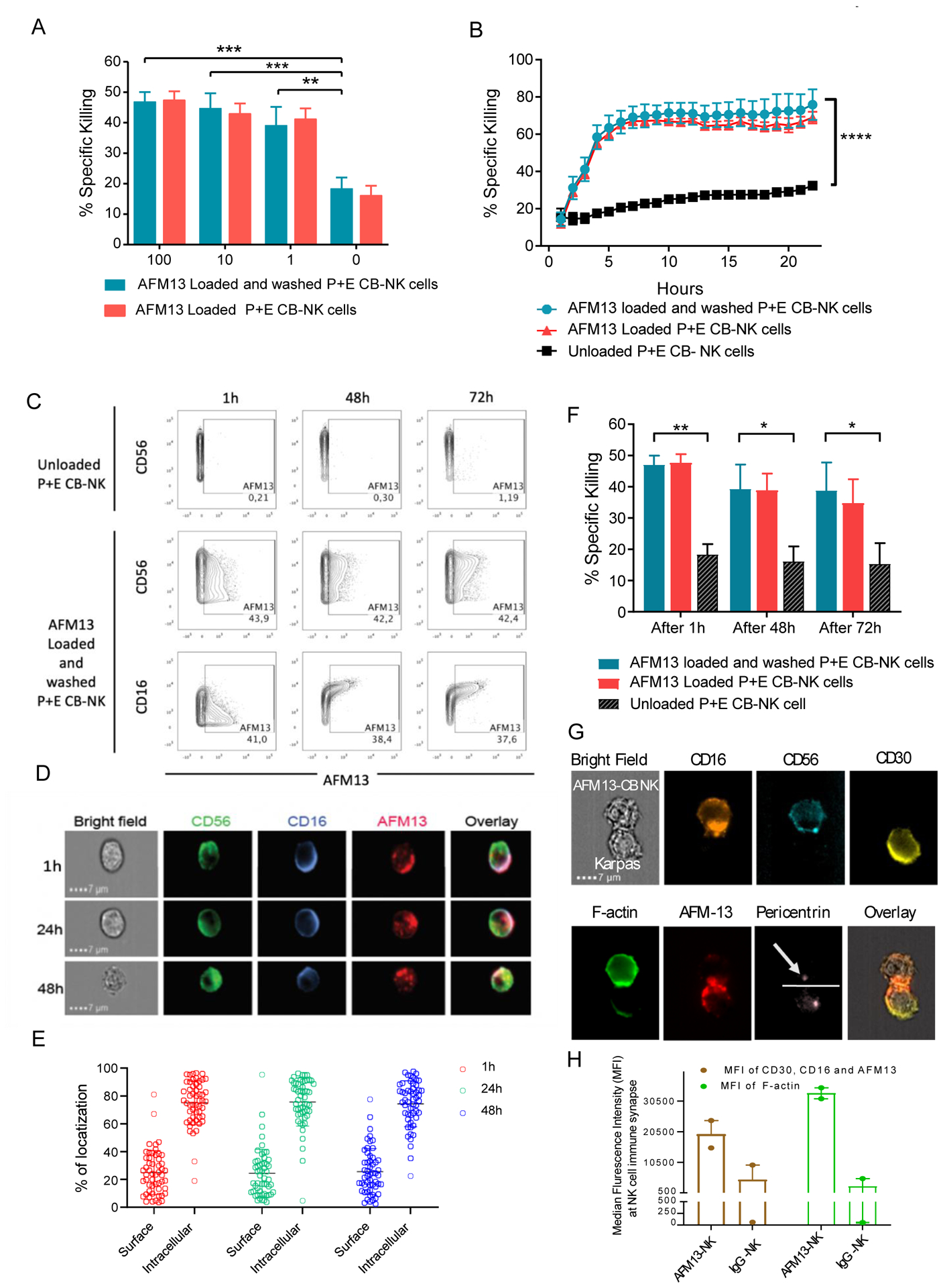
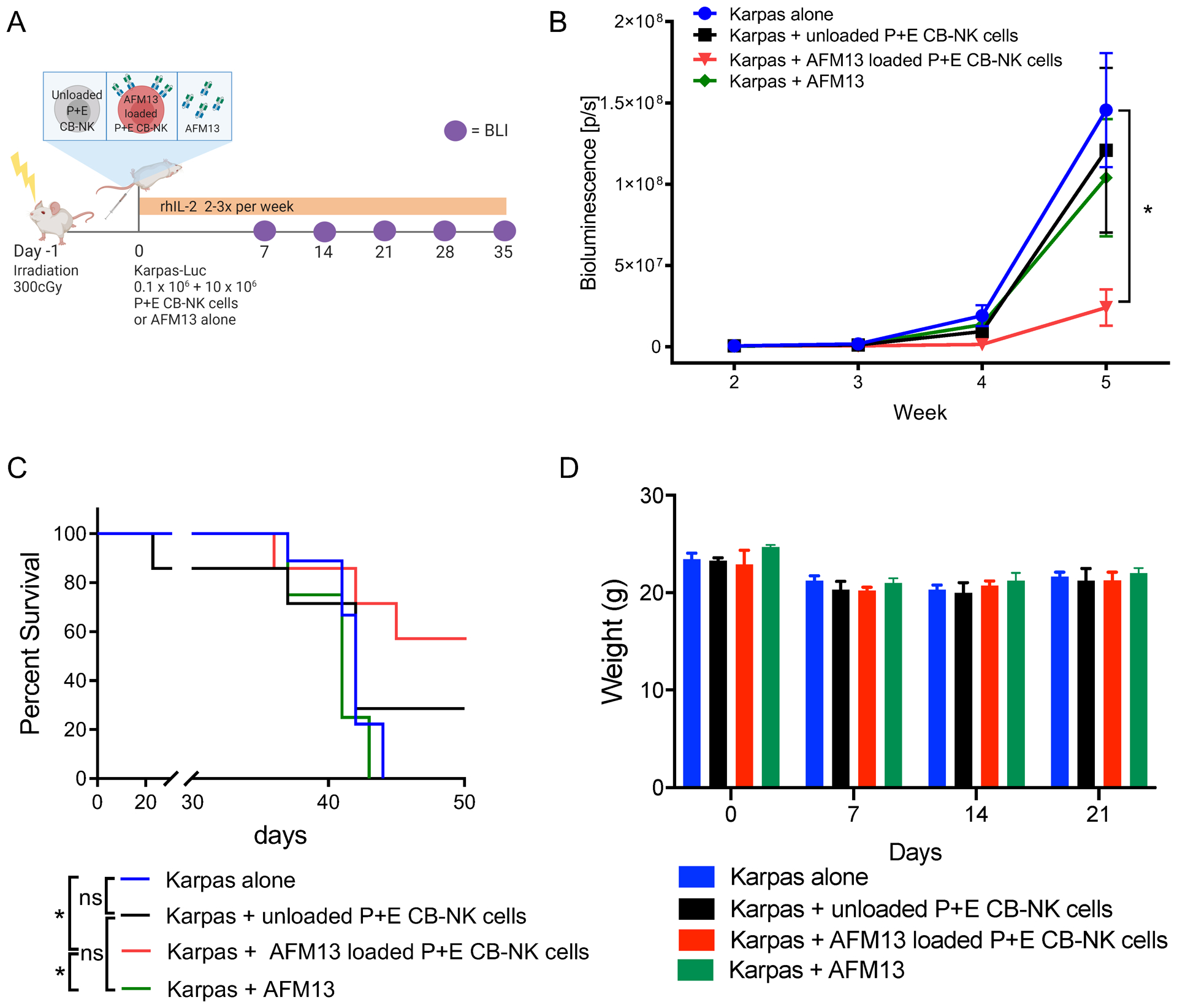
Results: We found heterogeneity within AFM13-directed conventional blood NK cell (cNK) responses, as well as consistent AFM13-directed polyfunctional activation of mature NK cells across donors. NK-cell source also impacted the AFM13 response, with cNK cells from healthy donors exhibiting superior responses to those from patients with Hodgkin lymphoma. IL12/15/18-induced memory-like NK cells from peripheral blood exhibited enhanced killing of CD30+ lymphoma targets directed by AFM13, compared with cNK cells. Cord-blood NK cells preactivated with IL12/15/18 and ex vivo expanded with K562-based feeders also exhibited enhanced killing with AFM13 stimulation via upregulation of signaling pathways related to NK-cell effector function. AFM13-NK complex cells exhibited enhanced responses to CD30+ lymphomas in vitro and in vivo.

Conclusions: We identify AFM13 as a promising combination with cytokine-activated adult blood or cord-blood NK cells to treat CD30+ hematologic malignancies, warranting clinical trials with these novel combinations.


Conflict of interest statement

Disclosure of Potential Conflict of Interest:

TAF and MMBE are consultants and have equity interest in Wugen, and may receive royalty income based on a technology developed by TAF and MMBE and licensed by Washington University to Wugen. JK and WF are employees of Affimed. MT is an employee of Arjuna Therapeutics. TAF has received research support from ImmunityBio, Compass Therapeutics, and HCW Biologics, and advises Kiadis, Nkarta, Indapta, and Orca Biosystems. LNK, RB, EL, SOA, EJS, KR and The University of Texas MD Anderson Cancer Center has an institutional financial conflict of interest with Affimed GmbH. This institutional financial conflict of interest is related to the research reported in this publication. Because MD Anderson is committed to the protection of human subjects and the effective management of its financial conflicts of interest in relation to its research activities, MD Anderson is implementing an Institutional Conflict of Interest Management and Monitoring Plan to manage and monitor the conflict of interest with respect to MD Anderson’s conduct of any other ongoing or future research related to this relationship. LNK, PPB, RB, MD, EL, SOA, REC, EJS, KR and The University of Texas MD Anderson Cancer Center have institutional financial conflict of interest with Takeda Pharmaceutical for the licensing of the technology related to CAR-NK cell research. MD Anderson has implemented an Institutional Conflict of Interest Management and Monitoring Plan to manage and monitor the conflict of interest with respect to MDACC’s conduct of any other ongoing or future research related to this relationship. KR participates on Scientific Advisory Board for GemoAb, AvengeBio, Virogin, GSK and Bayer. The remaining authors declare no competing financial interests.

Figures

Fig. 1.
Fig. 1.
AFM13 triggering induces cell activation and polyfunctional responses of human cNK cells, mostly with a mature phenotype. (A) Schema of cell stimulation used to evaluate NK cells upon AFM13 triggering by CyTOF. (B) Representative density viSNE maps show NK cells unstimulated and stimulated with Hut-78 cells, Hut-78 cells + AFM12 and Hut-78 cells + AFM13. Composite map shows a differential clustering of HuT-78 cells + AFM13. (C) ViSNE maps of a representative donor and summary data (D) show IFN-γ, TNF, MIP1α, CD107 and CD16 expression on NK cells unstimulated or stimulated with HuT-78, HuT-78 + AFM12 and HuT-78 + AFM13. (E) Percent of CD16, CD62L, CD25 and NKp44 positive NK cells unstimulated and stimulated with HuT-78 cells, HuT-78 + AFM12 or HuT-78 + AFM13. (F) Percent of NK cells single or multiple producers of IFN-γ, TNF, MIP1α or CD107 after stimulation with HuT-78 + AFM12 and HuT-78 + AFM13. Numbers under each pie represent the frequency of positive cells for 1, 2, 3 or all the 4 molecules. Bars represent mean + SEM. *p<0.05, **p<0.01, ***p<0.001, ****p<0.0001. One-way ANOVA with Tukey pots-hoc test. n=10.
Fig. 2.
Fig. 2.. AFM13-induced functional response is impacted by underlying NK cell subsets and NK repertoire.
(A) Schema of cell stimulation to evaluate functionality and phenotype of cNK cells responding to AFM13 triggering. Clustering channels included for SPADE analysis are shown. B-D) Representative examples of SPADE analysis from 3 different donors showing the expression of KIRs, CD57 and CDK2A/CD94 in the top 2 nodes with highest IFN-γ expression in response to AFM13. Numbers next to each node represent the node ID and color indicates IFN-g median expression. For each donor, selected nodes were backgated to their own viSNE plots to identify their phenotype. (B) IFN-γ production (nodes 1, 8) in this donor was restricted to mature CD57+KIR2DL2/DL3+ NK cells. In this donor, lack of HLA-Bw4 expression in HuT-78 cells correlates with a robust response of KIR3DL1/DS1+ NK cells. (C) The highest IFN-γ production was restricted to terminally matured CD57+KIR2DL2/3+NKG2A- NK cells in this donor. (D) IFN-γ (nodes 2, 3) was produced by both mature and immature NK cells (CD57+NKG2A+CD94+KIR+/−) NK cells in this donor. (E) Median expression of IFN-γ, TNF, MIP1α, CD107a and (F) NK cell markers associated with cell maturation or differentiation in nodes with the highest and lowest IFN-γ median expression per donor. In F, all donors were positive for the depicted KIR. Consolidated data of 10 donors. Bars represent mean ± SEM. *p<0.05, **p<0.01, ***p<0.001, ****p<0.0001. One-way ANOVA with Tukey pots-hoc test. n=10.
Fig. 3.
Fig. 3.. AFM13 binding triggers enhanced cytotoxicity and cytokine secretion of cytokine induced ML NK cells and pre-activated-expanded CB NK cells.
(A) Specific killing of NK cells from relapsed Hodgkin Lymphoma (HL), healthy donors (HD) or expanded cord blood (CB) against Karpas 299 targets in presence or absence of AFM13 (100 μg/ml). E:T ratio of 10:1. (B) Peripheral blood (PB) cNK but mainly ML NK cells from HD exhibit enhanced cytotoxicity against CD30+ HuT-78 lymphoma targets when triggered with AFM13. (C) Preactivated expanded (P+E) CB-NK cells shows enhanced cytotoxicity against Karpas 299 targets compared to expanded (Exp) CB-NK in presence of different concentrations of AFM13. E:T ratio 5:1. (D) PB cNK and ML NK cells were stimulated HuT-78 cells (5:1 E:T ratio) and the production of IFN-γ, TNF, and CD107a degranulation was evaluated by Flow Cytometry. PB ML NK cells exhibit enhanced cytokine production and degranulation in response HuT-78 cells compared to cNK. In both cNK and ML NK cells AFM13 triggering significantly upregulated the production of IFN-γ, TNF, and CD107 degranulation. (E) cytokine production (IFN-γ and TNF) and CD107a degranulation by AFM loaded (100 μg/ml) P+E CB-NK cells vs. AFM13 loaded (100 μg/ml) expanded (Exp) NK cells co-cultured with Karpas 299 cells for 6 hours. E:T ratio of 1:1 ratio. Bars represent mean ± SEM. Two-way ANOVA with (A, B, D, E) Tukey or (C) Sidak post-hoc test. *p<0.05, **p<0.01, ***p<0.001, ****p<0.0001. n=6–10.
Fig. 4.
Fig. 4.. Gene expression signature of IL-12/15/18 pre-activated and expanded CB-NK cells.
(A) Experimental schema describing the strategy to analyze gene expression of P+E CB NK cells. (B) Volcano plot showing significantly differentiated genes (red dots represent genes with higher expression; blue dots represent genes with lower expression) in P+E CB-NK cells compared to expanded CB-NK cells. (C) Heatmap of differentially expressed genes in P+E CB-NK cells comparing to Exp- CB-NK cells (Differences were identified at adjusted p-value <0.05 and absolute fold-change >1.5, -log10 adjusted p value in annotation padj). (D) Gene set enrichment analysis (GSEA) of differentially expressed genes between P+E CB-NK cells vs expanded CB-NK cells presented as a heatmap. Color scale indicates signal intensity, ranging from lower (blue) to higher (red) expression (for Hallmark). (E) GSEA plots showing enrichment of genes related to IFN-g response, TNF signaling, IL-2/STAT5 signaling, IL-6/JAK/STAT3 signaling and PI3K/AKT/MTOR signaling in P+E CB-NK cells compared to expanded NK cells. n=3
Fig. 5.
Fig. 5.. Retention of AFM13 on NK cell surface enhances cytotoxicity against CD30 positive Karpas 299.
(A) Cytotoxicity (4 hour 51Cr-release assay) of CB-NK cells loaded with AFM13 (0–100 μg/ml) that were either washed twice with PBS (blue bars) or left unwashed (red bars) against Karpas 299 cells at E:T ratio 10:1. (B) Karpas 299 killing over a 24 hour period by AFM13 (100 μg/ml) loaded and washed (blue lines), or unwashed (red lines), along with unloaded (black lines) CB-NK cells as measured by incuCyte. (C) For cell surface retention assays, 1×10⁶ human P+E CB-NK cells were labeled with 100 μg/ml of AFM13 at 37°C for one hour, cells were washed. At the indicated timepoints, the presence of AFM13 was measured using anti-AFM13 antibody followed by an APC-conjugated goat anti-rat IgG. The retention of AFM13 was evaluated by flow cytometry. (D) The retention of AFM13 on unwashed NK cell surface was observed by Image Stream at different timepoints. A representative example of NK cell from each of the time points stained for CD56 (green), CD16 (blue), and AFM13 (red) along with an overlay of all fluorescent channels and bright field is shown. Scale bar=7μm. (E) Percent distribution of total AFM13 fluorescent signal is summarized from the images of 50 cells, which were randomly sampled from 1 hour (red), 24 hours (green), and 48 hours (blue) time points. Each dot represents a cell, horizontal line the mean and error bars indicate +/− SD. (F) P+E CB-NK cells were cultured without or with AFM13 (100 ug/ml) for 1 hour at 37°C, followed by either a wash step or not. The cells were then cultured in media plus 100 IU/mL of rhIL-2 for 1 hour, 48 hours, or 72 hours and their cytotoxicity tested against Karpas 299 target cells at an E:T ratio of 5:1. (G) Bright field image showing AFM13 labelled NK cells were conjugated with Karpas 299 cells. Staining of the conjugates for CD16 (orange), CD56 (teal), CD30 (yellow), F-actin (green), AFM-13 (red), pericentrin (light pink) and an overlay of the BF images with AFM13, CD30 and CD16 are shown. The dotted line defines the synapse and the arrowhead is pointing to the pericentrin in NK cells. Conjugates were evaluated under a 60x objective in ImageStream; scale bar = 7mm. (H) Synaptic localization of CD30, CD16 and AFM13 (Brown), along with polymerization of F-actin (Green) at the immune synapse between NK cells and Karpas299 cells in the presence of AFM13 or IgG control antibody is shown. Horizontal bars represent the average and error bars indicate the range. Bars represent mean ± SEM. Two-way ANOVA with Sidak post-hoc test ** p ≤ 0.01, ***p ≤ 0.001, **** p ≤ 0.0001. n=3–4.
Fig. 6.
Fig. 6.. In vivo antitumor activity of AFM13 loaded P+E CB-NK cells.
(A) Experimental schema applied to evaluate in vivo activity of AFM13. (B) Bioluminescence imagining (BLI) was used to monitor the growth of FFluc-labeled Karpas 299 tumor cells in NSG mice. The plot summarizes the bioluminescence data from four groups of mice treated with Karpas 299 alone, Karpas 299 plus one dose of unloaded NK cells (10 × 106), Karpas 299 plus one dose of AFM13 loaded (100 μg/ml) and washed NK cells (AFM13-NK cell complex; 10 × 106), or Karpas 299 and an injection of AFM13 (100 μg/ml). (C) Kaplan–Meier plots showing the survival of mice described in panel A. Mice treated with a single dose of 10 × 106 AFM13-NK cell complex (blue line) had better survival than control groups. (D) Bar plots summarize the weight of the animals over time as a measure of toxicity. Bars represent mean ± SEM. Two-way ANOVA with Tukey post-hoc test, *p ≤ 0.05; ns, not significant. n=4–9 mice per group, 2 independent experiments.

References

    1. Farkona S, Diamandis EP, Blasutig IM. Cancer immunotherapy: the beginning of the end of cancer? BMC Med. 2016;14:73. - PMC - PubMed
    1. Rouce RH, Shaim H, Sekine T, Weber G, Ballard B, Ku S, et al. The TGF-β/SMAD pathway is an important mechanism for NK cell immune evasion in childhood B-acute lymphoblastic leukemia. Leukemia. 2016;30:800–11. - PMC - PubMed
    1. Wei SC, Duffy CR, Allison JP. Fundamental mechanisms of immune checkpoint blockade therapy. Cancer Discov. 2018. page 1069–86. - PubMed
    1. Garrido F, Cabrera T, Concha A, Glew S, Ruiz-Cabello F, Stern PL. Natural history of HLA expression during tumour development. Immunol. Today 1993. [Accessed 05/05/2020]. page 491–9. - PubMed
    1. Berrien-Elliott MM, Romee R, Fehniger TA. Improving natural killer cell cancer immunotherapy. Curr Opin Organ Transplant. 2015;20:671–80. - PMC - PubMed

Publication types