

Convexal Subarachnoid Hemorrhage Caused by Infective Endocarditis in a Patient with Advanced Human Immunodeficiency Virus (HIV): The Culprits and Bystanders
- PMID: 33986239
- PMCID: PMC8130978
- DOI: 10.12659/AJCR.931376
Convexal Subarachnoid Hemorrhage Caused by Infective Endocarditis in a Patient with Advanced Human Immunodeficiency Virus (HIV): The Culprits and Bystanders
Abstract
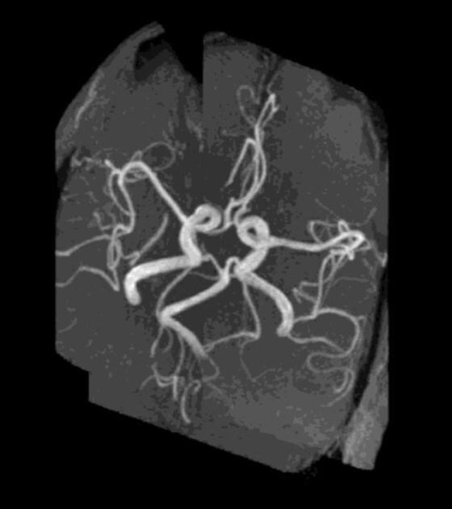
BACKGROUND Convexal subarachnoid hemorrhage (cSAH), a rare form of non-aneurysmal subarachnoid hemorrhage, is confined to cerebral convexities without extension into basal cisterns or ventricles. Typical presentation includes thunderclap/progressive headache or transient focal neurological symptoms; rare manifestations include seizures, intractable vomiting, or altered mental status. Here, we report the first case of convexal subarachnoid hemorrhage and multifocal ischemic lesions caused by infective endocarditis (IE) in a treatment-naïve advanced HIV patient. CASE REPORT A 52-year-old HAART-naïve, HIV-positive, African American man presented with altered mental status, shortness of breath, nonproductive cough, and generalized weakness. His past medical history was significant for congestive heart failure, chronic obstructive pulmonary disease, and end-stage renal disease (noncompliant with hemodialysis). Head computed tomography (CT) showed an isolated sulcal hemorrhage in the mid-left frontal lobe. Fluid-attenuated inversion recovery/gradient recalled echo sequences confirmed a hemorrhage in the left-mid-frontal sulcus, and diffusion-weighted imaging revealed multifocal bilateral ischemic lesions. Transesophageal echocardiography exhibited mitral valve vegetations. Multifocal ischemic lesions and cSAH caused by infectious endocarditis were confirmed. Initiation of intravenous vancomycin and piperacillin-tazobactam allowed the patient to have resolution of his altered mental status. A head CT 5 days later revealed the resolution of cSAH. CONCLUSIONS Infective endocarditis should be considered as an underlying etiology of cSAH, especially when present with multifocal ischemic lesions. Risk factors contributing to the development of cSAH in the IE patient population should be explored in future studies. HIV has not been previously reported in this subgroup and its prevalence should be considered. The prognosis for cSAH in relation to IE is generally favorable.
Conflict of interest statement
None.
Figures


References
-
- Beitzke M, Gattringer T, Enzinger C, et al. Clinical presentation, etiology, and long-term prognosis in patients with nontraumatic convexal subarachnoid hemorrhage. Stroke. 2011;42(11):3055–60. - PubMed
-
- Renou P, Tourdias T, Fleury O, Debruxelles S, et al. Atraumatic nonaneurysmal sulcal subarachnoid hemorrhages: A diagnostic workup based on a case series. Cerebrovasc Dis. 2012;34(2):147–52. - PubMed
-
- Dubosh NM, Bellolio MF, Rabinstein AA, Edlow JA. Sensitivity of early brain computed tomography to exclude aneurysmal subarachnoid hemorrhage: A systematic review and meta-analysis. Stroke. 2016;47(3):750–55. - PubMed
Publication types
MeSH terms
LinkOut - more resources
Full Text Sources
Other Literature Sources
Medical
Miscellaneous
