. 2021 May 13;12(1):2776.
doi: 10.1038/s41467-021-23086-z.

Role of neutrophil extracellular traps in radiation resistance of invasive bladder cancer

Affiliations

Role of neutrophil extracellular traps in radiation resistance of invasive bladder cancer

Surashri Shinde-Jadhav et al. Nat Commun. .

Abstract

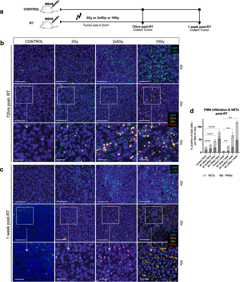
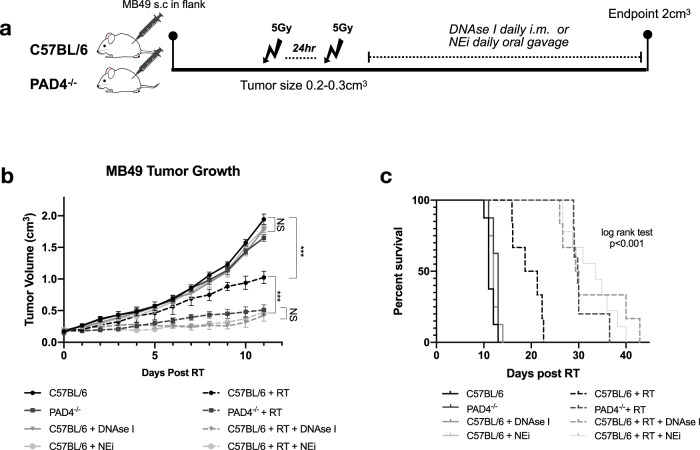
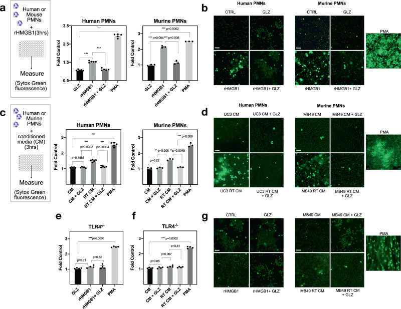
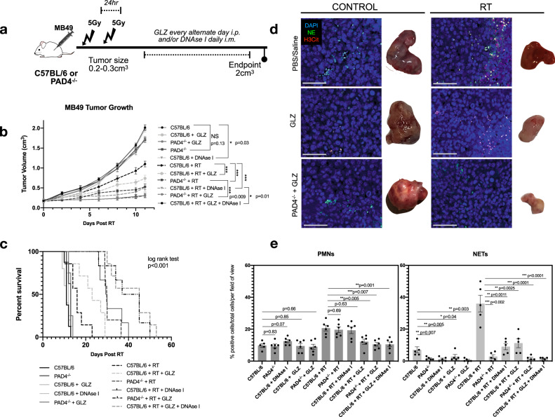
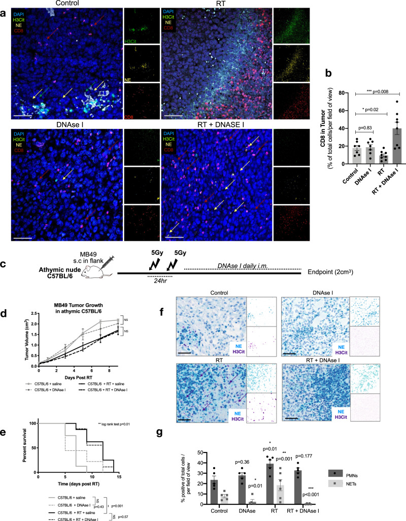
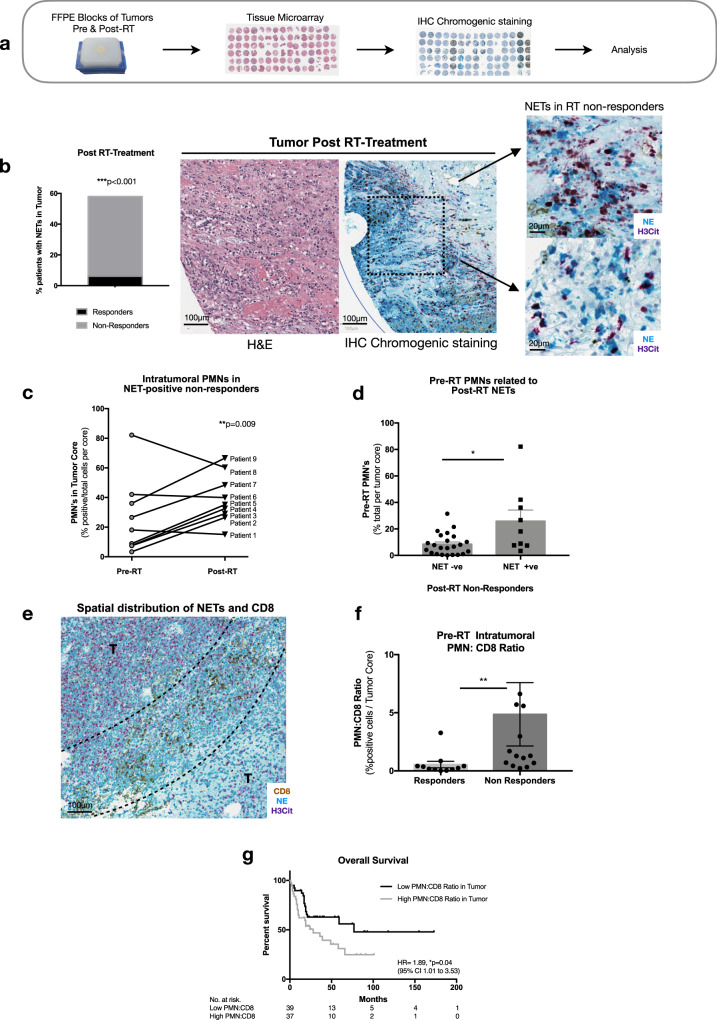
Radiation therapy (RT) is used in the management of several cancers; however, tumor radioresistance remains a challenge. Polymorphonuclear neutrophils (PMNs) are recruited to the tumor immune microenvironment (TIME) post-RT and can facilitate tumor progression by forming neutrophil extracellular traps (NETs). Here, we demonstrate a role for NETs as players in tumor radioresistance. Using a syngeneic bladder cancer model, increased NET deposition is observed in the TIME of mice treated with RT and inhibition of NETs improves overall radiation response. In vitro, the protein HMGB1 promotes NET formation through a TLR4-dependent manner and in vivo, inhibition of both HMGB1 and NETs significantly delays tumor growth. Finally, NETs are observed in bladder tumors of patients who did not respond to RT and had persistent disease post-RT, wherein a high tumoral PMN-to-CD8 ratio is associated with worse overall survival. Together, these findings identify NETs as a potential therapeutic target to increase radiation efficacy.


Conflict of interest statement

The authors declare no competing interests

Figures

Fig. 1
Fig. 1. Radiation induces NET formation in murine MB49 tumors.
a Schematic representation of time point experiment where tumors were irradiated with: 2 Gy, two fractions of 5 Gy (2 × 5 Gy) or 10 Gy. Tumors were collected 72 h and 1-week post-radiation (post-RT) for immunofluorescence analyses. b Representative confocal images of non-irradiated controls or irradiated tumors 72 h post-RT or c 1 week-post RT stained for PMNs (Ly6G-green, 20×, 63×) and NETs, representative from three independent experiments (H3Cit-red and NE-green, 20×, 63×), nuclei (blue). Scale bars are 50 μm for 20× images and 20 μm for 63× images. d Quantification of PMNs and NETs in confocal images using QuPath V6, expressed as percentage of positive cells per total cells per field of view. Data are expressed as mean ± SEM (n = 10 mice per group, n = 3). Unpaired two-tailed students t-test was used to assess statistical significance between groups (***p < 0.001 for NETs).
Fig. 2
Fig. 2. Inhibition and degradation of NETs improves radiation response.
a Schematic representation of tumor growth experiment, where C57BL/6 or PAD4−/− mice were injected subcutaneously (s.c) in the right flank with MB49 (500,000 cells). Mice were randomized into two groups: non-irradiated (control) or irradiated (RT). Tumors were irradiated with two fractions of 5 Gy and DNAse I was administered intramuscularly (i.m.) to degrade NETs or NEi was administered through oral gavage to inhibit NETs. b Tumor growth kinetics 11 days post-RT (n = 8 mice per arm). Data represented as mean ± SEM, two-way ANOVA with Bonferroni’s multiple comparison’s test was used to assess statistical significance. c Kaplan–Meier percent survival, log rank (Mantel-cox) test. NS = not significant p < 0.05 (C57BL/6 vs. PAD4−/− p = 0.28, C57BL/6 + DNAse I p = 0.07, C57BL/6 NEi p = 0.10, PAD4−/− RT vs. C57BL/6 RT + DNAse I p = 0.34, PAD4−/− RT vs. C57BL/6 RT + NEi p = 0.06, C57BL/6 RT + DNAse I vs. C57BL/6 RT + NEi p > 0.99), ***p < 0.001.
Fig. 3
Fig. 3. HMGB1 promotes NET formation through TLR4.
a Stimulation of human PMNs (N = 5 PMNs isolated from different healthy donors) or murine PMNs (N = 3) with rHMGB1 significantly induces NETs measured by Sytox green fluorescence ***p < 0.001. b Representative fluorescence images through confocal microscopy (10×), scale bar 100 μm. c Stimulation of human PMNs (n = 5 PMNs isolated from different healthy donors) with irradiated UM-UC3 conditioned media or murine PMNs (n = 3) with irradiated MB49 conditioned media significantly induces NETs and this is reversed through addition of GLZ. d Representative fluorescence images through confocal microscopy (10×), scale bar 100 μm. e Stimulation of murine PMNs from TLR4−/− mice stimulated with rHMGB1 (n = 3) or f irradiated MB49 conditioned media (n = 3), and g representative fluorescence images through confocal microscopy (10×), scale bar 100 μm. All data are represented as mean ± SEM of triplicates from individual experiments, paired-student’s t-test was used to assess statistical significance (a, c, e, f).
Fig. 4
Fig. 4. Inhibition of HMGB1 and NETs improves overall RT response.
a Schematic representation of tumor growth experiment where C57BL/6 or PAD4−/− mice were injected subcutaneously (s.c) in the right flank with MB49 (500,000 cells). Mice were randomized into two groups: non-irradiated (control) or irradiated (RT). Tumors were irradiated with two fractions of 5 Gy and DNAse I was administered intramuscularly (i.m.) to degrade NETs or GLZ was administered intraperitoneally (i.p) to inhibit extracellular HMGB1. b MB49 tumor growth 11 days post-RT (n = 8 mice per arm for all groups except n = 10 for C57BL/6 RT + DNAse I, n = 9 C57BL/6 + RT + GLZ + DNAse I). c Kaplan–Meier percent survival. d Immunofluorescence of tissues obtained at endpoint staining for PMNs and NETs through NE (green) and H3Cit (red) staining, nuclei (blue), 20×, scale bar 50 μm, representative of three independent experiments. e Quantification of PMN infiltration and NETs in tumors, data represented as percent positive of total cells per field of view (n = 6 mice). Data represented as mean ± SEM. Statistical significance was assessed using two-way ANOVA with Bonferroni’s multiple comparison’s test (b) log rank (Mantel-cox) test (c) unpaired two-tailed students t-test (e). NS not significant (compared to C57BL/6: C57BL/6 + GLZ p > 0.05, PAD4−/− + GLZ p = 0.053, PAD4−/− p = 0.13), ***p < 0.001.
Fig. 5
Fig. 5. DNAse I-treated mice show increased intratumoral CD8 infiltration post-RT.
a FFPE tumors were collected from C57BL/6 mice injected subcutaneously (s.c) with MB49 tumors from groups: non-irradiated control (Control), C57BL/6 mice treated with DNAse-treated mice (DNAse I), irradiated C57BL/6 (RT) and irradiated C57BL/6 treated with DNAse I (RT + DNAse I) at endpoint (3 weeks post-RT). Tissues were stained for immunofluorescence analyses with CD8 (red), NE (yellow), H3Cit (green), nuclei (blue). Representative confocal images from three independent experiments obtained with 20× objective, scale bar 50 μm. b Quantification of intratumoral CD8 T-cell infiltration using QuPathV6. Data represented as percentage CD8 T-cells per total cells per field (n = 7 mice per arm). c Schematic of tumor and survival experiment in athymic C57BL/6 mice. d Tumor growth kinetics of irradiated athymic mice injected s.c with MB49, treated with (n = 8 mice per group) or without DNAse I (n = 9 mice per group). e Kaplan–Meier percent survival. f Chromogenic immunohistochemical staining of athymic tumors, stained for PMNs and NETs (NE-blue, H3Cit-purple), scale bar 50 μm. g Quantification of PMN infiltration and NETs in tumors, data represented as percent positive of total cells per field of view (n = 5 mice per arm). Data represented as mean ± SEM, unpaired two tailed student’s t-test was used to assess statistical significance in (b, g) two way ANOVA with Bonferroni’s multiple comparison’s test (d), log rank (Mantel-cox) test (e). NS not significant (p > 0.05), *p < 0.05, **p < 0.01, ***p < 0.001.
Fig. 6
Fig. 6. NETs are present in human MIBC tumors and a high PMN to CD8 ratio is associated with worse overall survival.
a Schematic representation of retrospective study design. b Representative image of NETs staining in MIBC tumors, where NETs were observed in patients with persistent disease post-RT treatment compared to responders, two-sided Fischer’s exact test (n = 52% non responders, n = 6% responders). c Intratumoral PMN infiltration pre-RT and post-RT treatment in subset of patients whom were NET positive, unpaired two-tailed paired student t-test. d Pre-RT intratumoral PMN’s correlates with post-RT NETs, unpaired two-tailed Mann–Whitney test (p = 0.02) (NET + ve patients n = 9, NET−ve patients n = 23). e Representative image of the spatial distribution of NETs (H3Cit-purple, NE-blue) and CD8 (brown) observed in tumor of a non-responder, tumor region (T). f PMN to CD8 ratio pre-RT, data expressed as percentage of positive cells per tumor core, unpaired two-tailed Mann–Whitney test p = 0.0018 (n = 11 responders, n = 14 non-responders). g Overall survival of patients with low or high PMN to CD8 ratio, log rank test (Mantel-cox). Data represented as mean ± SEM, *p < 0.05, **p < 0.01, ***p < 0.001.

Comment in

References

    1. Sung, H. et al. Global cancer statistics 2020: GLOBOCAN estimates of incidence and mortality worldwide for 36 cancers in 185 countries. Cancer J. Clin. 68, 394–424 (2021). - PubMed
    1. Alfred Witjes J, et al. Updated 2016 EAU Guidelines on muscle-invasive and metastatic bladder cancer. Eur. Urol. 2017;71:462–475. doi: 10.1016/j.eururo.2016.06.020. - DOI - PubMed
    1. Stein JP, Skinner DG. Radical cystectomy for invasive bladder cancer: long-term results of a standard procedure. World J. Urol. 2006;24:296–304. doi: 10.1007/s00345-006-0061-7. - DOI - PubMed
    1. Gray PJ, et al. Use of potentially curative therapies for muscle-invasive bladder cancer in the United States: results from the National Cancer Data Base. Eur. Urol. 2013;63:823–829. doi: 10.1016/j.eururo.2012.11.015. - DOI - PubMed
    1. Gore JL, et al. Use of radical cystectomy for patients with invasive bladder cancer. J. Natl Cancer Inst. 2010;102:802–811. doi: 10.1093/jnci/djq121. - DOI - PMC - PubMed

MeSH terms

Grants and funding