. 2021 May 13;6(1):71.
doi: 10.1038/s41541-021-00331-6.

Highly conserved, non-human-like, and cross-reactive SARS-CoV-2 T cell epitopes for COVID-19 vaccine design and validation

Affiliations

Highly conserved, non-human-like, and cross-reactive SARS-CoV-2 T cell epitopes for COVID-19 vaccine design and validation

Lauren M Meyers et al. NPJ Vaccines. .

Abstract

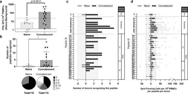
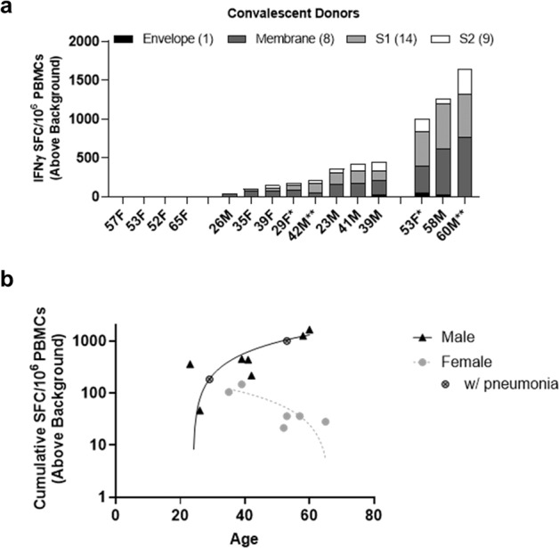
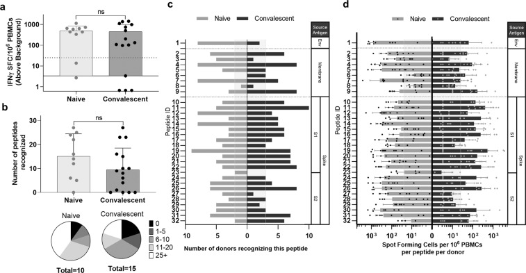
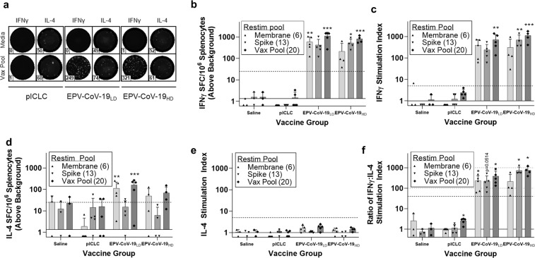
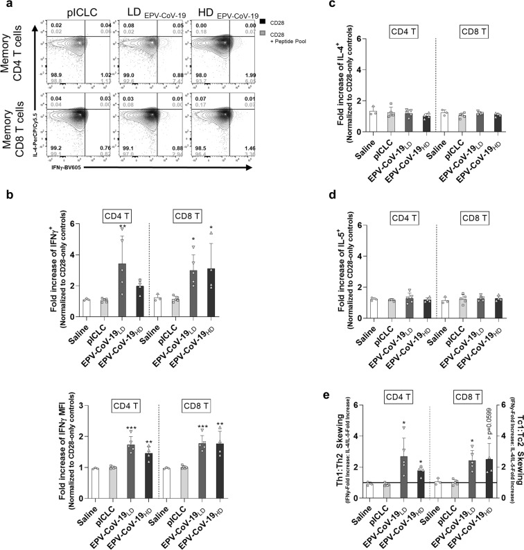
Natural and vaccine-induced SARS-CoV-2 immunity in humans has been described but correlates of protection are not yet defined. T cells support the SARS-CoV-2 antibody response, clear virus-infected cells, and may be required to block transmission. In this study, we identified peptide epitopes associated with SARS-CoV-2 T-cell immunity. Using immunoinformatic methods, T-cell epitopes from spike, membrane, and envelope were selected for maximal HLA-binding potential, coverage of HLA diversity, coverage of circulating virus, and minimal potential cross-reactivity with self. Direct restimulation of PBMCs collected from SARS-CoV-2 convalescents confirmed 66% of predicted epitopes, whereas only 9% were confirmed in naive individuals. However, following a brief period of epitope-specific T-cell expansion, both cohorts demonstrated robust T-cell responses to 97% of epitopes. HLA-DR3 transgenic mouse immunization with peptides co-formulated with poly-ICLC generated a potent Th1-skewed, epitope-specific memory response, alleviating safety concerns of enhanced respiratory disease associated with Th2 induction. Taken together, these epitopes may be used to improve our understanding of natural and vaccine-induced immunity, and to facilitate the development of T-cell-targeted vaccines that harness pre-existing SARS-CoV-2 immunity.


Conflict of interest statement

A.S.D.G. and W.D.M. are senior officers and shareholders, and L.M.M., A.H.G., C.M.B., F.T., B.G.M., and L.M. are employees of EpiVax, Inc., a privately owned biotechnology company located in Providence, RI. M.F.P. is an employee of EpiVax Therapeutics, Inc. Both EpiVax and EpiVax Therapeutics are developing COVID-19 vaccines. These authors acknowledge that there is a potential conflict of interest related to their relationship with EpiVax and attest that the work contained in this research report is free of any bias that might be associated with the commercial goals of the company. A.S. is a senior officer at Oncovir, Inc. Oncovir is developing a COVID-19 therapy. He acknowledges that there is a potential conflict of interest related to his relationship with Oncovir and attests that the work contained in this research report is free of any bias that might be associated with the commercial goals of the company.

Figures

Fig. 1
Fig. 1. Predicted SARS-CoV-2 T-cell epitopes are antigenic ex vivo in COVID-19 convalescent donors but not healthy donors.
a Convalescent (n = 15) and pre-SARS-CoV-2 pandemic donors (naive, n = 10) were stimulated with the total peptide pool consisting of our 32 predicted epitopes and IFNγ-producing cells were measured by Fluorospot assay. Open circles identify responses to low-dose restimulation at 0.313 μg/mL per peptide; closed circles are stimulations at 10 μg/mL per peptide. Dotted horizontal line indicates positivity criteria at SFC/106 splenocytes = 25; solid horizontal line indicates threshold of detection for assay (t = 2.058, d.f. = 23). b IFNγ responses to individual peptides were also assessed, identifying the breadth of response in individual donors (t = 2.113, d.f. = 23), c the frequency of responses to unique peptides within the cohort (vertical lines denote 20% of each cohort), d and the depth of response indicated by the frequency of IFNγ-producing, epitope-specific cells (vertical lines indicate positivity criteria at SFC/106 splenocytes = 25). Error bars depict SD. *p < 0.05.
Fig. 2
Fig. 2. SARS-CoV-2 experienced individuals exhibit variable immune recall responses ex vivo.
a Significant responses to individual peptides (combined per source antigen) identify three distinct immunotype cohorts within covalescent donors (donor notations: *pneumonia, **hospitalized, non-ICU). b Correlation of cumulative T-cell responses and age according to gender. Male cohort (n = 7, R2 = 0.7571, p = 0.0109). Female cohort (n = 8, R2 = 0.00001, p = 0.9934). Mild subset of female cohort (n = 6, R2 = 0.7034, p = 0.0368).
Fig. 3
Fig. 3. Antigen-specific T-cell expansion increases responses in COVID-19 convalescents and uncovers pre-existing SARS-CoV-2 immunity in healthy donors.
a PBMCs of convalescent (n = 15) and naive donors (n = 10) were restimulated with the total peptide pool following an 8-day expansion culture and IFNγ-producing cells were measured by Fluorospot assay. Dotted horizontal line indicates positivity criteria at SFC/106 splenocytes = 25, solid horizontal line indicates threshold of detection for assay (p = 0.8216, t = 0.2281, d.f. = 23). b IFNγ responses to individual peptides were also assessed, identifying the breadth of response in individual donors (p = 0.1424, t = 1.571, d.f. = 23), c the frequency of responses to unique peptides within the cohort (vertical lines denote 20% of each cohort), and d the depth of response indicated by the frequency of IFNγ-producing, epitope-specific cells (vertical lines indicate positivity criteria at SFC/106 splenocytes = 25). Error bars depict SD.
Fig. 4
Fig. 4. EPV-CoV-19 immunization stimulates strong type 1-skewed T-cell responses in HLA-DR3 transgenic mice.
Eight days post boost, murine splenocytes were isolated and assayed for epitope-specific recall responses. Cells were plated in dual IFNγ/IL-4 Fluorospot plates and restimulated with peptide pools for 48 h. a Representative images and spot counts are shown for both cytokines (n = 3). b IFNγ SFC counts were normalized to 1 × 106 cells and adjusted by background subtraction, and c IFNγ SI index was determined by calculating the fold change of individual restimulation replicates over background. d IL-4 SFC and e SI were similarly calculated. Dotted horizontal lines denote positivity criteria of SFC > 25 and SI > 5, respectively. Solid horizontal lines denote threshold of detection for assay. f From the reported IFNγ and IL-4 stimulation indexes, we calculated the IFNγ : IL-4 ratio of each restimulation replicate to model the overall skewing of the immune response, identifying a sharply type 1 skewed phenotype in all vaccinated animals. Dotted horizontal lines identify 40, 100, and 1000-fold skewing of type 1/type 2 response. Solid horizontal line identifies hypothetical balanced response (Th1 = Th2). Error bars depict SD. Saline n = 3, pICLC n = 5, EPV-CoV-19LD n = 5, EPV-CoV-19HD n = 4, *p < 0.05, **p < 0.01, ***p < 0.001.
Fig. 5
Fig. 5. EPV-CoV-19 immunization stimulates type 1-skewed memory CD4+ and CD8+ T cells in HLA-DR3 transgenic mice.
Splenocytes were restimulated with a vaccine-matched peptide pool for 6 h in the presence of brefeldin A and monensin. Following incubation, cells were stained for surface markers, fixed and permeabilized, stained for intracellular markers, and expression of markers was recorded by flow cytometry. Memory CD4+ T cells and CD8+ T cells were assessed for IFNγ, IL-4, or IL-5-production (both frequency in parent T-cell population and mean fluorescence intensity (MFI) of cytokines). a Representative images of type 1 and type 2 skewed, epitope-specific memory T-cell populations are shown. b The fold increase of epitope-specific responses (over anti-CD28 stimulated controls) identify vaccine-specific induction of IFNγ, but not c IL-4 or d IL-5. e From ICS generated data, we calculated the fold increase of IFNγ or IL-4- and/or IL-5-producing cells with peptide restimulation, and used the ratio of type 1-to-type 2 responses to model Th-skewing and Tc-skewing in vaccinated animals. Error bars depict SD. Saline n = 3, pICLC n = 5, EPV-CoV-19LD n = 5, EPV-CoV-19HD n = 4, *p < 0.05, *p < 0.01, ***p < 0.001.

References

    1. Sette A, Crotty S. Adaptive immunity to SARS-CoV-2 and COVID-19. Cell. 2021;184:861–880. doi: 10.1016/j.cell.2021.01.007. - DOI - PMC - PubMed
    1. Huang AT, et al. A systematic review of antibody mediated immunity to coronaviruses: kinetics, correlates of protection, and association with severity. Nat. Commun. 2020;11:4704. doi: 10.1038/s41467-020-18450-4. - DOI - PMC - PubMed
    1. Choe PG, et al. Waning antibody responses in asymptomatic and symptomatic SARS-CoV-2 infection. Emerg. Infect. Dis. 2021;27:327–329. doi: 10.3201/eid2701.203515. - DOI - PMC - PubMed
    1. Perreault, J. et al. Waning of SARS-CoV-2 RBD antibodies in longitudinal convalescent plasma samples within four months after symptom onset. Blood. 10.1182/blood.2020008367 (2020). - PMC - PubMed
    1. Ogega, C. O. et al. Durable SARS-CoV-2 B cell immunity after mild or severe disease. J. Clin. Invest. 10.1172/jci145516 (2021). - PMC - PubMed