

The use of magnetic resonance imaging technique and 3D printing in order to develop a three-dimensional fistula model for patients with Crohn's disease: personalised medicine
- PMID: 33986892
- PMCID: PMC8112265
- DOI: 10.5114/pg.2020.101629
The use of magnetic resonance imaging technique and 3D printing in order to develop a three-dimensional fistula model for patients with Crohn's disease: personalised medicine
Abstract
Introduction: Preoperative evaluation of magnetic resonance (MR) images may not be sufficient for the precise planning of anal fistula surgery or for stem cell therapy. Three-dimensional (3D) printing allows one to obtain spatial structures in a 1 : 1 scale with unprecedented precision.
Aim: To combine magnetic resonance imaging (MRI) with 3D printing for more precise visualisation of perianal Crohn's disease.
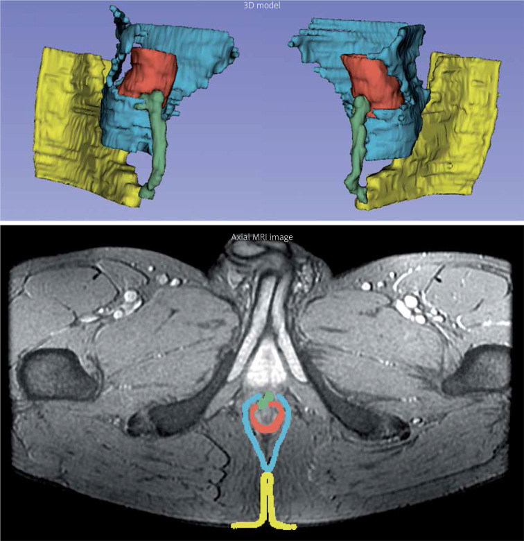
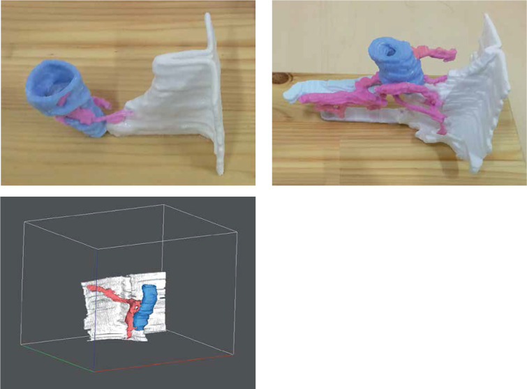
Material and methods: MRI at 1.5T and a 3D printer were used. DICOM (Digital imaging and communications in medicine) images were imported into 3D Slicer v.4.8.0. Firstly, anal fistula was modelled on the basis of axial images. Fistula locations, the anus and anal canal, were marked with different coloured markers. The last step was to mark the skin that was connected to the anus and contact areas of the fistula with the skin. The prepared models were then exported to an STL format file. The anal fistula model was printed using a 3D printer. The development of the model, including printing, took approximately 6 h.
Results and conclusions: The accessibility of a rotatable 3D model before surgery allows for a more precise detection of the location and the degree of perianal disease. Moreover, this may also lower the inter-observer bias connected with interpretation of complex MR imaging before planned surgery. Development of MRI image transfer to 3D printing and the decreasing cost of 3D printers suggests a promising future of this technology in medical applications.
Keywords: 3D printing; anal fistula 3D model; magnetic resonance imaging.
Copyright © 2021 Termedia.
Conflict of interest statement
The authors declare no conflict of interest.
Figures
References
-
- Schwartz DA, Loftus EV Jr, Tremaine WJ, et al. The natural history of fistulizing Crohn’s disease in Olmsted County, Minnesota. Gastroenterology 2007; 122: 875-80. - PubMed
-
- Andreani SM, Dang HH, Grondona P, et al. Rectovaginal fistula in Crohn’s disease. Dis Colon Rectum 2007; 50: 2215-22. - PubMed
-
- Farmer RG, Hawk WA, Turnbull RB Jr. Clinical patterns in Crohn’s disease: a statistical study of 615 cases. Gastroenterology 1975; 68: 627635. - PubMed
LinkOut - more resources
Full Text Sources
Other Literature Sources