. 2021 Apr 30:9:e11367.
doi: 10.7717/peerj.11367. eCollection 2021.

The hypoferremic response to acute inflammation is maintained in thalassemia mice even under parenteral iron loading

Affiliations

The hypoferremic response to acute inflammation is maintained in thalassemia mice even under parenteral iron loading

Chanita Sanyear et al. PeerJ. .

Abstract

Background: Hepcidin controls iron homeostasis by inducing the degradation of the iron efflux protein, ferroportin (FPN1), and subsequently reducing serum iron levels. Hepcidin expression is influenced by multiple factors, including iron stores, ineffective erythropoiesis, and inflammation. However, the interactions between these factors under thalassemic condition remain unclear. This study aimed to determine the hypoferremic and transcriptional responses of iron homeostasis to acute inflammatory induction by lipopolysaccharide (LPS) in thalassemic (Hbbth3 /+) mice with/without parenteral iron loading with iron dextran.

Methods: Wild type and Hbbth3 /+ mice were intramuscularly injected with 5 mg of iron dextran once daily for two consecutive days. After a 2-week equilibration, acute inflammation was induced by an intraperitoneal injection of a single dose of 1 µg/g body weight of LPS. Control groups for both iron loading and acute inflammation received equal volume(s) of saline solution. Blood and tissue samples were collected at 6 hours after LPS (or saline) injection. Iron parameters and mRNA expression of hepcidin as well as genes involved in iron transport and metabolism in wild type and Hbbth3 /+ mice were analyzed and compared by Kruskal-Wallis test with pairwise Mann-Whitney U test.

Results: We found the inductive effects of LPS on liver IL-6 mRNA expression to be more pronounced under parenteral iron loading. Upon LPS administration, splenic erythroferrone (ERFE) mRNA levels were reduced only in iron-treated mice, whereas, liver bone morphogenetic protein 6 (BMP6) mRNA levels were decreased under both control and parenteral iron loading conditions. Despite the altered expression of the aforementioned hepcidin regulators, the stimulatory effect of LPS on hepcidin mRNA expression was blunt in iron-treated Hbbth3 /+ mice. Contrary to the blunted hepcidin response, LPS treatment suppressed FPN1 mRNA expression in the liver, spleen, and duodenum, as well as reduced serum iron levels of Hbbth3 /+ mice with parenteral iron loading.

Conclusion: Our study suggests that a hypoferremic response to LPS-induced acute inflammation is maintained in thalassemic mice with parenteral iron loading in a hepcidin-independent manner.

Keywords: Hepcidin; Iron loading; Iron transporters; Lipopolysaccharide; Thalassemic mice.


Conflict of interest statement

The authors declare that they have no competing interests.

Figures
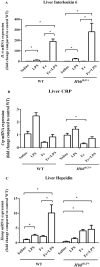
Figure 1
Figure 1. Effects of LPS on the mRNA expression of interleukin 6, C-reactive protein and hepcidin in the liver of wild type and thalassemic mice with/without parenteral iron loading.
The mRNA expression of (A) interleukin 6 (IL-6), (B) C-reactive protein (CRP) and (C) hepcidin in the liver of wild type (WT) and thalassemic (Hbbth3/+) mice treated with iron dextran/saline followed by LPS/saline administration. Tissue samples were collected at 6 hours after LPS/saline injection. Gene expression was normalized to β-actin (Actb) expression. Data are presented as mean and SEM of the fold change compared to saline-treated WT mice (WT-Saline) (n = 5 per group). Statistical analysis was performed using Kruskal–Wallis test with pairwise Mann–Whitney U test. The acquired P values were subsequently adjusted using the Bonferroni correction (*adjusted P-value < 0.01).
Figure 2
Figure 2. Effects of LPS on the mRNA expression of upstream regulators of hepcidin in wild type and thalassemic mice with/without parenteral iron loading.
The mRNA expression of (A) spleen ERFE, (B) liver BMP6 and (C) liver TMPRSS6 in wild type (WT) and thalassemic (Hbbth3/+) mice treated with iron dextran/saline followed by LPS/saline administration. Tissue samples were collected at 6 hours after LPS/saline injection. Gene expression was normalized to β-actin (Actb) expression. Data are presented as mean and SEM of the fold change compared to saline-treated WT mice (WT-Saline) (n = 5 per group). Statistical analysis was performed using Kruskal–Wallis test with pairwise Mann–Whitney U test. The acquired P values were subsequently adjusted using the Bonferroni correction (*adjusted P-value < 0.01).
Figure 3
Figure 3. Effects of LPS on the mRNA expression of DMT1 and FPN1 in the liver and spleen of wild type and thalassemic mice with/without parenteral iron loading.
The mRNA expression of (A) liver DMT1, (B) liver ferroportin (FPN1), (C) spleen DMT1 and (D) spleen ferroportin (FPN1) in wild type (WT) and thalassemic (Hbbth3/+) mice treated with iron dextran/saline followed by LPS/saline administration. Tissue samples were collected at 6 hours after LPS/saline injection. Gene expression was normalized to β-actin (Actb) expression. Data are presented as mean and SEM of the fold change compared to saline-treated WT mice (WT-Saline) (n = 4–5 per group). Statistical analysis was performed using Kruskal–Wallis test with pairwise Mann–Whitney U test. The acquired P values were subsequently adjusted using the Bonferroni correction (*adjusted P-value < 0.01).
Figure 4
Figure 4. Effects of LPS on the mRNA expression of iron transport molecules in the duodenum of wild type and thalassemic mice with/without parenteral iron loading.
The mRNA expression of (A) DCYTB, (B) DMT1 and (C) ferroportin (FPN1) in the duodenum of wild type (WT) and thalassemic (Hbbth3/+) mice treated with iron dextran/saline followed by LPS/saline administration. Tissue samples were collected at 6 hours after LPS/saline injection. Gene expression was normalized to β-actin (Actb) expression. Data are presented as mean and SEM of the fold change compared to saline-treated WT mice (WT-Saline) (n = 5 per group). Statistical analysis was performed using Kruskal–Wallis test with pairwise Mann–Whitney U test. The acquired P values were subsequently adjusted using the Bonferroni correction (*adjusted P-value < 0.01).

Similar articles

References

    1. Abboud S, Haile DJ. A novel mammalian iron-regulated protein involved in intracellular iron metabolism. Journal of Biological Chemistry. 2000;275(26):19906–19912. doi: 10.1074/jbc.M000713200. - DOI - PubMed
    1. Agoro R, Taleb M, Quesniaux VFJ, Mura C. Cell iron status influences macrophage polarization. PLOS ONE. 2018;13(5):e0196921. doi: 10.1371/journal.pone.0196921. - DOI - PMC - PubMed
    1. Andriopoulos B, Jr, Corradini E, Xia Y, Faasse SA, Chen S, Grgurevic L, Knutson MD, Pietrangelo A, Vukicevic S, Lin HY, Babitt JL. BMP6 is a key endogenous regulator of hepcidin expression and iron metabolism. Nature Genetics. 2009;41(4):482–487. doi: 10.1038/ng.335. - DOI - PMC - PubMed
    1. Arezes J, Jung G, Gabayan V, Valore E, Ruchala P, Gulig PA, Ganz T, Nemeth E, Bulut Y. Hepcidin-induced hypoferremia is a critical host defense mechanism against the siderophilic bacterium Vibrio vulnificus. Cell Host & Microbe. 2015;17:47–57. - PMC - PubMed
    1. Atanasova B, Mudway IS, Laftah AH, Latunde-Dada GO, McKie AT, Peters TJ, Tzatchev KN, Simpson RJ. Duodenal ascorbate levels are changed in mice with altered iron metabolism. Journal of Nutrition. 2004;134(3):501–505. doi: 10.1093/jn/134.3.501. - DOI - PubMed

LinkOut - more resources