. 2021 Apr 27:11:664242.
doi: 10.3389/fonc.2021.664242. eCollection 2021.

miR-1301-3p Promotes Cell Proliferation and Facilitates Cell Cycle Progression via Targeting SIRT1 in Gastric Cancer

Affiliations

miR-1301-3p Promotes Cell Proliferation and Facilitates Cell Cycle Progression via Targeting SIRT1 in Gastric Cancer

Dakui Luo et al. Front Oncol. .

Abstract

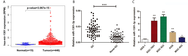
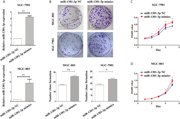
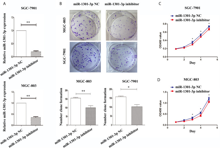
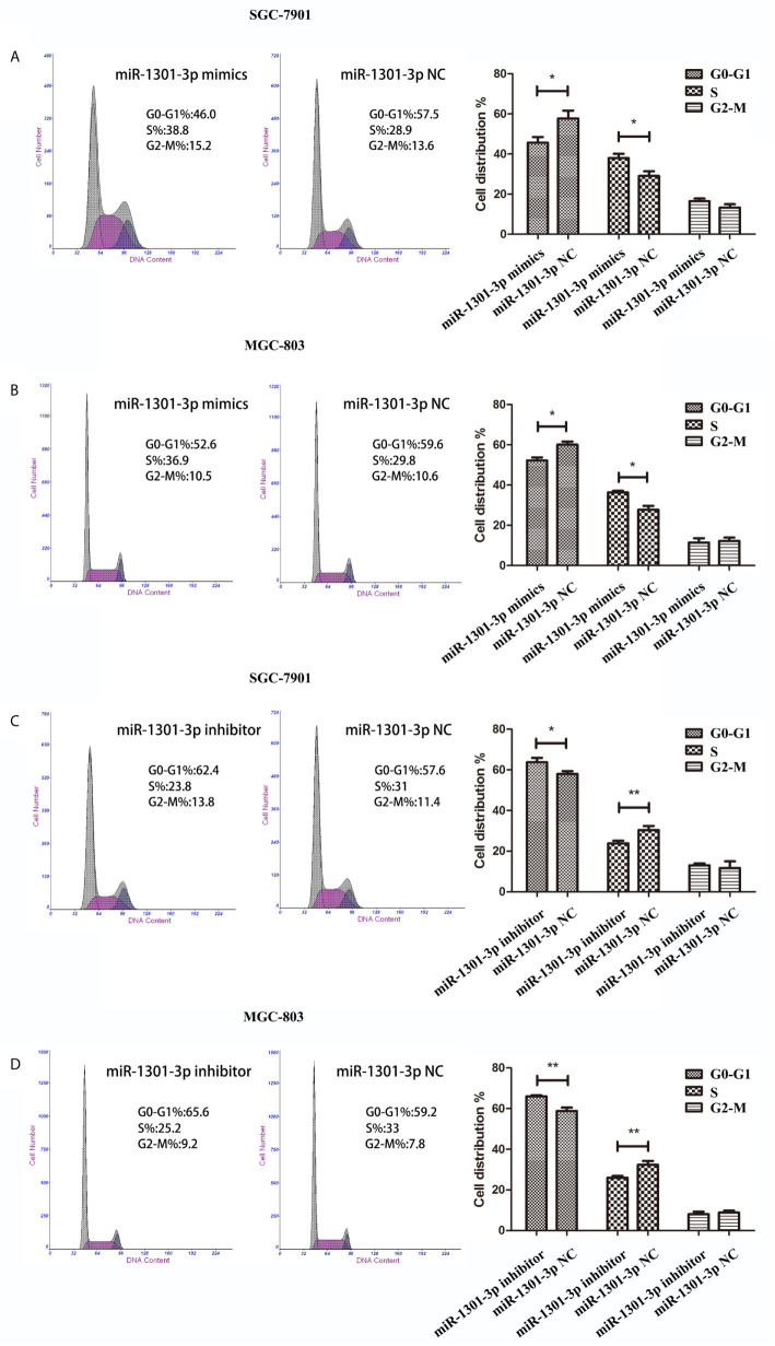
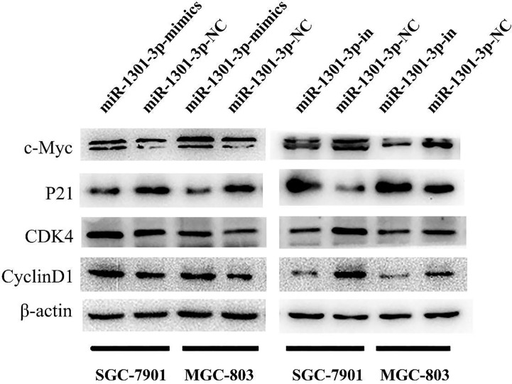
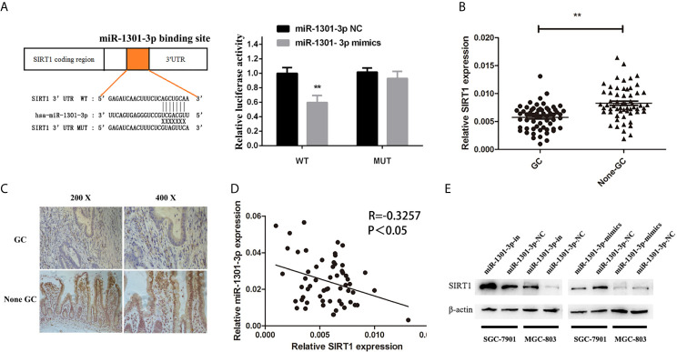
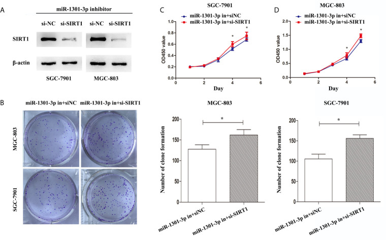
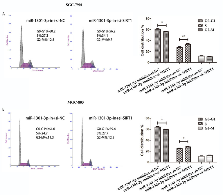
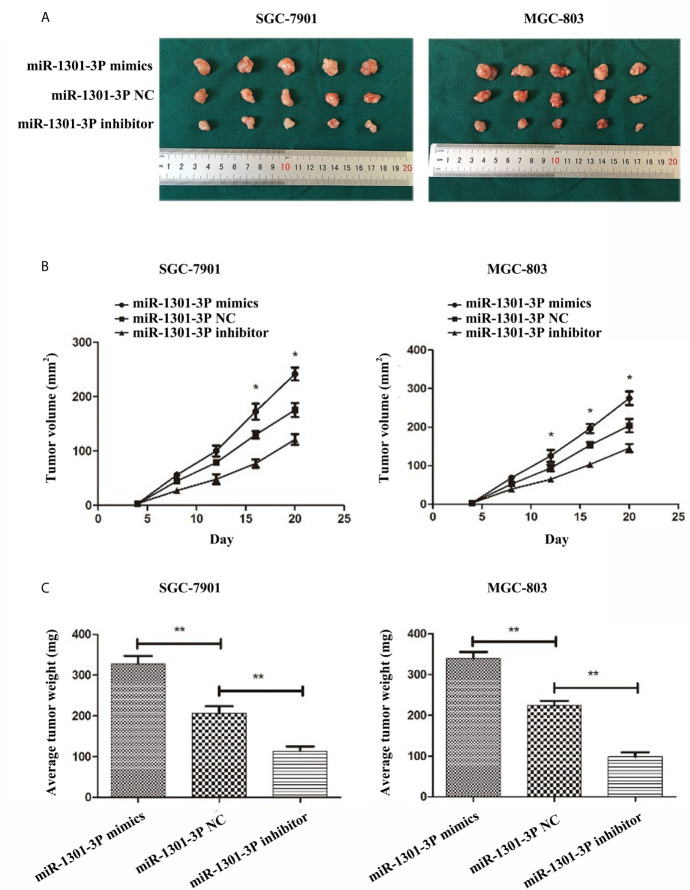
So far, many existing evidences indicate that microRNAs (miRNA) are closely associated with the tumorigenesis and progression of various tumors. It has been reported that miR-1301-3p is abnormally expressed in several malignant tumors. However, the role of miR-1301-3p in gastric cancer (GC) remains unclear and is worth studying. Through qRT-PCR, the expression of miR-1301-3p and SIRT1 were detected in GC tissues and cells. The cell proliferation and cell cycle were measured through CCK-8 assay and clone formation assay. Dual luciferase reporter assay was used to determine the target of miR-1301-3p. Though tumorigenesis assay, we monitored the effect of miR-1301-3p on GC cell growth in vivo. miR-1301-3p was upregulated in GC tissues and cells in our study. Overexpression of miR-1301-3p accelerated GC cell proliferation, cell cycle progression and tumorigenesis. Notably, altering the expression miR-1301-3p caused deregulation of Cyclin D1, CDK4, c-Myc and P21. Furthermore, SIRT1 was the direct target of miR-1301-3p by luciferase reporter assay. After transfecting with miR-1301-3p inhibitor, we found that knockdown of SIRT1 could enhance the ability of proliferation. Our results identify miR-1301-3p as a novel potential therapeutic target that is associated with the tumorigenesis and progression of gastric cancer.

Keywords: SIRT1; cell cycle; cell proliferation; gastric cancer; miR-1301-3p.


Conflict of interest statement

The authors declare that the research was conducted in the absence of any commercial or financial relationships that could be construed as a potential conflict of interest.

Figures

Figure 1
Figure 1
Expression of miR-1301 in human gastric tissues and cells. (A) The expression levels of miR-1301 in TCGA database. (B) The expression levels of miR-1301-3p in 60 pairs of GC and adjacent normal tissues. (C) The expression levels of miR-1301-3p in GES-1 and GC cells. *p < 0.05, **p < 0.01, ***p < 0.001.
Figure 2
Figure 2
Overexpression of miR-1301-3 promotes the proliferation of GC cells. (A) qRT-PCR was utilized to verify the efficacy of transfection. (B) Colony formation assay demonstrated overexpression of miR-1301-3p enhanced the proliferation of GC cells. (C, D) CCK-8 assay demonstrated that overexpression of miR-1301-3p promoted the proliferation of GC cells. *p < 0.05, **p < 0.01.
Figure 3
Figure 3
Knockdown of miR-1301-3p inhibited the proliferation of GC cells. (A) qRT-PCR was utilized to verify the efficacy of transfection. (B) Colony formation assay demonstrated knockdown of miR-1301-3p inhibited the proliferation of GC cells. (C, D) CCK-8 assay demonstrated that knockdown of miR-1301-3p inhibited the proliferation of GC cells. *p < 0.05, **p < 0.01.
Figure 4
Figure 4
miR-1301-3p facilitated cell cycle progression. (A, B) miR-1301-3p mimics resulted in a decrease of the proportion in the G0/G1 phase and an increase of the proportion in the S phase compared with the control. (C, D) miR-1301-3p inhibitor increased the proportion in the G0/G1 phase and decreased the proportion in the S phase compared with the control. *p < 0.05, **p < 0.01.
Figure 5
Figure 5
Expression of key proteins for G1/S transition. Overexpression of miR-1301-3p elevated the Cyclin D1, CDK4, c-Myc expression and inhibited P21 expression, knockdown of miR-1301-3p inhibited Cyclin D1, CDK4, c-Myc expression and elevated P21 expression.
Figure 6
Figure 6
SIRT1 was a direct target of miR-1301-3p in GC. (A) Luciferase reporter assay proved that SIRT1 was a direct target of miR-1301-3p. (B) The expression levels of SIRT1 in 60 pairs of GC and adjacent normal tissues. (C) IHC investigations showed decreased SIRT1 expression in GC tissues compared with that in the paired adjacent normal tissues. (D) There was a negative correlation between the expression of miR-1301-3p and SIRT1 in GC specimens. (E) Overexpression of miR-1301-3p inhibited SIRT1 protein expression, knockdown of miR-1301-3p could promote SIRT1 protein expression. **p < 0.01.
Figure 7
Figure 7
Knockdown of SIRT1 in gastric cancer cells transfected with miR-1301-3p inhibitor enhanced the ability of proliferation. (A) Western blotting was utilized to verify the efficacy of transfection. (B) Colony formation assay demonstrated that knockdown of SIRT1 and miR-1301-3p simultaneously promoted cell proliferation in gastric cancer. (C, D) CCK-8 assay demonstrated that knockdown of SIRT1 and miR-1301-3p simultaneously promoted cell proliferation in gastric cancer. *p < 0.05.
Figure 8
Figure 8
Cell cycle assay demonstrated that knockdown of SIRT1 and miR-1301-3p simultaneously promoted cell cycle progression. (A, B) Cell cycle assay in SGC-7901 and MGC-803 cells transfected with miR-1301-3p-in+si-NC and miR-1301-3p-in+si-SIRT1. *p < 0.05, **p < 0.01.
Figure 9
Figure 9
miR-1301-3p facilitates tumor growth of GC cells in vivo. (A) GC cells SGC-7901 and MGC-803 transfected with miR-1301-3p mimics facilitated the tumor formation in the flank of nude mice, GC cells SGC-7901 and MGC-803 transfected with miR-1301-3p inhibitor retarded the tumor formation in the flank of nude mice. (B, C) The graphs represented the growth tendency of tumors 3 weeks after inoculation. The volume and weight of tumors were calculated. *p < 0.05, **p < 0.01.

Similar articles

Cited by

References

    1. Torre LA, Bray F, Siegel RL, Ferlay J, Lortet-Tieulent J, Jemal A. Global Cancer Statistics, 2012. CA Cancer J Clin (2015) 65(2):87–108. 10.3322/caac.21262 - DOI - PubMed
    1. Karimi P, Islami F, Anandasabapathy S, Freedman ND, Kamangar F. Gastric Cancer: Descriptive Epidemiology, Risk Factors, Screening, and Prevention. Cancer Epidemiol Biomarkers Prev (2014) 23(5):700–13. 10.1158/1055-9965.EPI-13-1057 - DOI - PMC - PubMed
    1. Ambros V. microRNAs: Tiny Regulators With Great Potential. Cell (2001) 107(7):823–6. 10.1016/s0092-8674(01)00616-x - DOI - PubMed
    1. Rupaimoole R, Calin GA, Lopez-Berestein G, Sood AK. Mirna Deregulation in Cancer Cells and the Tumor Microenvironment. Cancer Discovery (2016) 6(3):235–46. 10.1158/2159-8290.CD-15-0893 - DOI - PMC - PubMed
    1. Han TS, Hur K, Xu G, Choi B, Okugawa Y, Toiyama Y, et al. . MicroRNA-29c Mediates Initiation of Gastric Carcinogenesis by Directly Targeting ITGB1. Gut (2015) 64(2):203–14. 10.1136/gutjnl-2013-306640 - DOI - PMC - PubMed