Artemisia vulgaris extract causes precocious acrosome reaction and viability loss but low rate of membrane damage in mouse spermatozoa
- PMID: 33987584
- PMCID: PMC7882851
- DOI: 10.5187/jast.2021.e8
Artemisia vulgaris extract causes precocious acrosome reaction and viability loss but low rate of membrane damage in mouse spermatozoa
Abstract
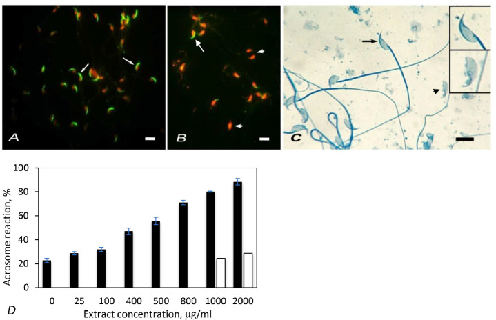
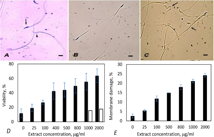
Several herbs including Artemisia are known to possess conceptive property. In the present study, mouse spermatozoa were incubated with ethanol extract of Artemisia vulgaris leaves. The effect of extract on acrosome exocytosis was studied by labeling spermatozoa with fluorescein isothiocyanate (FITC) peanut agglutinin and by staining with Coomassie blue. Viability and membrane integrity were studied by Trypan-blue staining and hypo-osmotic swelling test. Artemisia extract at very low concentration caused precocious acrosome reaction and loss of sperm viability. Acrosome reaction increased remarkably from 22.63% to 88.42% with increasing extract concentration from 0 to 2,000 µg/mL. However, the viability loss of spermatozoa was increased from 11.71% in control to 63.73% in samples treated, evaluated by Trypan-blue staining method. Membrane damage caused by the extract, evaluated by hypo-osmotic swelling test was even low, ranging from 2.27% to only 24.23%. These results indicate that Artemisia extract might block fertilization by causing precocious acrosome exocytosis in spermatozoa. A direct contraceptive effect was tested by injecting the plant extract into the vagina of female mice and then allowing them to mate with normal males. The treated female mice delivered significantly fewer litters in comparison to the control.
Keywords: Acrosome; Artemisia; Membrane damage; Spermatozoa; Viability.
© Copyright 2021 Korean Society of Animal Science and Technology.
Conflict of interest statement
No potential conflict of interest relevant to this article was reported.
Figures
Similar articles
-
Evaluation of the acrosome reaction and viability in buffalo spermatozoa using two staining methods: the effects of heparin and calcium ionophore A23187.Int J Androl. 2002 Aug;25(4):215-22. doi: 10.1046/j.1365-2605.2002.00350.x. Int J Androl. 2002. PMID: 12121571
-
Effect of storage on sperm membrane integrity and other functional characteristics of canine spermatozoa: In vitro bioassay for canine semen.Theriogenology. 1994;41(7):1355-66. doi: 10.1016/0093-691x(94)90187-n. Theriogenology. 1994. PMID: 16727490
-
Use of peanut agglutinin to assess the acrosomal status and the zona pellucida-induced acrosome reaction in stallion spermatozoa.J Androl. 1996 Nov-Dec;17(6):674-82. J Androl. 1996. PMID: 9016398
-
Evaluation of acrosomal status and sperm viability in fresh and cryopreserved specimens by the use of fluorescent peanut agglutinin lectin in conjunction with hypo-osmotic swelling test.Int Braz J Urol. 2007 May-Jun;33(3):364-74; discussion 375-6. doi: 10.1590/s1677-55382007000300009. Int Braz J Urol. 2007. PMID: 17626653
-
Hydrogen hexachloroplatinate induces the acrosome reaction in human spermatozoa.Int J Androl. 1995 Dec;18(6):321-5. doi: 10.1111/j.1365-2605.1995.tb00569.x. Int J Androl. 1995. PMID: 8719848
Cited by
-
Effects of Essential Oils as Antioxidant and Cryoprotective Agents in Improving Frozen and Thawed Human Sperm Criteria.Antioxidants (Basel). 2025 Jan 10;14(1):75. doi: 10.3390/antiox14010075. Antioxidants (Basel). 2025. PMID: 39857409 Free PMC article.
References
-
- Singh A, Kala S, Kapoor DN. Reversible contraceptive efficacy and safety evaluation of ethanolic extract of Tinospora cordifolia in animal model. Arch Appl Sci Res. 2011;3:587–92.
-
- Dehghan MH, Martin T, Dehghanan R. Antifertility effect of Iranian neem seed alcoholic extract on epididymal sperm of mice. J Reprod BioMed. 2005;3:83–9.
LinkOut - more resources
Full Text Sources
Other Literature Sources