. 2021 Jan;63(1):114-124.
doi: 10.5187/jast.2021.e12. Epub 2021 Jan 31.

Wheat phytase can alleviate the cellular toxic and inflammatory effects of lipopolysaccharide

Affiliations

Wheat phytase can alleviate the cellular toxic and inflammatory effects of lipopolysaccharide

Jeongmin An et al. J Anim Sci Technol. 2021 Jan.

Abstract

The objective of this study was to characterize the enzymatic hydrolysis of lipopolysaccharide (LPS) by wheat phytase and to investigate the effects of wheat phytase-treated LPS on in vitro toxicity, cell viability and release of a pro-inflammatory cytokine, interleukin (IL)-8 by target cells compared with the intact LPS. The phosphatase activity of wheat phytase towards LPS was investigated in the presence or absence of inhibitors such as L-phenylalanine and L-homoarginine. In vitro toxicity of LPS hydrolyzed with wheat phytase in comparison to intact LPS was assessed. Cell viability in human aortic endothelial (HAE) cells exposed to LPS treated with wheat phytase in comparison to intact LPS was measured. The release of IL-8 in human intestinal epithelial cell line, HT-29 cells applied to LPS treated with wheat phytase in comparison to intact LPS was assayed. Wheat phytase hydrolyzed LPS, resulting in a significant release of inorganic phosphate for 1 h (p < 0.05). Furthermore, the degradation of LPS by wheat phytase was nearly unaffected by the addition of L-phenylalanine, the inhibitor of tissue-specific alkaline phosphatase or L-homoarginine, the inhibitor of tissue-non-specific alkaline phosphatase. Wheat phytase effectively reduced the in vitro toxicity of LPS, resulting in a retention of 63% and 54% of its initial toxicity after 1-3 h of the enzyme reaction, respectively (p < 0.05). Intact LPS decreased the cell viability of HAE cells. However, LPS dephosphorylated by wheat phytase counteracted the inhibitory effect on cell viability. LPS treated with wheat phytase decreased IL-8 secretion from intestinal epithelial cell line, HT-29 cell to 14% (p < 0.05) when compared with intact LPS. In conclusion, wheat phytase is a potential therapeutic candidate and prophylactic agent for control of infections induced by pathogenic Gram-negative bacteria and associated LPS-mediated inflammatory diseases in animal husbandry.

Keywords: Inflammatory diseases; Lipopolysaccharide; Prophylactic agent; Therapeutic candidate; Wheat phytase.


Conflict of interest statement

No potential conflict of interest relevant to this article was reported.

Figures

Fig. 1.
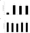
Fig. 1.. Phosphatase activity of wheat phytase against LPS at different enzyme reaction times (A) or enzyme units (B).
(A) LPS (100 μg/mL) was treated with wheat phytase (28.6 mU/mL) in acetate buffer (pH 5.0) at 37°C for the given duration. I: absence of enzyme reaction, II: enzyme reaction for 15 min, III: enzyme reaction for 1 h. Data were presented as mean and standard errors from three experiments. a,bMeans lacking common superscripts differ significantly (p < 0.05). (B) LPS (100 μg/mL) was treated with different units of wheat phytase in acetate buffer (pH 5.0) at 37°C for 1 h. I: absence of enzyme reaction, II: the enzyme (14.3 mU/mL), III: the enzyme (57.2 mU/mL). Data were presented as mean and standard errors from three experiments. a–cMeans lacking common superscripts differ significantly (p < 0.05). LPS, lipopolysaccharide.
Fig. 2.
Fig. 2.. Effect of enzyme inhibitors (10 mM) such as L-phenylalanine and L-homoarginine on dephosphorylation of LPS (100 μg/mL) by wheat phytase (28.6 mU/mL) (A) and the phosphatase activity of the enzyme against the substrate with different concentrations (5 and 20 mM) of the inhibitors (B).
(A) I: absence of enzyme reaction, II: enzyme reaction for 1 h in the absence of enzyme inhibitors, III: enzyme reaction for 1 h in the presence of L-phenylalanine, IV: enzyme reaction for 1 h in the presence of L-homoarginine. Data were presented as mean and standard errors from three experiments. a,bMeans lacking common superscripts differ significantly (p < 0.05). (B) I: enzyme reaction for 1 h in the absence of enzyme inhibitors, II: enzyme reaction for 1 h in the presence of 5 mM L-phenylalanine, III: enzyme reaction for 1 h in the presence of 20 mM L-phenylalanine, IV: enzyme reaction for 1 h in the presence of 5 mM L-homoarginine, V: enzyme reaction for 1 h in the presence of 20 mM L-homoarginine. Data were presented as mean and standard errors from three experiments. a,bMeans lacking common superscripts differ significantly (p < 0.05). LPS, lipopolysaccharide.
Fig. 3.
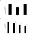
Fig. 3.. Effect of wheat phytase (11.44 × 10⁻³ mU/mL) on in vitro toxicity of LPS (20 ng/mL) (A) and the change of the toxicity at different levels of LPS (10 and 40 ng/mL) treated with the enzyme (B).
(A) I: absence of enzymatic dephosphorylation of LPS, II: enzymatic dephosphorylation of LPS for 1 h, III: enzymatic dephosphorylation of LPS for 3 h. Data were presented as mean and standard errors from three experiments. a–cMeans lacking common superscripts differ significantly (p < 0.05). (B) I: (closed bar); absence of enzymatic dephosphorylation of LPS (10 ng/mL), (open bar); enzymatic dephosphorylation of LPS (10 ng/mL) for 3 h, II: (closed bar); absence of enzymatic dephosphorylation of LPS (40 ng/mL), (open bar); Enzymatic dephosphorylation of LPS (40 ng/mL) for 3 h. Data were presented as mean and standard errors from three experiments. a–cMeans lacking common superscripts differ significantly (p < 0.05). LPS, lipopolysaccharide.
Fig. 4.
Fig. 4.. Cell viability of HAE cells exposed to LPS treated with wheat phytase (A) and the change of cell viabilities at different levels of LPS treated with the enzyme (B).
(A) I: no addition, II: addition of intact LPS (200 μg/mL), III: addition of LPS (200 μg/mL) dephosphorylated by wheat phytase (28.6 mU/mL). Data were presented as mean and standard errors from three experiments. a,bMeans lacking common superscripts differ significantly (p < 0.05). (B) I: no addition, II: (closed bar); addition of intact LPS (100 μg/mL), (open bar); addition of LPS (100 μg/mL) dephosphorylated by wheat phytase (28.6 mU/mL), III: (closed bar); addition of intact LPS (200 μg/mL), (open bar); addition of LPS (200 μg/mL) dephosphorylated by wheat phytase (28.6 mU/mL), IV: (closed bar); addition of intact LPS (400 μg/mL), (open bar); addition of LPS (400 μg/mL) dephosphorylated by wheat phytase (28.6 mU/mL). Data were presented as mean and standard errors from three experiments. a–dMeans lacking common superscripts differ significantly (p < 0.05). HAE, human aortic endothelial; LPS, lipopolysaccharide.
Fig. 5.
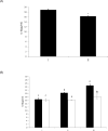
Fig. 5.. Effect of LPS treated with wheat phytase on IL-8 secretion in HT-29 cells (A) and IL-8 release from the cells exposed at different levels of LPS treated with the enzyme (B).
(A) I: addition of intact LPS (10 μg/mL), II: addition of LPS (10 μg/mL) dephosphorylated by wheat phytase (286 mU/mL). Data were presented as mean and standard errors from three experiments. (*p < 0.05: Student’s t-test). (B) I: (closed bar); addition of intact LPS (5 μg/mL), (open bar); addition of LPS (5 μg/mL) dephosphorylated by wheat phytase (286 mU/mL), II: (closed bar); addition of intact LPS (10 μg/mL), (open bar); addition of LPS (10 μg/mL) dephosphorylated by wheat phytase (286 mU/mL), III: (closed bar); addition of intact LPS (20 μg/mL), (open bar); addition of LPS (20 μg/mL) dephosphorylated by wheat phytase (286 mU/mL). Data were presented as mean and standard errors from three experiments. a–cMeans lacking common superscripts differ significantly (p < 0.05). LPS, lipopolysaccharide.

Similar articles

Cited by

References

    1. van Gucht S, van Reeth K, Pensaert M. Interaction between porcine reproductive-respiratory syndrome virus and bacterial endotoxin in the lungs of pigs: potentiation of cytokine production and respiratory disease. J Clin Microbiol. 2003;41:960–6. doi: 10.1128/JCM.41.3.960-966.2003. - DOI - PMC - PubMed
    1. Estaki M, DeCoffe D, Gibson DL. Interplay between intestinal alkaline phosphatase, diet, gut microbes and immunity. World J Gastroenterol. 2014;20:15650–6. doi: 10.3748/wjg.v20.i42.15650. - DOI - PMC - PubMed
    1. Lallès JP. Intestinal alkaline phosphatase: novel functions and protective effects. Nutr Rev. 2014;72:82–94. doi: 10.1111/nure.12082. - DOI - PubMed
    1. Xie H, Rath NC, Huff GR, Huff WE, Balog JM. Effects of Salmonella typhimurium lipopolysaccharide on broiler chickens. Poult Sci. 2000;79:33–40. doi: 10.1093/ps/79.1.33. - DOI - PubMed
    1. Rauber RH, Perlin VJ, Fin CD, Mallmann AL, Miranda DP, Giacomini LZ, et al. Interference of Salmonella typhimurium lipopolysaccharide on performance and biological parameters of broiler chickens. Brazilian J Poultry Sci. 2014;16:77–81. doi: 10.1590/S1516-635X2014000100011. - DOI