COVID-19 Endothelial Dysfunction Can Cause Erectile Dysfunction: Histopathological, Immunohistochemical, and Ultrastructural Study of the Human Penis
- PMID: 33988001
- PMCID: PMC8255400
- DOI: 10.5534/wjmh.210055
COVID-19 Endothelial Dysfunction Can Cause Erectile Dysfunction: Histopathological, Immunohistochemical, and Ultrastructural Study of the Human Penis
Abstract
Purpose: A pilot study to describe histopathological features of penile tissue of patients who recovered from symptomatic COVID-19 infection and subsequently developed severe erectile dysfunction (ED).
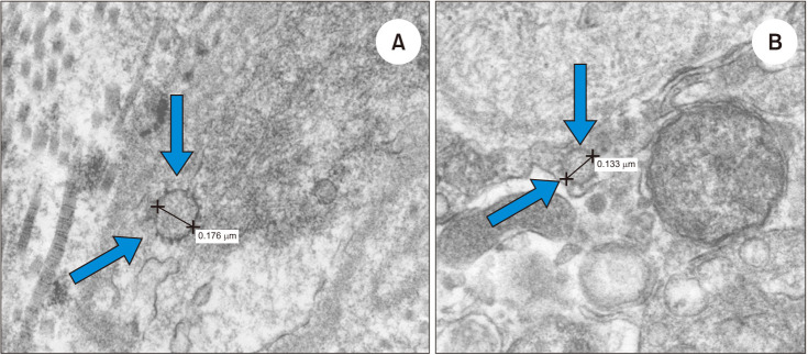
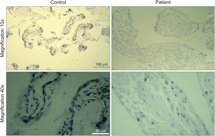
Materials and methods: Penile tissue was collected from patients undergoing surgery for penile prosthesis for severe ED. Specimens were obtained from two men with a history of COVID-19 infection and two men with no history of infection. Specimens were imaged with TEM and H&E staining. RT-PCR was performed from corpus cavernosum biopsies. The tissues collected were analyzed for endothelial Nitric Oxide Synthase (eNOS, a marker of endothelial function) and COVID-19 spike-protein expression. Endothelial progenitor cell (EPC) function was assessed from blood samples collected from COVID-19 (+) and COVID-19 (-) men.
Results: TEM showed extracellular viral particles ~100 nm in diameter with peplomers (spikes) near penile vascular endothelial cells of the COVID-19 (+) patients and absence of viral particles in controls. PCR showed presence of viral RNA in COVID-19 (+) specimens. eNOS expression in the corpus cavernosum of COVID-19 (+) men was decreased compared to COVID-19 (-) men. Mean EPC levels from the COVID-19 (+) patients were substantially lower compared to mean EPCs from men with severe ED and no history of COVID-19.
Conclusions: Our study is the first to demonstrate the presence of the COVID-19 virus in the penis long after the initial infection in humans. Our results also suggest that widespread endothelial cell dysfunction from COVID-19 infection can contribute to ED. Future studies will evaluate novel molecular mechanisms of how COVID-19 infection leads to ED.
Keywords: COVID-19; Endothelium; Erectile dysfunction; Histopathology; Immunohistochemistry; SARS-CoV-2.
Copyright © 2021 Korean Society for Sexual Medicine and Andrology.
Conflict of interest statement
The authors have nothing to disclose.
Figures
Comment in
-
COVID-19 and Erectile Dysfunction: Endothelial Dysfunction and Beyond.World J Mens Health. 2021 Oct;39(4):820-821. doi: 10.5534/wjmh.210081. Epub 2021 Jun 7. World J Mens Health. 2021. PMID: 34184433 Free PMC article. No abstract available.
References
LinkOut - more resources
Full Text Sources
Other Literature Sources
Miscellaneous