. 2021 May 14:38:E008.
doi: 10.1017/S0952523821000079.

Functional connexin35 increased in the myopic chicken retina

Affiliations

Functional connexin35 increased in the myopic chicken retina

Seema Banerjee et al. Vis Neurosci. .

Erratum in

Abstract

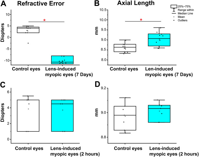
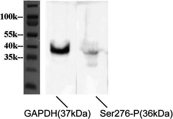
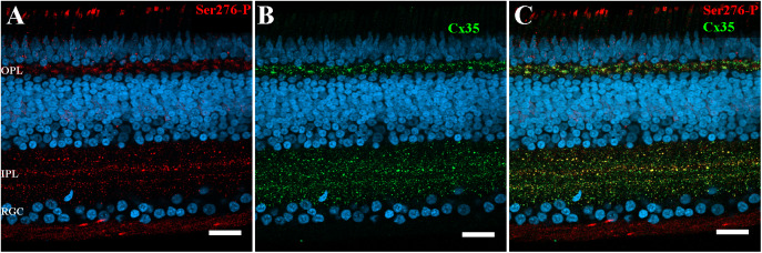
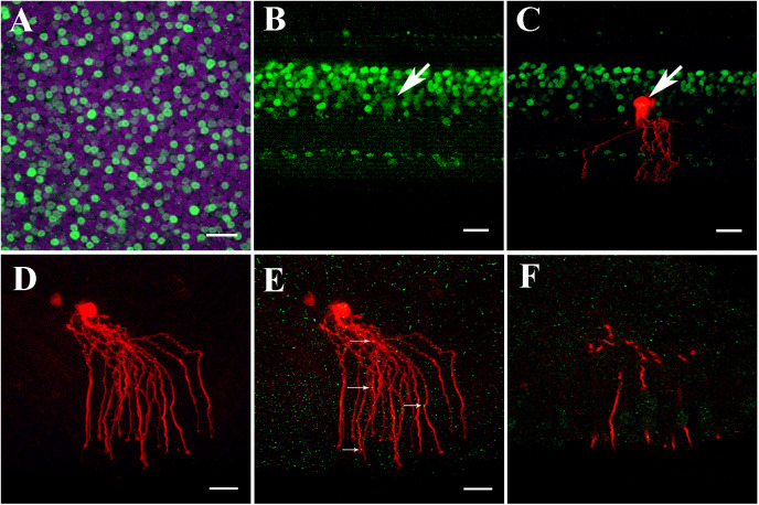
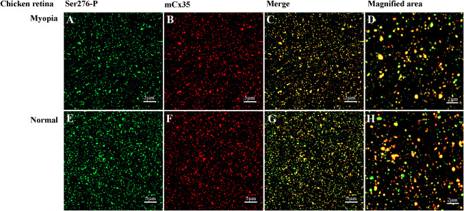
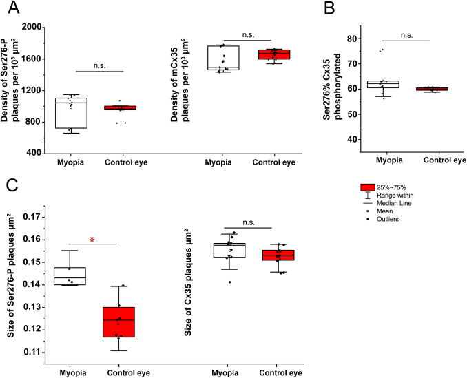
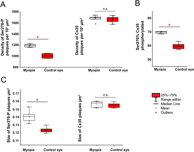
Our previous research showed that increased phosphorylation of connexin (Cx)36 indicated extended coupling of AII amacrine cells (ACs) in the rod-dominant mouse myopic retina. This research will determine whether phosphorylation at serine 276 of Cx35-containing gap junctions increased in the myopic chicken, whose retina is cone-dominant. Refractive errors and ocular biometric dimensions of 7-days-old chickens were determined following 12 h and 7 days induction of myopia by a -10D lens. The expression pattern and size of Cx35-positive plaques were examined in the early (12 h) and compensated stages (7 days) of lens-induced myopia (LIM). At the same time, phosphorylation at serine 276 (functional assay) of Cx35 in strata 5 (S5) of the inner plexiform layer was investigated. The axial length of the 7 days LIM eyes was significantly longer than that of non-LIM controls (P < 0.05). Anti-phospho-Ser276 (Ser276-P)-labeled plaques were significantly increased in LIM retinas at both 12 h and 7 days. The density of Ser276-P of Cx35 was observed to increase after 12 h LIM. In the meanwhile, the areas of existing Cx35 plaques did not change. As there was more phosphorylation of connexin35 at Ser276 at both the early and late stages (12 h) and 7 days of LIM chicken retinal activity, the coupling with ACs could be increased in myopia development of the cone-dominated chicken retina.

Keywords: amacrine cell; gap junction; myopia; retina.


Figures

Fig. 1.
Fig. 1.
Measurement of Lens-induced refractive errors in chicken eyes by streak retinoscopy and A-scan ultrasonography. (A) Refractive errors of chicken eyes were measured by streak retinoscopy. LIM method was employed for 7 days to produce precise and consistent myopia in chicken eyes (−10.08 ± 0.89 D, mean ± s.e.m., P < 0.05, n = 10). (B) AL of the chicken eye was measured from the cornea’s surface to the RPE layer by A-scan ultrasonography. The AL of LIM eyes was 9.06 ± 0.12 mm, in contrast to 8.63 ± 0.08 mm (mean ± s.e.m., P < 0.05, n = 10), of the contralateral control eyes, an average increase in AL of 0.29 ± 0.12 mm. (C) 12 h LIM did not affect refractive errors of chicken eyes by streak retinoscopy (control, 3.86 ± 0.75 D; 12 h LIM, 3.71 ± 0.71 D, P = 0.36, n = 7). (D) AL of the chicken eye had no significant change after 12 h LIM. The AL of control eyes was 8.97 ± 0.04 mm, in contrast to the AL of 12 h LIM (9.01 ± 0.03 mm, P = 0.06, n = 7).
Fig. 2.
Fig. 2.
Specificity of the phospho-Cx35/36 antibodies. Western blot analysis of anti-phospho-Ser276 (Ser276-P) of Cx35/36 antibody in the chicken retina (36 kDa). Anti-GAPDH antibody (1:5000, 37 kDa) was used as the loading control.
Fig. 3.
Fig. 3.
Ser276-P antibody labeling pattern in the chicken retina after 7 days −10D defocus. (A–C) Labeling pattern of anti-phospho-Ser276 antibody in the 7 days LIM chicken retina. (A) Ser276-P antibody labeled(red) abundant punctate structures in both IPL and OPL. The labeling is also observed in the optic fiber layer, in the retinal pigment epithelium layer, and photoreceptor inner/outer segments. (B) Labeling with polyclonal Cx35 antibody (green) shows labeling of Cx35 also in the OPL and IPL. (C) The merged image of A and B shows multiple plaques of Ser276-P co-localized with Cx35 antibody labeling (yellow). Scale bar is 20 μm.
Fig. 4.
Fig. 4.
AII-like ACs in chicken retina expressed Cx35. (A) Prox1 immunostaining in the chicken retina. Prox1-immunoreactive AII-like AC bodies and bipolar cells in the INL in a whole-mounted chicken retina. (B) Prox1 immunoreactivity in AII AC and bipolar cell bodies in INL in the vertical section. Prox1-immunoreactive AC bodies are present at the bottom of the INL. (C) Prox1-immunoreactive AII-like AC’s cell body co-localized with neurobiotin injection. The AII-like AC was similar to AII AC’s morphology in the mouse retina, having thick lobular appendages in the OFF sublamina of the IPL and descending arboreal processes to the ON sublamina of the IPL. (D) Cy3-labeled AII-like AC in the chicken retina (200 μm slice) was targeted and injected with neurobiotin in the INL to show single AII-like AC’s soma–dendritic morphology in Z-stack by three-dimensional reconstruction image. The cell had a round cell body with distinct dendritic trees. The proximal dendrites near soma had lobular appendages. The distal processes overlapped along central-to-peripheral axes. In addition, the injected cell is coupled with the nearby cells. (E) AII-like AC double-labeled with anti-Cx35 (green) puncta located predominately on the arboreal dendrites (as indicated by white arrows). (F) Single plane of the confocal image showing Cx35 puncta on segments of AII like AC dendrites. A–C: Scale bar is 20 μm; D–F: Scale bar is 5 μm.
Fig. 5.
Fig. 5.
Phospho-Ser276 antibody recognizes Cx35 in the whole mount chicken retina of 7 days LIM model. (A–D [−10D treated]) Confocal stack sections in stratum 5 of the IPL in a myopic chicken retina after 7 days LIM treatment: Cx35, labeled in red, and its phosphorylated form, Ser276-P (green), are present with similar punctate labeling. The magnified areas showed the merged images of phosphorylated Ser-276 and Cx35, reflected by yellow color. (E–H [non-LIM]) Ser276-P antibody recognizes Cx35 in the non-LIM control chicken retina. Images are 2 μm deep stacks.
Fig. 6.
Fig. 6.
Quantification of phosphorylation Ser276 of Cx35 gap junctions in 7 days lens-induced chicken myopic retinas. The size of Ser276-P plaques (C) was significantly increased in the myopic retina compared to that of control retinas in 7 days LIM-treated eyes. The density (plaques number per 103 μm2) of phosphorylation reflected by detectable Ser276-P labeling (A), the density (plaques number per 103 μm2) of Cx35 labeling (A), the percentage of Ser276-P of Cx35 phosphorylated rate (B), and the size of Cx35 plaque (C) did not differ between retinas of LIM and non-LIM eyes. The data are presented as averages; error bars are s.e.m.. Significance is based on Wilcoxon Signed Ranks Test, where * indicates 0.01 < P < 0.05 and n.s. indicates P > 0.05.
Fig. 7.
Fig. 7.
Quantification of phosphorylation of Cx35 gap junctions in 12 h lens-induced chicken myopic retinas. In the 12 h LIM chicken eye, the density (plaques number per 103 μm2) of phosphorylation reflected by detectable Ser276-P labeling (A), the percentage of Ser276-P of Cx35 phosphorylated rate (B), and the size of Ser276-P plaque (C) were significantly increased in the LIM retina compared to non-LIM control retinas. The density of Cx35 labeling (A) and the size of Cx35 plaque (C) had no significant difference between the LIM retinas compared to non-LIM control retinas. The data are presented as averages; error bars are s.e.m. Significance is based on Wilcoxon Signed Ranks Test, where * indicates 0.01 < P < 0.05 and n.s. indicates P > 0.05.

Similar articles

Cited by

References

    1. Banerjee, S., Wang, Q., So, C.H. & Pan, F. (2020). Defocused images change multineuronal firing patterns in the mouse retina. Cells 9, pii:E530. - PMC - PubMed
    1. Banerjee, S., Wang, Q., Zhao, F., Tang, G., So, C., Tse, D., To, C.H., Feng, Y., Zhou, X. & Pan, F. (2020). Increased Connexin36 phosphorylation in AII Amacrine cell coupling of the mouse myopic retina. Frontiers in Cellular Neuroscience 14, 124. - PMC - PubMed
    1. Baylor, D.A. & Hodgkin, A.L. (1973). Detection and resolution of visual stimuli by turtle photoreceptors. The Journal of Physiology 234, 163–198. - PMC - PubMed
    1. Bloomfield, S.A. & Volgyi, B. (2004). Function and plasticity of homologous coupling between AII amacrine cells. Vision Research 44, 3297–3306. - PubMed
    1. Bloomfield, S.A. & Volgyi, B. (2009). The diverse functional roles and regulation of neuronal gap junctions in the retina. Nature Reviews. Neuroscience 10(Suppl. 7), 495–506. - PMC - PubMed

Publication types