. 2021 Jun 15;134(12):jcs258540.
doi: 10.1242/jcs.258540. Epub 2021 Jun 28.

Chlamydomonas FAP70 is a component of the previously uncharacterized ciliary central apparatus projection C2a

Affiliations

Chlamydomonas FAP70 is a component of the previously uncharacterized ciliary central apparatus projection C2a

Yuqing Hou et al. J Cell Sci. .

Abstract

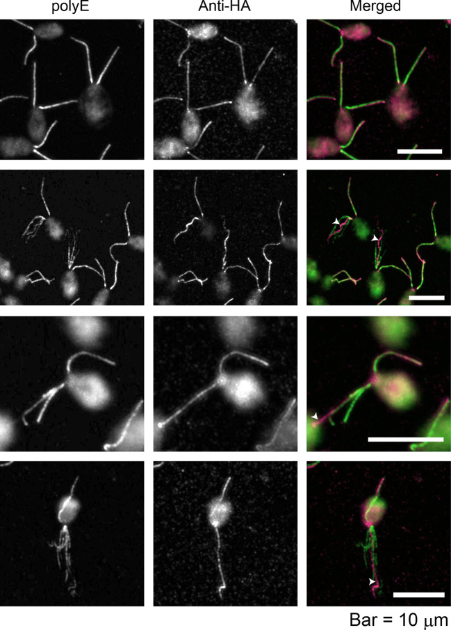
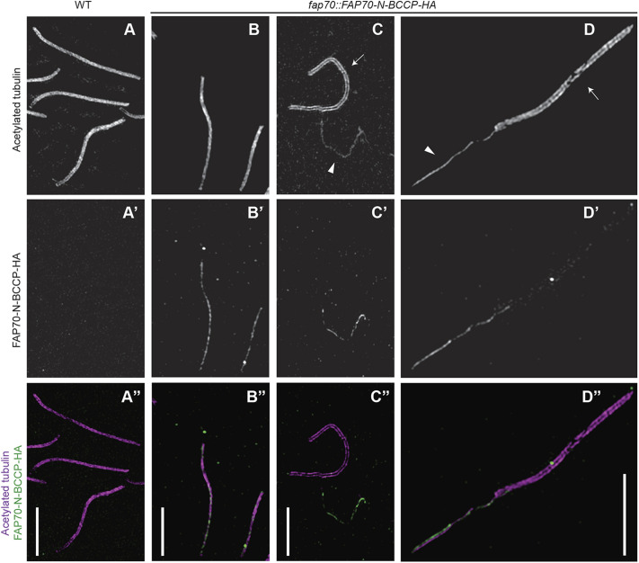
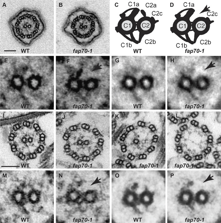
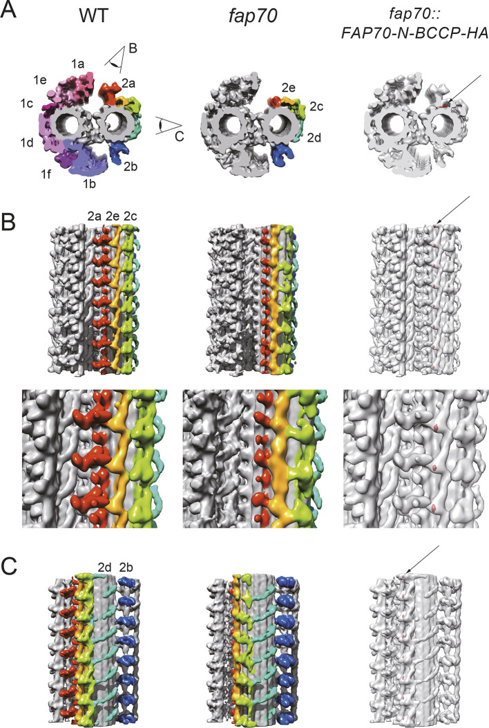
Cilia are essential organelles required for cell signaling and motility. Nearly all motile cilia have a '9+2' axoneme composed of nine outer doublet microtubules plus two central microtubules; the central microtubules together with their projections are termed the central apparatus (CA). In Chlamydomonas reinhardtii, a model organism for studying cilia, 30 proteins are known CA components, and ∼36 more are predicted to be CA proteins. Among the candidate CA proteins is the highly conserved FAP70 (CFAP70 in humans), which also has been reported to be associated with the doublet microtubules. Here, we determined by super-resolution structured illumination microscopy that FAP70 is located exclusively in the CA, and show by cryo-electron microscopy that its N-terminus is located at the base of the C2a projection of the CA. We also found that fap70-1 mutant axonemes lack most of the C2a projection. Mass spectrometry revealed that fap70-1 axonemes lack not only FAP70 but two other conserved candidate CA proteins, FAP65 (CFAP65 in humans) and FAP147 (MYCBPAP in humans). Finally, FAP65 and FAP147 co-immunoprecipitated with HA-tagged FAP70. Taken together, these data identify FAP70, FAP65 and FAP147 as the first defining components of the C2a projection.

Keywords: ASH domains; Axonemal central apparatus; CFAP70; FAP147; FAP174; FAP65; Flagella; MYCBP; MYCBPAP.


Conflict of interest statement

Competing interests The authors declare no competing or financial interests.

Figures

Fig. 1.
Fig. 1.
FAP70 localizes to only one microtubule bundle in frayed axonemes. fap70::FAP70-N-BCCP-HA cells were double labeled with anti-HA antibody and anti-polyE antibody, which exclusively labels DMTs. The upper row shows cells with intact axonemes; the other rows show cells with frayed axonemes. In the merged images, green is anti-polyE labeling, and magenta is anti-HA labeling. As reported previously, there is a gradient of polyE labeling of intact axonemes, with the base being more strongly labeled than the tip (upper row); this is less apparent in frayed axonemes (Kubo et al., 2010). Anti-HA labeling is more uniform; in those frayed axonemes in which it is brighter toward the tip (third row), it may reflect partial extrusion of the CA, or increased antibody accessibility to the tip region, where axonemal fraying begins. The anti-HA antibody brightly labels only one microtubule bundle in each frayed axoneme (white arrowheads).
Fig. 2.
Fig. 2.
FAP70 localizes exclusively to the CA, as demonstrated by super-resolution structured-illumination microscopy. (A-D) Axonemes from wild-type (WT, column A) or fap70::FAP70-N-BCCP-HA (columns B to D) cells were double labeled with anti-acetylated tubulin antibody (upper row, A-D) and anti-HA antibody (middle row, A′-D′). The lower row (A″-D″) shows merged images with labeling by anti-acetylated tubulin in magenta and anti-HA in green. In column A, wild-type axonemes are intact with the CA still inside the axonemes. As expected, the acetylated tubulin label covers the entire width of the axoneme, and there is no HA signal. (Column B) Two intact fap70::FAP70-N-BCCP-HA axonemes. The HA signal colocalizes with the acetylated tubulin signal but is thinner, suggesting that it emanates from the CA. (Column C) fap70::FAP70-N-BCCP-HA axoneme (arrow in panel C) from which the CA has been completely extruded, and a nearby extruded CA (arrowhead in panel C). The HA signal is associated exclusively with the CA. Only the edges of the axoneme are labeled by the antibody to acetylated tubulin, revealing the void that was formerly occupied by the CA. (Column D) fap70::FAP70-N-BCCP-HA axoneme (arrow in panel D) from which the CA (arrowhead in panel D) has been partially extruded. Anti-HA strongly labels the extruded CA but not the DMTs in the portion of the axoneme from which the CA has been extruded. Therefore, FAP70 is located exclusively in the CA. Scale bars: 5 µm.
Fig. 3.
Fig. 3.
Loss of FAP70 causes ablation of the C2a projection. (A,B) Representative cross-sections of intact in situ flagella of wild-type (WT) and fap70-1 cells, respectively, as imaged by conventional TEM. All flagella of both strains had axonemes with nine DMTs and two central microtubules. (C,D) Diagrams of cross-sections of CAs of wild type and fap70-1, respectively, illustrating the C1 and C2 microtubules connected by bridge structures and with major projections (C1a, C1b, C2a, C2c and C2b) normally visible by conventional TEM labeled. The fap70-1 CA appears to lack the C2a projection (arrow in D). (E,F) Enlargement of CAs from panels A and B, respectively. The C2a projection appears to be absent from the fap70-1 CA (arrow in F). (G,H) Image averages based on six wild-type (G) and six fap70-1 (H) CAs of intact in situ flagella showing the apparent absence of the C2a projection in the fap70-1 flagellum (arrow in H). (I-L) Representative cross-sections of isolated axonemes from wild type (I) and fap70-1 (J-L). Some cross-sections of fap70-1 axonemes lacked one (K) or both (L) central microtubules. (M,N) Enlargement of CAs from panels I and J, respectively. The C2a projection appears to be absent from the fap70-1 CA (arrow in N). (O,P) Image averages based on six wild-type (O) and six fap70-1 (P) CAs of isolated axonemes showing the apparent absence of the C2a projection in the fap70-1 axoneme (arrow in P). Scale bars: 100 nm.
Fig. 4.
Fig. 4.
Cryo-electron tomography confirms ablation of the C2a projection in fap70 axonemes and localizes FAP70 within the CA. (A-C) Averaged subtomograms of the CA. Wild-type (WT) and fap70-1 CAs are shown in cross-sectional (A, left and middle subtomograms, respectively) and longitudinal (B and C, left and middle subtomograms, respectively) views. In A, the CA is viewed from base to tip. In B and C, the proximal end of the CA is at the bottom. Most of the C2a projection is absent in the fap70-1 CA. Red, C2a; yellow, C2e; green, C2c; cyan, C2d; and blue, C2b. Eye symbols indicate the directions of the views in B and C. (A-C, right) Three-dimensional-localization of the N-terminus of FAP70-N-BCCP-HA. Tag densities (red, arrows) were visualized by comparing the streptavidin-treated wild-type and streptavidin-labeled fap70::FAP70-N-BCCP-HA CAs. The N-terminus of FAP70 is located at the base of the C2a projection below the C2e arcade. The top row of panel B shows seven 16-nm repeat units of the C2a, C2e and C2c projections. The bottom row of panel B shows magnified views of three of the repeat units. Panel C shows seven 16-nm repeats of the C2d and C2b projections.

References

    1. Anvarian, Z., Mykytyn, K., Mukhopadhyay, S., Pedersen, L. B. and Christensen, S. T. (2019). Cellular signalling by primary cilia in development, organ function and disease. Nat. Rev. Nephrol. 15, 199-219. 10.1038/s41581-019-0116-9 - DOI - PMC - PubMed
    1. Bernstein, M., Beech, P. L., Katz, S. G. and Rosenbaum, J. L. (1994). A new kinesin-like protein (Klp1) localized to a single microtubule of the Chlamydomonas flagellum. J. Cell Biol. 125, 1313-1326. 10.1083/jcb.125.6.1313 - DOI - PMC - PubMed
    1. Beurois, J., Martinez, G., Cazin, C., Kherraf, Z. E., Amiri-Yekta, A., Thierry-Mieg, N., Bidart, M., Petre, G., Satre, V., Brouillet, S.et al. (2019). CFAP70 mutations lead to male infertility due to severe astheno-teratozoospermia. A Case Report. Hum. Reprod 34, 2071-2079. 10.1093/humrep/dez166 - DOI - PubMed
    1. Brown, J. M. and Witman, G. B. (2014). Cilia and diseases. Bioscience 64, 1126-1137. 10.1093/biosci/biu174 - DOI - PMC - PubMed
    1. Brown, J. M., DiPetrillo, C. G., Smith, E. F. and Witman, G. B. (2012). A FAP46 mutant provides new insights into the function and assembly of the C1d complex of the ciliary central apparatus. J. Cell Sci. 125, 3904-3913. 10.1242/jcs.107151 - DOI - PMC - PubMed

Publication types