Review
. 2021 Jun 14;7(6):2106-2149.
doi: 10.1021/acsbiomaterials.1c00217. Epub 2021 May 14.

Potential Use of Exosomes as Diagnostic Biomarkers and in Targeted Drug Delivery: Progress in Clinical and Preclinical Applications

Affiliations
Review

Potential Use of Exosomes as Diagnostic Biomarkers and in Targeted Drug Delivery: Progress in Clinical and Preclinical Applications

Md Nurul Huda et al. ACS Biomater Sci Eng. .

Abstract

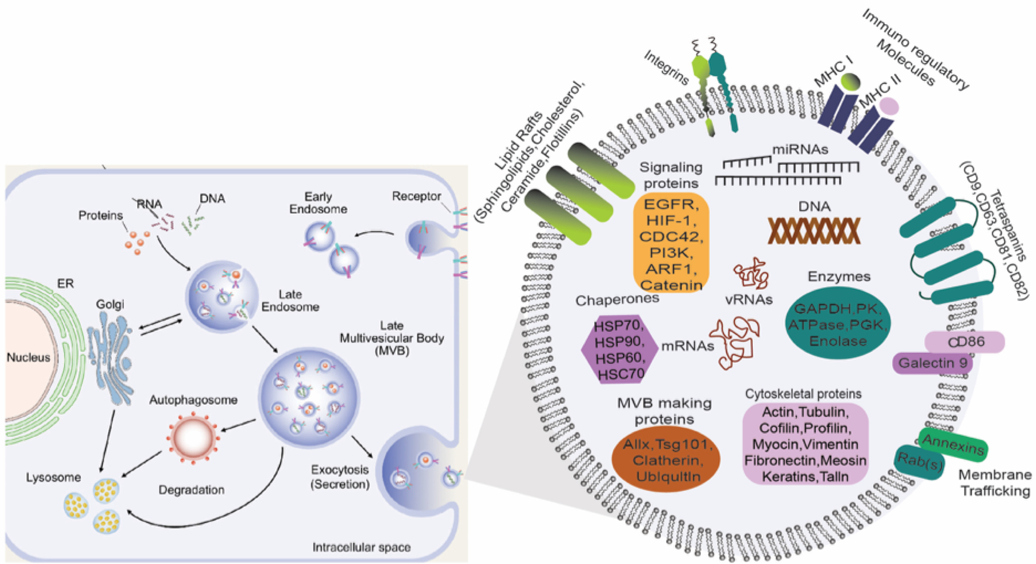
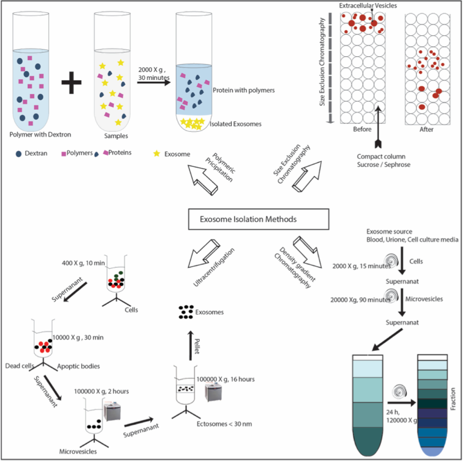
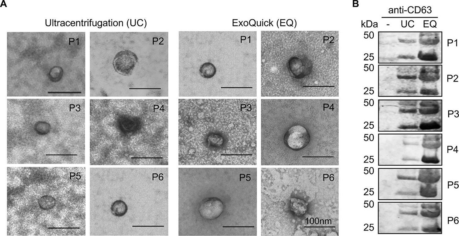
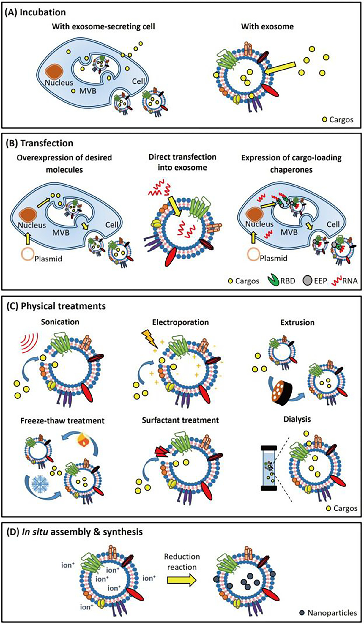
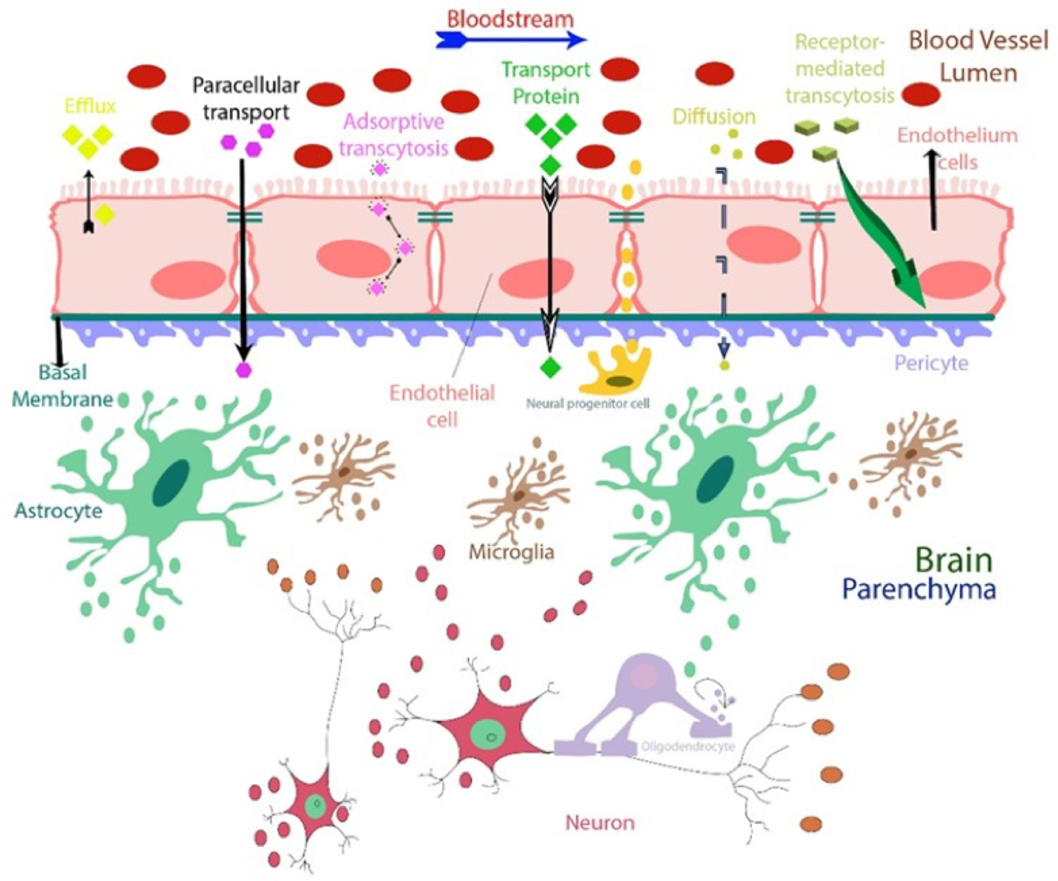
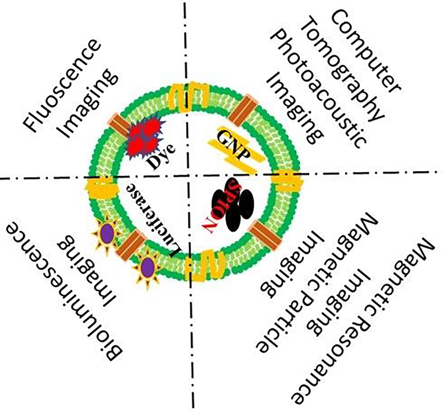
Exosomes are cell-derived vesicles containing heterogeneous active biomolecules such as proteins, lipids, mRNAs, receptors, immune regulatory molecules, and nucleic acids. They typically range in size from 30 to 150 nm in diameter. An exosome's surfaces can be bioengineered with antibodies, fluorescent dye, peptides, and tailored for small molecule and large active biologics. Exosomes have enormous potential as a drug delivery vehicle due to enhanced biocompatibility, excellent payload capability, and reduced immunogenicity compared to alternative polymeric-based carriers. Because of active targeting and specificity, exosomes are capable of delivering their cargo to exosome-recipient cells. Additionally, exosomes can potentially act as early stage disease diagnostic tools as the exosome carries various protein biomarkers associated with a specific disease. In this review, we summarize recent progress on exosome composition, biological characterization, and isolation techniques. Finally, we outline the exosome's clinical applications and preclinical advancement to provide an outlook on the importance of exosomes for use in targeted drug delivery, biomarker study, and vaccine development.

Keywords: Exosome; biomarker; clinical translation; diagnosis; drug delivery; vaccine.


Conflict of interest statement

Conflict of Interest: Authors declare no conflict of interest.

Figures

Figure 1.
Figure 1.
Exosome biogenesis begins with the formation of intraluminal vesicles (ILs) in late endosomes following cargo sorting. Both ESCRT dependent and ESCRT independent lipid-driven pathways are involved in creating multivesicular bodies. Exocytic MVCs fuse with the plasma membrane in Rab GTPases regulated miRNAs; exosome content depends on cell type and cells’ physiological and pathological conditions. Here we illustrate the components of exosomes identified in multiple proteomic studies and different cell content. Adapted from Gurunath et al. (2019) , Copyright @ 2019 MDPI and modified to accompany our review on exosome biogenesis and composition.
Figure 2.
Figure 2.
Schematic summary of standard laboratory methods for exosome purification. Four different isolation techniques are demonstrated here: Polymeric precipitation, (top left), column for size exclusion chromatography, (top right), density gradient chromatography, (bottom right) and differential ultracentrifugation, (bottom left). Temperature maintained at 4°C for most of the protocol.
Figure 3:
Figure 3:
Validation of exosome enrichment from human cell-free sera. (A) TEM micrographs of exosomes in ultracentrifugation (UC) and ExoQuick (EQ) preparations. Data for 6 independent patient samples are shown (P1–6). Exosomes confirmed by size (30–100nm) and appearance. Scale bar in each image represents 100 nm. (B) Immunoblot of CD63 in unprocessed cell-free serum alone (−), UC, and EQ exosome preparations. Adapted from Prendergast et al. (2018), Copyright @ 2018, PLOS.
Figure 4:
Figure 4:
Exosome drug loading techniques, (A) Exosome-secreting cells or exosomes incubated with desired cargos. Cargos diffuse across the cell and exosomal membrane and are subsequently packaged within the exosomes. (B) Desired nucleic acids can be loaded into exosomes via a transfection-based strategy. Transfected with vectors, the donor cell generates RNAs/proteins and packages these products into exosomes using endogenous expression and sorting machinery of donor cell, respectively. Exosomes can be directly transfected with small RNAs for cargo loading purposes. (C) Cargos can be loaded into exosomes directly through physical treatments. Electroporation, sonication, and surfactant treatment generate pores on the exosomal membranes that facilitate cargo loading. Freeze-thaw treatment, extrusion, and dialysis enhance cargo loading into exosomes during membrane recombination processes. Adapted from Shengyang Fu et al.(2020), Copyright @ ELSEVIER.
Figure 5.
Figure 5.
Recent studies confirm that exosomes can pass through the blood-brain barrier (BBB) in both directions. This means that specific exosomes detected in the cerebrospinal fluid (CSF), or in the blood from the brain can release into the bloodstream and vice versa. Each cell type releases a specific type of exosome(s) that are released and communicates with neighboring cells, acting as the messenger. This characteristic makes exosomes attractive as new sources of biomarkers and therapeutic targets suitable for use in clinical practice, such as liquid biopsy that could replace current invasive diagnostic methods. Exosomes also have a potential role in drug delivery for brain disease models, and their membrane markers can be used to identify their cellular origin.
Figure 6:
Figure 6:
LPS injection induces changes in extracellular vehicles (exosomes) and miRNAs in the cerebrospinal fluid (CSF) A. Representative transmission electron microscope (TEM) image showing the presence of EVs in the CSF in two independent experiments. B. NanoSight quantification of the number of particles in the CSF at 0, 1, 2, 4, and 6 h after i.p. LPS injection (n = 3–5). C. Size distribution of the EVs in vivo in the CSF before (black; n = 5) and 6 h after (gray; n = 3) LPS treatment determined by NanoSight analysis. D–G. Quantitative real-time polymerase chain reaction analysis of miR-1a (D), miR-9 (E), miR-146a (F), and miR-155 (G) (n = 4). RNA was isolated from pooled CSF (50 μl) from different mice (n = 3). Data information: Data in (B, D-G) are displayed as mean ± SEM and analyzed by Student’s t-test. Significance levels are indicated on the graphs: *0.01 ≤ P < 0.05; **0.001 ≤ P < 0.01. Adapted from Balusu et al. (2016) , Copyright @ EMBO Press.
Figure 7:
Figure 7:
Systemic inflammation activates the exosomal machinery in the choroid plexus. A. Representative confocal images of CD63, RAB5, and ANXA2 (red) in the choroid plexus (CP) at 0, 4, and 8 h after LPS treatment. Hoechst (blue) was used to stain the nucleus. The dotted line indicates the ependymal cells that line the ventricle, and the square boxes indicate the zoomed insert images displayed at the right corner of each image. Scale bars, 100 μm. B, C. Representative TEM images showed the presence of MVBs in the CPE cells before (B) and 6 h after (C) LPS administration in vivo. Black arrowheads point to exosomes present in MVBs. Scale bars, 9 μm. D–F. Quantification of number of MVBs per cell section (D), number of exosomes per MVB (E), and number of exosomes per cell section (F), based on TEM analysis of several adjacent cells (0 h, n = 20; 3 h, n = 21; 4 h, n = 13; 6 h, n = 23). G–J. Quantitative real-time polymerase chain reaction (qPCR) analysis of miR-1a (G), miR-9 (H), miR-146a (I), and miR-155 (J). Data is presented as relative expression normalized with housekeeping miRs by TaqMan qPCR assay (0 h, n = 4; 1 h, n = 5; 6 h, n = 5; 24 h, n = 3). K. NanoSight analysis of CSF isolated from LPS-injected mice followed by icv injection of vehicle or GW4869, a neutral sphingomyelinase inhibitor that inhibits exosome secretion (n = 8). L. qPCR analysis of the expression of miR-1a, miR-9, miR-146a, and miR-155 in the choroid plexus of mice injected with LPS and then icv injected with vehicle (black) or GW4869 (gray) (n = 4). M. NanoSight analysis of the supernatant of choroid plexus explants from PBS- or LPS-injected mice (n = 6). Data information: Data in (D–M) are displayed as mean ± SEM and analyzed by Student’s t-test. Significance levels are indicated on the graphs: *0.01 ≤ P < 0.05; **0.001 ≤ P < 0.01; ***0.0001 ≤ P < 0.001; ****P < 0.0001. Adapted from Balusu et al. (2016), Copyright @ EMBO Press.
Figure 8.
Figure 8.
Exosome visualization using various imaging modalities. Fluorescence dye-labeled or luciferase-expressing exosomes visualize the biodistribution or tissue uptake under optical imaging systems (fluorescence imaging or bioluminescence imaging). Gold nanoparticles (GNPs) labeling exosomes observe the whole-body tracking in deep tissues under computer tomography (CT) or photoacoustic imaging (PAI). Superparamagnetic iron oxide nanoparticles (SPIONs) labeled exosomes show the active cell migration or homing to target regions in vivo under magnetic resonance imaging (MRI) or magnetic particle imaging (MPI).
Figure 9:
Figure 9:
In vitro and in vivo Photoacoustic imaging and Magnetic Resonance Imaging of exosome loaded nanocomposites (V2C-TAT@Ex-RGD) (A) In vitro PAI images and the quantitative curve of PA intensity of the V2C-TAT at different concentrations. (B) In vivo PA images of mice 12 h after intravenous injection of PBS, V2C-TAT (10 mg/kg), and V2C-TAT@Ex-RGD (V2C-TAT, 10 mg/kg). (C) Quantification of the PA signals from the tumor sites from different groups treated with (1) PBS, (2) V2C-TAT (10 mg/kg), and (3) nanocomposites (V2C-TAT, 10 mg/kg). (D, E) In vivo PA images and the responding signal intensities of mice at different times after intravenous injection of nanocomposites (V2C-TAT, 10 mg/kg). (F) T1-weighted MR images of mice 24 h after intravenous injection of PBS, V2C-TAT (V2C-TAT, 10 mg/kg), and nanocomposites (V2C-TAT, 10 mg/kg). (G) Quantification of the MR signals from the tumor sites from different groups. Adapted from Cai et al (2019) , Copyright @2019, American Chemical Society.

References

    1. Zhang Y; Liu Y; Liu H; Tang WH Exosomes: Biogenesis, Biologic Function and Clinical Potential. Cell Biosci. 2019, 9 (1), 19. 10.1186/s13578-019-0282-2. - DOI - PMC - PubMed
    1. Lin J; Li J; Huang B; Liu J; Chen X; Chen X-M; Xu Y-M; Huang L-F; Wang X-Z Exosomes: Novel Biomarkers for Clinical Diagnosis. Sci. World J. 2015, 2015, 657086. 10.1155/2015/657086. - DOI - PMC - PubMed
    1. Lu M; Yuan S; Li S; Li L; Liu M; Wan S The Exosome-Derived Biomarker in Atherosclerosis and Its Clinical Application. J. Cardiovasc. Transl. Res. 2019, 12 (1), 68–74. 10.1007/s12265-018-9796-y. - DOI - PubMed
    1. Lakhal S; Wood MJA Exosome Nanotechnology: An Emerging Paradigm Shift in Drug Delivery. BioEssays 2011, 33 (10), 737–741. 10.1002/bies.201100076. - DOI - PubMed
    1. He C; Zheng S; Luo Y; Wang B Exosome Theranostics: Biology and Translational Medicine. Theranostics 2018, 8 (1), 237–255. 10.7150/thno.21945. - DOI - PMC - PubMed

Publication types

LinkOut - more resources