. 2021 Jul 1;321(1):H1-H14.
doi: 10.1152/ajpheart.00885.2020. Epub 2021 May 14.

Aerobic exercise training reduces cardiac function and coronary flow-induced vasodilation in mice lacking adiponectin

Affiliations

Aerobic exercise training reduces cardiac function and coronary flow-induced vasodilation in mice lacking adiponectin

Jacob T Caldwell et al. Am J Physiol Heart Circ Physiol. .

Abstract

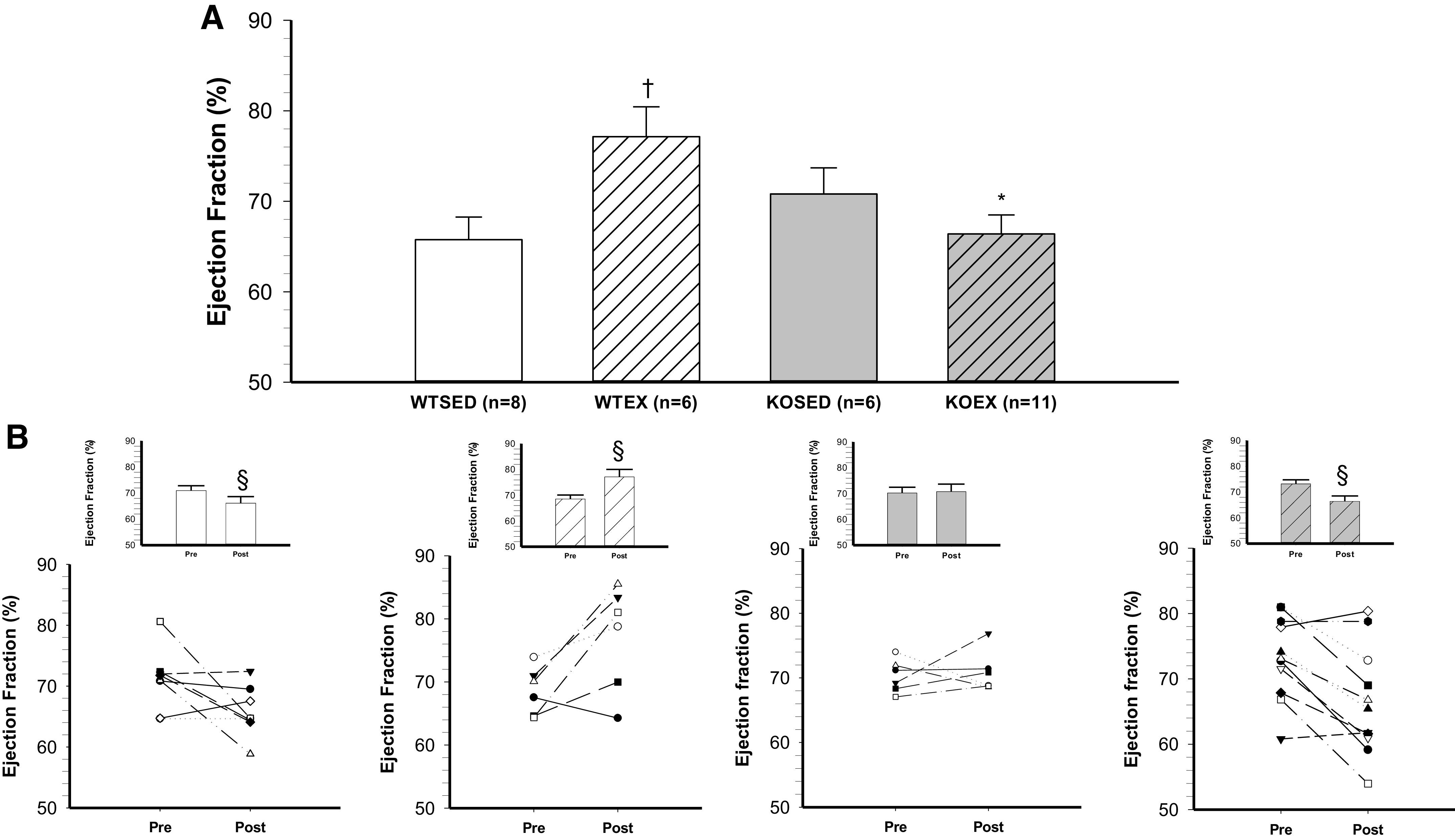
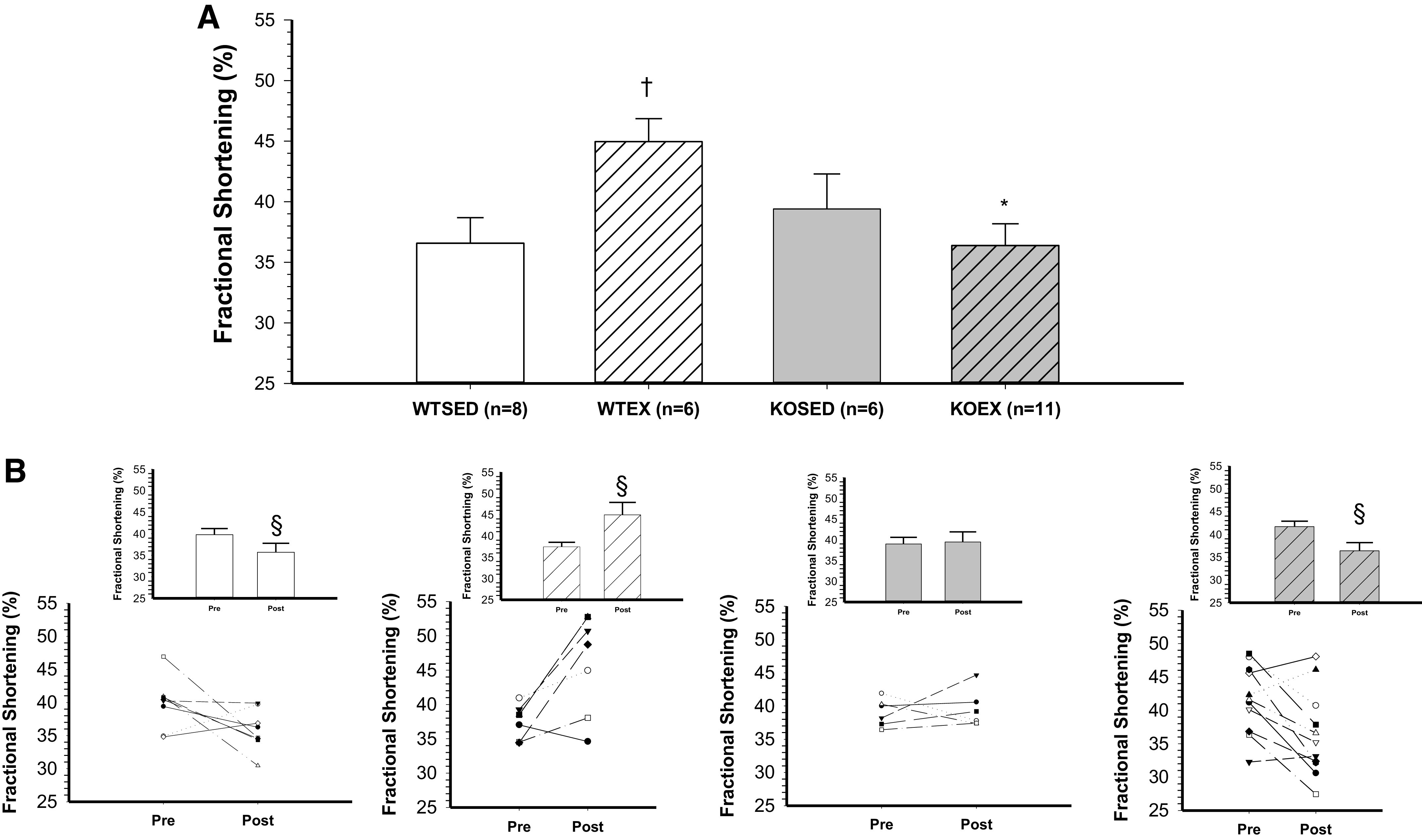
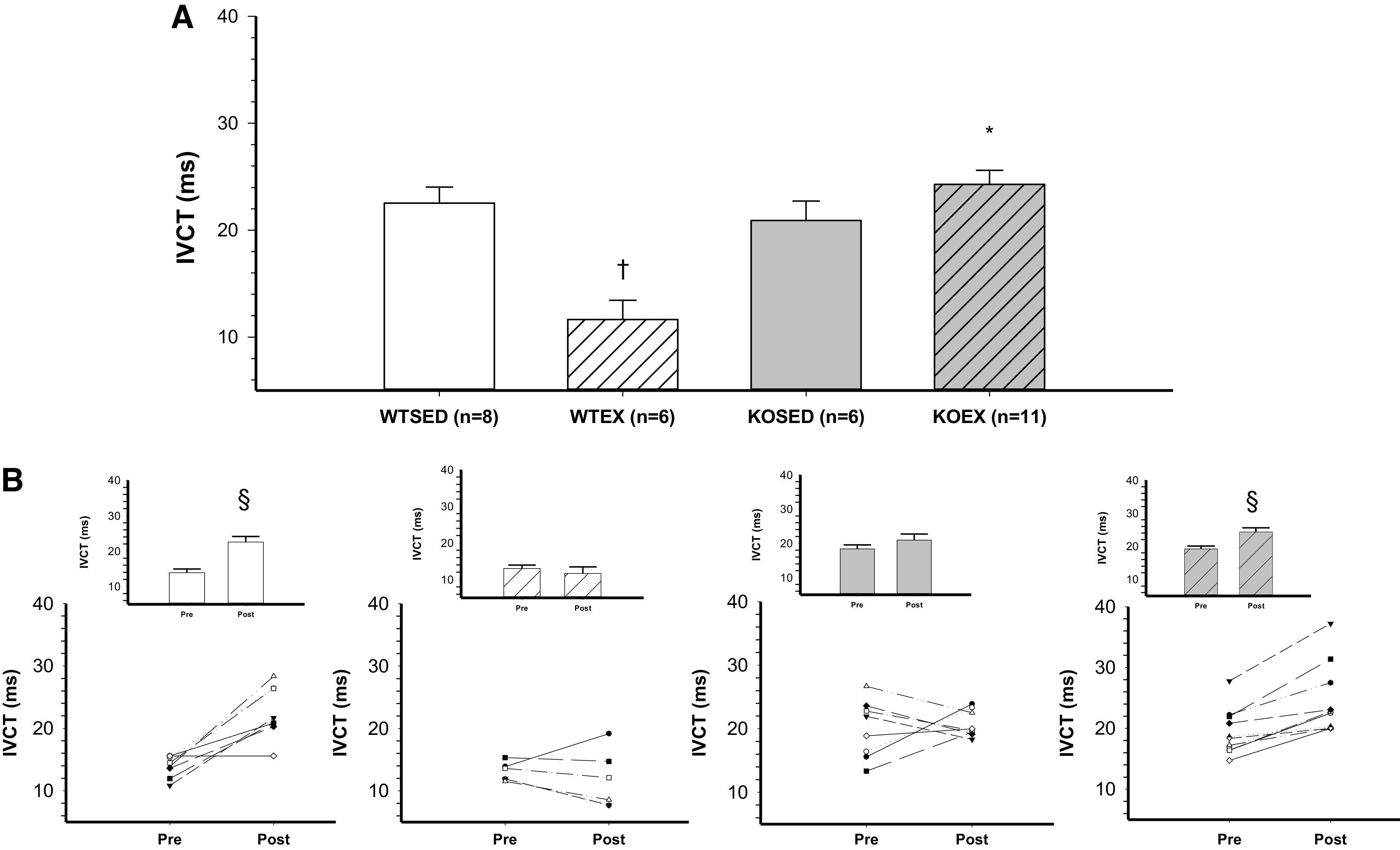
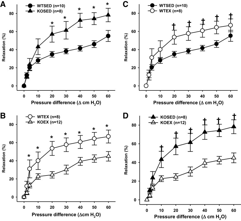
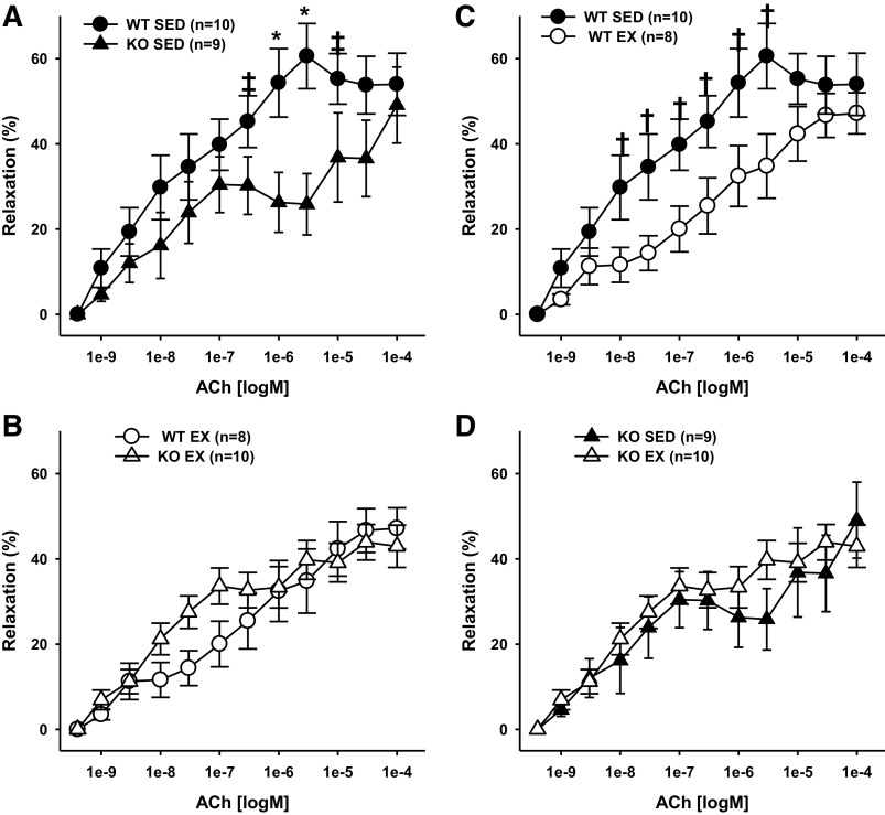
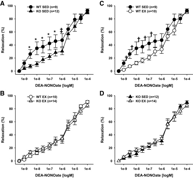
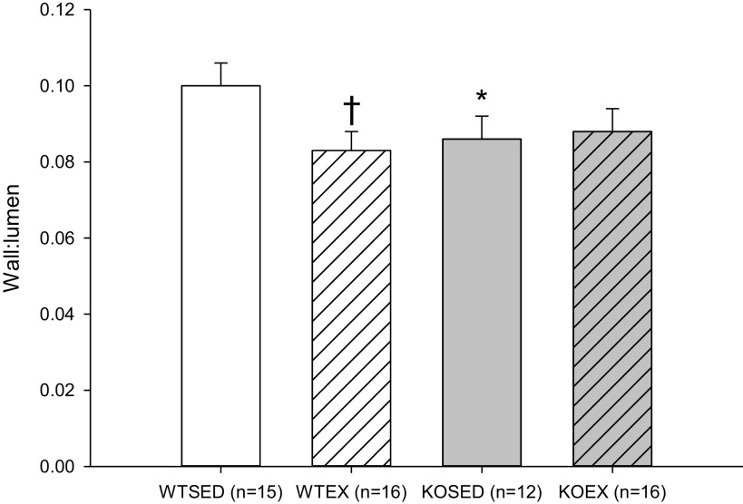
We tested the hypothesis that adiponectin deficiency attenuates cardiac and coronary microvascular function and prevents exercise training-induced adaptations of the myocardium and the coronary microvasculature in adult mice. Adult wild-type (WT) or adiponectin knockout (adiponectin KO) mice underwent treadmill exercise training or remained sedentary for 8-10 wk. Systolic and diastolic functions were assessed before and after exercise training or cage confinement. Vasoreactivity of coronary resistance arteries was assessed at the end of exercise training or cage confinement. Before exercise training, ejection fraction and fractional shortening were similar in adiponectin KO and WT mice, but isovolumic contraction time was significantly lengthened in adiponectin KO mice. Exercise training increased ejection fraction (12%) and fractional shortening (20%) with no change in isovolumic contraction time in WT mice. In adiponectin KO mice, both ejection fraction (-9%) and fractional shortening (-12%) were reduced after exercise training and these decreases were coupled to a further increase in isovolumic contraction time (20%). In sedentary mice, endothelium-dependent dilation to flow was higher in arterioles from adiponectin KO mice as compared with WT mice. Exercise training enhanced dilation to flow in WT mice but decreased flow-induced dilation in adiponectin KO mice. These data suggest that compensatory mechanisms contribute to the maintenance of cardiac and coronary microvascular function in sedentary mice lacking adiponectin; however, in the absence of adiponectin, cardiac and coronary microvascular adaptations to exercise training are compromised.NEW & NOTEWORTHY We report that compensatory mechanisms contribute to the maintenance of cardiac and coronary microvascular function in sedentary mice in which adiponectin has been deleted; however, when mice lacking adiponectin are subjected to the physiological stress of exercise training, beneficial coronary microvascular and cardiac adaptations are compromised or absent.

Keywords: ejection fraction; endothelium; fractional shortening; heart; vasodilation.


Conflict of interest statement

No conflicts of interest, financial or otherwise, are declared by the authors.

Figures

Figure 1.
Figure 1.
Effect of genotype and exercise training on ejection fraction. A: ejection fraction in wild-type sedentary (WTSED), wild-type trained (WTEX), adiponectin KO sedentary (adiponectin KOSED), and adiponectin KO trained (adiponectin KOEX). B: individual values before (pre) and after 8-wk exercise training or cage confinement. Values are means ± SE. Three-way ANOVA (genotype, exercise training, time) with one repeated measure (time). *Genotype effect; †exercise training effect; §significant pre/post difference, P ≤ 0.05. KO, knockout.
Figure 2.
Figure 2.
Effect of genotype and exercise training on fractional shortening. A: fractional shortening in wild-type sedentary (WTSED), wild-type trained (WTEX), adiponectin KO sedentary (adiponectin KOSED), and adiponectin KO trained (adiponectin KOEX). B: individual values before (pre) and after 8-wk exercise training or cage confinement. Values are means ± SE. Three-way ANOVA (genotype, exercise training, time) with one repeated measure (time). *Genotype effect; †exercise training effect; §significant pre/post difference, P ≤ 0.05. KO, knockout.
Figure 3.
Figure 3.
Effect of genotype and exercise training on isovolumic contraction time. A: isovolumic contraction time in wild-type sedentary (WTSED), wild-type trained (WTEX), adiponectin KO sedentary (adiponectin KOSED), and adiponectin KO trained (adiponectin KOEX). B: individual values before (pre) and after 8-wk exercise training or cage confinement. Values are means ± SE. Three-way ANOVA (genotype, exercise training, time) with one repeated measure (time). *Genotype effect; †exercise training effect; §significant pre/post difference, P ≤ 0.05. KO, knockout.
Figure 4.
Figure 4.
Endothelium-dependent flow-induced vasodilation of coronary resistance arteries from sedentary wild-type and adiponectin KO (WTSED and adiponectin KOSED, respectively) and exercise trained (WTEX and adiponectin KOEX, respectively). A and B: effect of genotype on endothelium-dependent vasodilation. C and D: effect of exercise training on endothelium-independent vasodilation. Values are means ± SE. Three-way ANOVA (genotype, exercise training, time) with one repeated measure (time). *Genotype effect; †exercise training effect; P ≤ 0.05. KO, knockout.
Figure 5.
Figure 5.
Endothelium-dependent acetylcholine-induced vasodilation of coronary resistance arteries from sedentary wild-type and adiponectin KO (WTSED and adiponectin KOSED, respectively) and exercise trained (WTEX and adiponectin KOEX, respectively). A and B: effect of genotype on endothelium-dependent vasodilation. C and D: effect of exercise training on endothelium-dependent vasodilation. Values are means ± SE. Three-way ANOVA (genotype, exercise training, time) with one repeated measure (time). *Genotype effect; †exercise training effect; P ≤ 0.05. ‡indicates genotype effect; P < 0.10. KO, knockout.
Figure 6.
Figure 6.
Endothelium-independent DEA-NONOate-induced vasodilation of coronary resistance arteries from sedentary wild-type and adiponectin KO (WTSED and adiponectin KOSED, respectively) and exercise trained (WTEX and adiponectin KOEX, respectively). A and B: effect of genotype on endothelium-independent vasodilation. C and D: effect of exercise training on endothelium-independent vasodilation. Values are means ± SE. Three-way ANOVA (genotype, exercise training, time) with one repeated measure (time). *Genotype effect; †exercise training effect; P ≤ 0.05. ‡indicates genotype effect; P < 0.10. KO, knockout.
Figure 7.
Figure 7.
Wall-to-lumen ratio (Wall:Lumen) in coronary resistance arteries from sedentary wild-type and adiponectin KO (WTSED and adiponectin KOSED, respectively) and exercise trained (WTEX and adiponectin KOEX, respectively). Three-way ANOVA (genotype, exercise training, time) with one repeated measure (time). *Genotype effect; †exercise training effect; P ≤ 0.05. KO, knockout.

Similar articles

Cited by

References

    1. Zarzour A, Kim HW, Weintraub NL. Understanding obesity-related cardiovascular disease: it's all about balance. Circulation 138: 64–66, 2018. doi:10.1161/CIRCULATIONAHA.118.034454. - DOI - PMC - PubMed
    1. Patel JV, Abraheem A, Dotsenko O, Creamer J, Gunning M, Hughes EA, Lip GY. Circulating serum adiponectin levels in patients with coronary artery disease: relationship to atherosclerotic burden and cardiac function. J Intern Med 264: 593–598, 2008. doi:10.1111/j.1365-2796.2008.02007.x. - DOI - PubMed
    1. Kenchaiah S, Evans JC, Levy D, Wilson PW, Benjamin EJ, Larson MG, Kannel WB, Vasan RS. Obesity and the risk of heart failure. N Engl J Med 347: 305–313, 2002. doi:10.1056/NEJMoa020245. - DOI - PubMed
    1. Hall JE. The kidney, hypertension, and obesity. Hypertension 41: 625–633, 2003. doi:10.1161/01.HYP.0000052314.95497.78. - DOI - PubMed
    1. Ding M, Carrao AC, Wagner RJ, Xie Y, Jin Y, Rzucidlo EM, Yu J, Li W, Tellides G, Hwa J, Aprahamian TR, Martin KA. Vascular smooth muscle cell-derived adiponectin: a paracrine regulator of contractile phenotype. J Mol Cell Cardiol 52: 474–484, 2012. doi:10.1016/j.yjmcc.2011.09.008. - DOI - PMC - PubMed

Publication types

LinkOut - more resources