. 2021 May 14;16(5):e0250381.
doi: 10.1371/journal.pone.0250381. eCollection 2021.

The number of neurons in Drosophila and mosquito brains

Affiliations

The number of neurons in Drosophila and mosquito brains

Joshua I Raji et al. PLoS One. .

Abstract

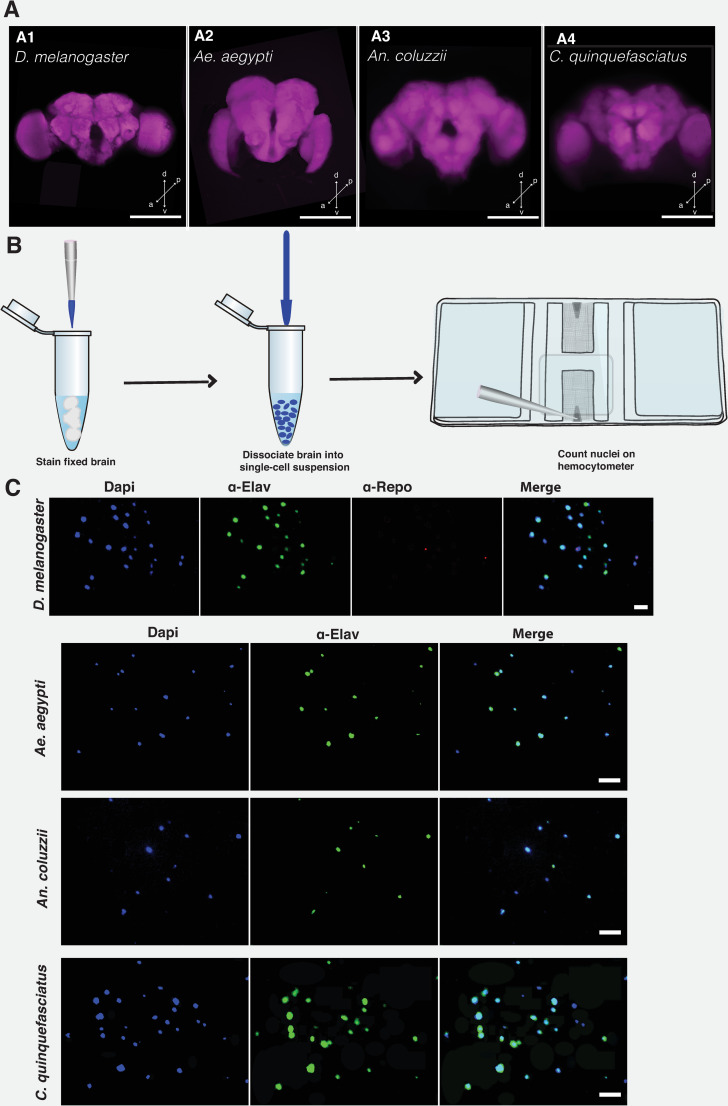
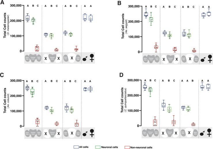
Various insect species serve as valuable model systems for investigating the cellular and molecular mechanisms by which a brain controls sophisticated behaviors. In particular, the nervous system of Drosophila melanogaster has been extensively studied, yet experiments aimed at determining the number of neurons in the Drosophila brain are surprisingly lacking. Using isotropic fractionator coupled with immunohistochemistry, we counted the total number of neuronal and non-neuronal cells in the whole brain, central brain, and optic lobe of Drosophila melanogaster. For comparison, we also counted neuronal populations in three divergent mosquito species: Aedes aegypti, Anopheles coluzzii and Culex quinquefasciatus. The average number of neurons in a whole adult brain was determined to be 199,380 ±3,400 cells in D. melanogaster, 217,910 ±6,180 cells in Ae. aegypti, 223,020 ± 4,650 cells in An. coluzzii and 225,911±7,220 cells in C. quinquefasciatus. The mean neuronal cell count in the central brain vs. optic lobes for D. melanogaster (101,140 ±3,650 vs. 107,270 ± 2,720), Ae. aegypti (109,140 ± 3,550 vs. 112,000 ± 4,280), An. coluzzii (105,130 ± 3,670 vs. 107,140 ± 3,090), and C. quinquefasciatus (108,530 ±7,990 vs. 110,670 ± 3,950) was also estimated. Each insect brain was comprised of 89% ± 2% neurons out of its total cell population. Isotropic fractionation analyses did not identify obvious sexual dimorphism in the neuronal and non-neuronal cell population of these insects. Our study provides experimental evidence for the total number of neurons in Drosophila and mosquito brains.


Conflict of interest statement

The authors have declared that no competing interests exist.

Figures

Fig 1
Fig 1. Estimating brain cell population using isotropic fractionator method.
Confocal Z-stacks of the whole brain of D. melanogaster (A1), Ae. aegypti (A2), An. coluzzii (A3), and C. quinquefasciatus (A4). Brain was counterstained with nc82 antibody that labels the pre-synaptic active zone protein, Bruchpilot. Whole brain was imaged on a LSM 700 Zeiss confocal microscope at 512 × 512 pixel resolution, with 1.04 μm pixel size. Scale bar represents 50 μm. Arrow directions represent the dorsal (d), ventral (v), posterior (p), and anterior (a), respectively. (B). Cartoon showing the steps for performing isotropic fractionator. Brain tissue is fixed and stained with the appropriate dye or antibody. Tissue is dissociated into a single-cell suspension, which is then counted on a Neubauer counting chamber or hemocytometer. (C) Images of nuclei are stained with DAPI (4’, 6-diamidino-2-phenylindole dihydrochloride), α-elav (embryonic lethal abnormal vision), and α-repo (Reversed polarity protein) to label nuclei, neuronal and non-neuronal cells, respectively. Staining of glial cells with α-repo was possible only for Drosophila brain cells. Scale bar represents 10 μm. For illustration purposes, all images were processed on ImageJ software and Adobe Illustrator.
Fig 2
Fig 2. Quantification of the cell population in vinegar fly and mosquito brains.
Box plot showing total cell counts, neuronal, and non-neuronal cell population in the whole brain, central brain, and optic lobe of D. melanogaster (2A), Ae. aegypti (2B), An. coluzzii (2C), and C. quiquefasciatus (2D). Blue plots represent all cells, while green and red plots represent neuronal and non-neuronal cells, respectively. Different groups (whole brain, central brain, optic lobes, and sex) are separated by dashed lines. Each data point represents an average of three brains. The number of trials for whole brain (n = 15), central brain (n = 10), optic lobes (n = 10), male (n = 8), and female (n = 7) is consistent for all animals. Within each group, different letters are statistically different (P < 0.05) when analyzed by one-way ANOVA with Tukey’s post hoc test. Statistical comparisons between male and female total cell was performed by T-Test. Data analysis was performed using the GraphPad Prism 9 software package (GraphPad Software, San Diego, CA, USA).

References

    1. Lent R, Azevedo FAC, Andrade-Moraes CH, Pinto AVO. How many neurons do you have? Some dogmas of quantitative neuroscience under revision: Neuroscience dogmas and brain cell numbers. Eur J Neurosci. 2012;35: 1–9. 10.1111/j.1460-9568.2011.07923.x - DOI - PubMed
    1. Andersen BB, Gundersen HJG, Pakkenberg B. Aging of the human cerebellum: a stereological study. J Comp Neurol. 2003;466: 356–365. 10.1002/cne.10884 - DOI - PubMed
    1. Andersen K, Andersen BB, Pakkenberg B. Stereological quantification of the cerebellum in patients with Alzheimer’s disease. Neurobiol Aging. 2012;33: 197.e11–197.e20. 10.1016/j.neurobiolaging.2010.06.013 - DOI - PubMed
    1. Storks L, Powell BJ, Leal M. Peeking Inside the Lizard Brain: Neuron Numbers in Anolis and Its Implications for Cognitive Performance and Vertebrate Brain Evolution. Integr Comp Biol. 2020. [cited 14 Jan 2021]. 10.1093/icb/icaa129 - DOI - PubMed
    1. Oliveira-Pinto AV, Santos RM, Coutinho RA, Oliveira LM, Santos GB, Alho ATL, et al. Sexual Dimorphism in the Human Olfactory Bulb: Females Have More Neurons and Glial Cells than Males. PLOS ONE. 2014;9: e111733. 10.1371/journal.pone.0111733 - DOI - PMC - PubMed

Publication types

LinkOut - more resources