Review
. 2021 Sep 1;1863(9):183644.
doi: 10.1016/j.bbamem.2021.183644. Epub 2021 May 11.

Nanopore sensing: A physical-chemical approach

Affiliations
Review

Nanopore sensing: A physical-chemical approach

Joseph W F Robertson et al. Biochim Biophys Acta Biomembr. .

Abstract

Protein nanopores have emerged as an important class of sensors for the understanding of biophysical processes, such as molecular transport across membranes, and for the detection and characterization of biopolymers. Here, we trace the development of these sensors from the Coulter counter and squid axon studies to the modern applications including exquisite detection of small volume changes and molecular reactions at the single molecule (or reactant) scale. This review focuses on the chemistry of biological pores, and how that influences the physical chemistry of molecular detection.

Keywords: DNA sequencing; Ion channel; Nanopore sensor; Peptide detection; Porin.


Figures

Figure 1:
Figure 1:
The development of nanopore sensors have their origin in the discovery of the propagation of electrical signals in squid axon (a) and the Coulter method (b). (a) Hodgkin, Huxley and Katz’s single axon measurement apparatus [22], and (b) the cell-counting unit from an early version of the Coulter counter [25].
Figure 2:
Figure 2:
Nanopore-based biosensors fall in two major categories characterized by how the analyte interacts with the pore. (a) In the most common method, the analyte, or analyte and co-analyte partition into the central cavity of the pore causing current interruptions, and (b) the analyte induces conformational changes in the pore causing the channel to gate. The images were adapted from Chavis et al. [29] and Perez-Rathke et al. [30], respectively.
Figure 3:
Figure 3:
Structure of various biological pores used for nanopore sensing. A. Ribbon diagram of the heptameric α-hemolysin pore where the left image shows its side view and the right image shows its top-down view [43] B. The side view of the aerolysin nanopore (left) and the top view (right) derived from electron microscopy [44] C. The outer surface side view (left) and top down view (right) of the MspA porin along with the inner channel highlighted with black lining. Green and yellow colors depict the polar and nonpolar regions of the surface respectively [45] D. Side view (left) and a cut-through view (right) of a CsgG nonamer channel [46] E. Cross-section view of Wildtype FraC (WtFraC) (left). The blue and red colors indicate the positive and negative charged residue along the pore surface, respectively. The image on the right is the top view of WtFraC (top) and D10R/K159E FraC (ReFraC) nanopore (bottom) [47] F. Histogram of single-channel conductance for both WtFraC and ReFraC in a 1 mol/L NaCl, 15 mmol/L Tris-HCl, pH 7.5 solution under a +50 mV transmembrane potential. The current trace was recorded at a sampling rate of 10 kHz and filtered with a 2 kHz low-pass Bessel filter.
Figure 4:
Figure 4:
Nanopore sensors operate according to a simple reaction scheme that requires capture, retention and release either in the forward or reverse direction. The magnitude of the free energy barriers, which can be entropic, enthalpic or both dictate the efficiency and effectiveness of the sensor. In the simplified scheme presented here, a polymer reorganizes to cross a barrier for entry into the pore and is held in the pore by barriers at either end. The event is complete when the polymer exits in either direction. The barriers are dependent on the chemical details of the molecule and the pore. Understanding and manipulating these barriers is a major focus for biosensor development.
Figure 5:
Figure 5:
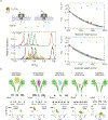
Nanopores as size-selective sensors. Blockade depth histograms show the evolution of mass resolution of PEG. (a) Recast data from [51] shows the full range of size discrimination of PEG from n = 18 to n = 72. (b) increasing the residence time with a pore-modifying gold cluster improves the resolution (red) with respect to the cluster free αHL pore (black) due to higher escape barriers and an order of magnitude increase in residence time [178]. (c) Aerolysin pores alter the balance of electroosmotic and electrophoretic transport and possibly the dynamics of PEG inside the pore and further improve molecular resolution [97].
Figure 6:
Figure 6:
Size selectivity for peptides in a nanopore sensor shows that volume (mass) of the peptides serves as a reliable discriminant. (a) Small peptides in an αHL pore show that peptide size scales well with PEG blockade depth suggesting a common volume dependent mechanism [29]. (b) This selectivity is preserved with the more complex pore geometry of FraC, which provides a gradient selectivity governed by the unique shape of the pore [116]. See text for further details.
Figure 7:
Figure 7:
Protein nanopores have been used to follow a number of different chemical interactions and transformations while trapped inside the pore. These are as varied as (a) observing ligand exchange on a gold nanocluster [205], (b) pH dependent base-flipping of mismatched DNA [52], and (c) conformational changes of an enzyme during its reaction cycle [212].

References

    1. Behrends JC, Evolution of the Ion Channel Concept: The Historical Perspective, Chemical Reviews. 112 (2012) 6218–6226. 10.1021/cr300349g. - DOI - PubMed
    1. Montal M, Mueller P, Formation of bimolecular membranes from lipid monolayers and a study of their electrical properties., Proceedings of the National Academy of Sciences. 69 (1972) 3561–3566. - PMC - PubMed
    1. Sackmann E, Supported membranes: scientific and practical applications., Science. 271 (1996) 43–48. - PubMed
    1. Hladky SB, Haydon DA, Ion transfer across lipid membranes in the presence of gramicidin A. I. Studies of the unit conductance channel., Biochimica Et Biophysica Acta. 274 (1972) 294–312. 10.1016/0005-2736(72)90178-2. - DOI - PubMed
    1. Bechinger B, Structure and functions of channel-forming peptides: magainins, cecropins, melittin and alamethicin., The Journal of Membrane Biology. 156 (1997) 197–211. - PubMed

Publication types