. 2021;12(3):857-871.
doi: 10.1016/j.jcmgh.2021.05.006. Epub 2021 May 11.

Sodium Butyrate Supplementation Inhibits Hepatic Steatosis by Stimulating Liver Kinase B1 and Insulin-Induced Gene

Affiliations

Sodium Butyrate Supplementation Inhibits Hepatic Steatosis by Stimulating Liver Kinase B1 and Insulin-Induced Gene

Ze-Hua Zhao et al. Cell Mol Gastroenterol Hepatol. 2021.

Abstract

Background and aims: Butyric acid is an intestinal microbiota-produced short-chain fatty acid, which exerts salutary effects on alleviating nonalcoholic fatty liver disease (NAFLD). However, the underlying mechanism of butyrate on regulating hepatic lipid metabolism is largely unexplored.

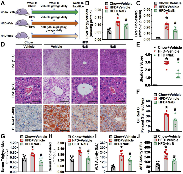
Methods: A mouse model of NAFLD was induced with high-fat diet feeding, and sodium butyrate (NaB) intervention was initiated at the eighth week and lasted for 8 weeks. Hepatic steatosis was evaluated and metabolic pathways concerning lipid homeostasis were analyzed.

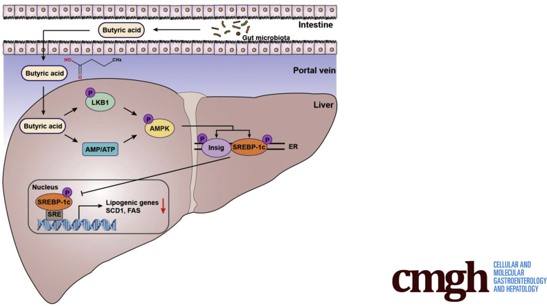
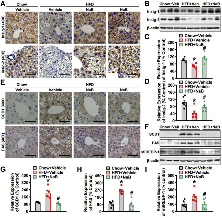
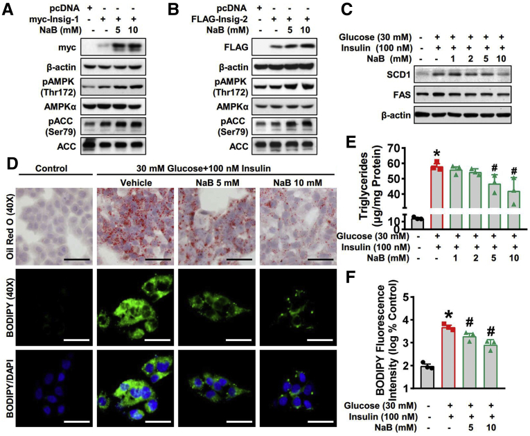
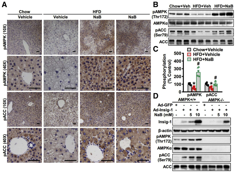
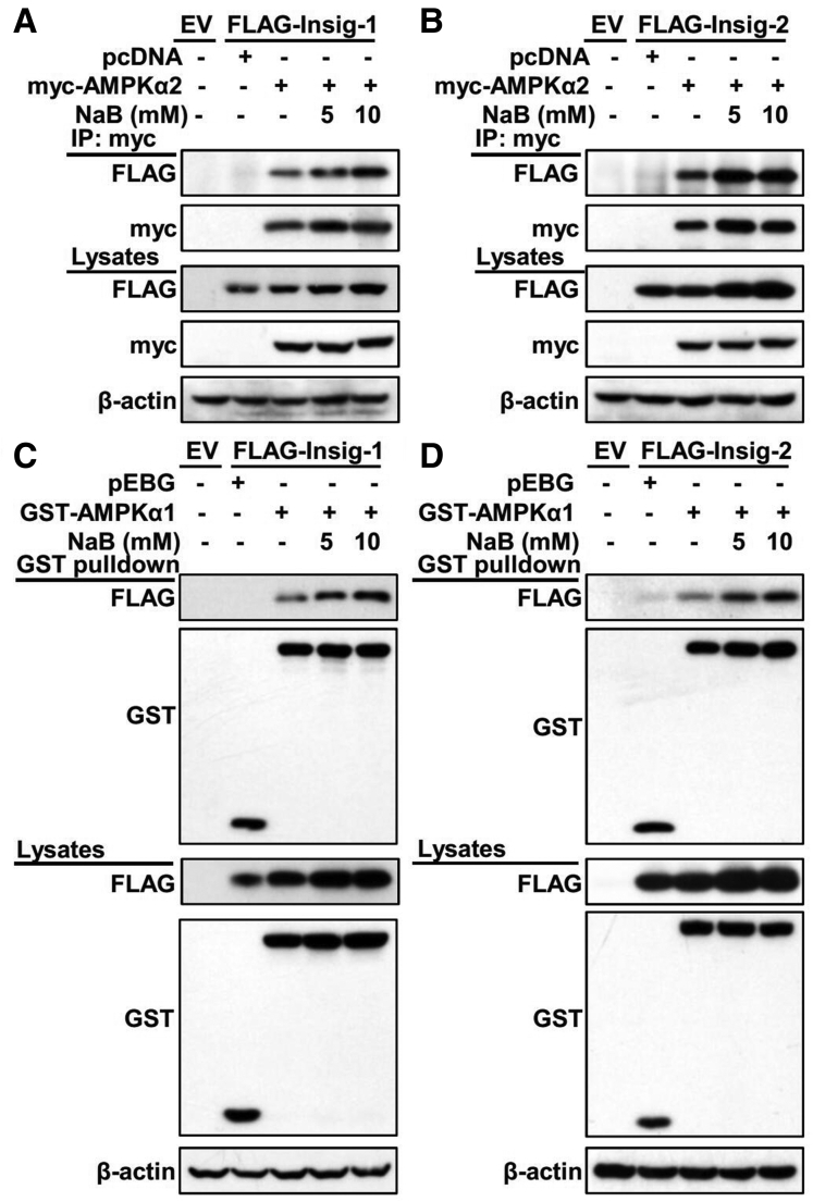
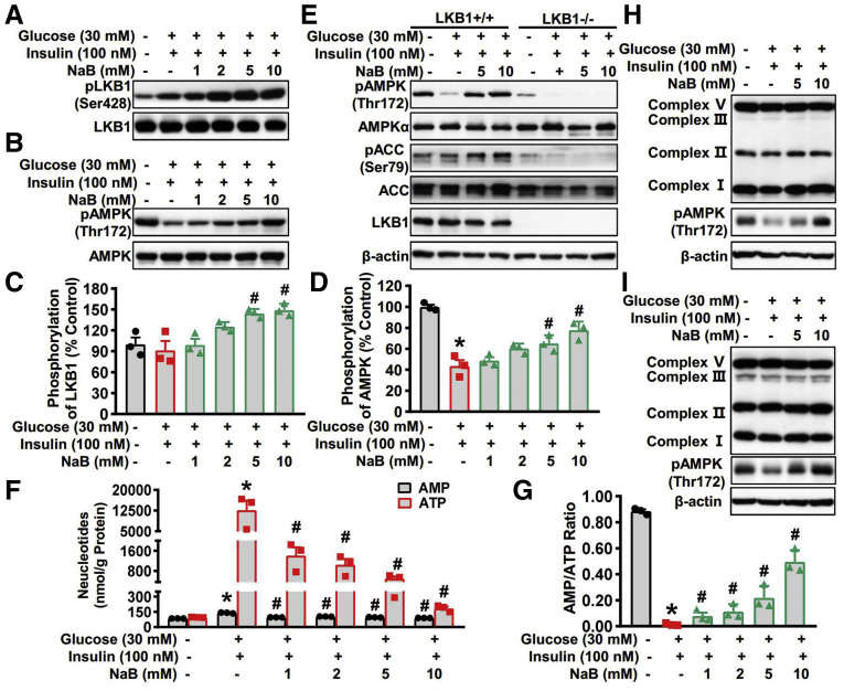
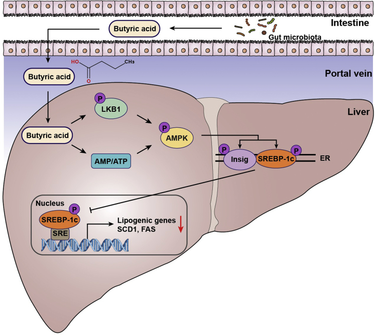
Results: Here, we report that administration of NaB by gavage once daily for 8 weeks causes an augmentation of insulin-induced gene (Insig) activity and inhibition of lipogenic gene in mice fed with high-fat diet. Mechanistically, NaB is sufficient to enhance the interaction between Insig and its upstream kinase AMP-activated protein kinase (AMPK). The stimulatory effects of NaB on Insig-1 activity are abolished in AMPKα1/α2 double knockout (AMPK-/-) mouse primary hepatocytes. Moreover, AMPK activation by NaB is mediated by LKB1, as evidenced by the observations showing NaB-mediated induction of phosphorylation of AMPK, and its downstream target acetyl-CoA carboxylase is diminished in LKB1-/- mouse embryonic fibroblasts.

Conclusions: These studies indicate that NaB serves as a negative regulator of hepatic lipogenesis in NAFLD and that NaB attenuates hepatic steatosis and improves lipid profile and liver function largely through the activation of LKB1-AMPK-Insig signaling pathway. Therefore, NaB has therapeutic potential for treating NAFLD and related metabolic diseases.

Keywords: Hepatic Lipogenesis; Insulin-Induced Gene; LKB1; NAFLD; Sodium Butyrate.


Figures

None
Graphical abstract
Figure 1
Figure 1
Beneficial effects of NaB on lowering hepatic steatosis in mice fed with HFD. Eight-week-old male C57BL/6 mice were fed with a HFD for 8 weeks, and then gavaged with NaB (1 g/kg/d) or vehicle (phosphate-buffered saline) for 8 weeks. Mice were sacrificed at the end of 16th week and livers were harvested. (A) Schematic illustration showing the design of the in vivo experiment. (B) Hepatic triglycerides and (C) cholesterol levels were assessed in mice (n = 6–8). (D) Representative hematoxylin and eosin (H&E) and Oil Red O stainings of the liver sections are shown (scale bar = 50 μm). (E) Steatosis score based on the SAF score algorithm is measured (n = 6). (F) Quantification of Oil Red O–stained areas is shown (n = 6). Serum levels of (G) triglycerides, (H) cholesterol, (I) alanine aminotransferase, and (J) aspartate aminotransferase are detected (n = 5–6). The data are presented as the mean ± SEM, unpaired 2-tailed Student’s t test, n = 5–8. ∗P < .05 vs mice fed with chow diet; #P < .05 vs mice fed with HFD.
Figure 2
Figure 2
Administration of NaB stimulates the activity of Insig to inhibit lipogenic gene expression in mice fed with HFD. (A–C) The protein levels of Insig-1 and Insig-2 are increased in the liver of mice treated with NaB. (A) Representative immunohistochemical staining of the liver sections with Insig-1 and Insig-2 antibodies (scale bar = 50 μm). (B) Immunoblots were performed, and the band intensity of (C) Insig-1 and (D) Insig-2 was quantified by densitometry (n = 6). (E–I) The protein levels of lipogenic genes are decreased in the liver of mice treated with NaB. (E) Representative immunohistochemical staining of the liver sections with SCD1 and FAS antibodies (scale bar = 50 μm). (F) Immunoblots were performed, and the band intensity of (G) SCD1, (H) FAS, and (I) nuclear SREBP-1 was quantified by densitometry (n = 6). The data are presented as the mean ± SEM, unpaired 2-tailed Student’s t test, n = 6. ∗P < .05 vs mice fed with chow diet; #P < .05 vs mice fed with HFD.
Figure 3
Figure 3
NaB inhibits lipogenic gene expression and lipid accumulation by enhancing Insig activity in hepatocytes. (A, B) NaB treatment increases protein levels of Insig. HEK293 cells were transfected with pcDNA, Myc-tagged Insig-1, or FLAG-tagged Insig-2 for 24 hours, followed by treatment with 5 mM or 10 mM NaB as indicated. Immunoblots were performed. (C–F) NaB treatment decreases protein levels of lipogenic enzymes and alleviates lipid accumulation in human HepG2 cells exposed to high glucose plus insulin. HepG2 cells were fasted for 24 hours, followed by 30 mM glucose and 100 nM insulin treatment, together with NaB (1–10 mM) or vehicle (phosphate-buffered saline) treatment. (C) Immunoblots show that NaB treatment decreases protein levels of SCD-1 and FAS in a dose-dependent manner. (D–F) Triglycerides accumulation is attenuated with 5 mM and 10 mM NaB treatment. (D) Representative oil red O staining (scale bar = 50 μm) and (E) the quantification of triglycerides are shown (n = 3). (D) Representative BODIPY staining (scale bar = 50 μm) and (F) the quantification are shown (n = 3). The data are presented as the mean ± SEM, unpaired 2-tailed Student’s t test, n = 3. ∗P < .05 vs vehicle; #P < .05 vs treatment with high glucose plus insulin.
Figure 4
Figure 4
AMPK is required for Insig-1 induction in response to NaB treatment. (A–C) NaB increases the expression levels of phosphorylated AMPK in mice fed with HFD. (A) Representative immunohistochemical staining of the liver sections with phosphorylated AMPK (pAMPK) and phosphorylated ACC (pACC) antibodies (scale bar = 50 μm). (B) Immunoblots were performed, and (C) the band intensity of pAMPK and pACC was quantified by densitometry (n = 6). (D) AMPK is required for Insig-1 induction in response to NaB treatment. AMPK+/+ or AMPKα1/α2 double knockout (AMPK−/−) mouse primary hepatocytes were infected with adenoviruses encoding Insig-1 (Ad-Insig-1) or green fluorescent protein (GFP) for 24 hours, followed by treatment with 5 mM or 10 mM NaB for 24 hours. The data are presented as the mean ± SEM, unpaired 2-tailed Student’s t test, n = 6. ∗P < .05 vs mice fed with chow diet; #P < .05 vs mice fed with HFD.
Figure 5
Figure 5
Administration of NaB enhances the interaction between Insig and its upstream kinase AMPK. (A, B) NaB enhances the association of AMPKα2 subunit with Insig in a dose-dependent manner. FLAG-tagged (A) Insig-1 or (B) Insig-2 was co-transfected with Myc-tagged AMPKα2 in HEK293 cells for 24 hours, followed by treatment with 5 mM or 10 mM NaB for 24 hours. The cell lysates were incubated with Myc antibodies and purified with protein A/G-Sepharose beads. The precipitates and lysates were individually immunoblotted with indicated antibodies. (C, D) NaB enhances the association of AMPKα1 subunit with Insig in a dose-dependent manner. FLAG-tagged (D) Insig-1 or (D) Insig-2 was co-transfected with GST-tagged AMPKα1 in HEK293 cells for 24 hours, followed by treatment with 5 mM or 10 mM NaB for 24 hours. The cell lysates were then purified with GST Sepharose beads. The precipitates and lysates were immunoblotted with antibodies against FLAG or GST. EV, empty vector; IP, immunoprecipitation.
Figure 6
Figure 6
The stimulatory effects of NaB on AMPK activity are mediated by LKB1 phosphorylation and intracellular energy levels. (A–D) NaB increases levels of phosphorylated LKB1 and phosphorylated AMPK in a dose-dependent manner in HepG2 cells. HepG2 cells were fasted for 24 hours, followed by 30 mM glucose and 100 nM insulin treatment, together with NaB (1–10 mM) or vehicle (phosphate-buffered saline) treatment. (A) Immunoblots show that NaB treatment increases phosphorylation of LKB1 in a dose-dependent manner. (C) The band intensity of phosphorylated LKB1 was quantified by densitometry (n = 3). (B) Immunoblots show that NaB treatment increases phosphorylation of AMPK in a dose-dependent manner. (D) The band intensity of phosphorylated AMPK (pAMPK) was quantified by densitometry (n = 3). (E) LKB1 is required for AMPK activation in response to NaB treatment. LKB1−/− mouse embryonic fibroblasts were fasted for 24 hours, followed by 30 mM glucose and 100 nM insulin treatment, together with NaB (5 mM and 10 mM) or vehicle (phosphate-buffered saline) treatment. (F, G) NaB treatment regulates the levels of AMP and ATP and increases AMP-to-ATP ratio in HepG2 cells exposed to high glucose plus insulin. HepG2 cells were fasted for 24 hours, followed by 30 mM glucose and 100 nM insulin treatment, together with different doses of NaB (1–10 mM) or vehicle (phosphate-buffered saline) treatment, and then the lysates were subjected to AMP and ATP detection. (F) The levels of AMP and ATP were quantified and (G) the AMP-to-ATP ratio were calculated. (H, I) Protein levels of mitochondrial electron transport chain complex are not altered by NaB treatment. Immunoblots were performed in (H) human HepG2 cells and (I) mouse primary hepatocytes. The data are presented as the mean ± SEM, unpaired 2-tailed Student’s t test, n = 3. ∗P < .05 vs vehicle; #P < .05 vs treatment with high glucose plus insulin. pACC, phosphorylated acetyl-CoA carboxylase.
Figure 7
Figure 7
The proposed model for butyric acid–mediated inhibition of lipogenesis via LKB1-AMPK-Insig signaling pathway. Gut-derived metabolite butyric acid exerts beneficial effects on hepatocytes, enhancing LKB1 phosphorylation and elevating AMP-to-ATP ratio, which facilitates AMPK activation. Subsequently, interaction between AMPK and Insig is strengthened by butyric acid, resulting in phosphorylation of Insig at Thr222 and decline of proteasome-mediated degradation. The increased Insig restrains the processing and translocation of SREBP-1c and suppresses its transcriptional activity. The activation of AMPK by butyric acid also facilitates SREBP-1c phosphorylation at Ser372 and suppresses its proteolytic cleavage. These mechanisms by which butyric acid inhibits de novo lipogenesis in hepatocytes provide therapeutic potential for hepatic steatosis management. ER, endoplasmic reticulum.

Similar articles

Cited by

References

    1. Fan J.G., Kim S.U., Wong V.W. New trends on obesity and NAFLD in Asia. J Hepatol. 2017;67:862–873. - PubMed
    1. Paik J.M., Golabi P., Younossi Y., Mishra A., Younossi Z.M. Changes in the global burden of chronic liver diseases from 2012 to 2017: The growing impact of nonalcoholic fatty liver disease. Hepatology. 2020;72:1605–1616. - PubMed
    1. Samuel V.T., Shulman G.I. Nonalcoholic fatty liver disease as a nexus of metabolic and hepatic diseases. Cell Metab. 2018;27:22–41. - PMC - PubMed
    1. Smith G.I., Shankaran M., Yoshino M., Schweitzer G.G., Chondronikola M., Beals J.W., Okunade A.L., Patterson B.W., Nyangau E., Field T., Sirlin C.B., Talukdar S., Hellerstein M.K., Klein S. Insulin resistance drives hepatic de novo lipogenesis in nonalcoholic fatty liver disease. J Clin Invest. 2020;130:1453–1460. - PMC - PubMed
    1. Goedeke L., Bates J., Vatner D.F., Perry R.J., Wang T., Ramirez R., Li L., Ellis M.W., Zhang D., Wong K.E., Beysen C., Cline G.W., Ray A.S., Shulman G.I. Acetyl-CoA carboxylase inhibition reverses nafld and hepatic insulin resistance but promotes hypertriglyceridemia in rodents. Hepatology. 2018;68:2197–2211. - PMC - PubMed

MeSH terms