. 2021 May 14;21(1):84.
doi: 10.1186/s12862-021-01818-5.

Adaptive evolution of Moniliophthora PR-1 proteins towards its pathogenic lifestyle

Affiliations

Adaptive evolution of Moniliophthora PR-1 proteins towards its pathogenic lifestyle

Adrielle A Vasconcelos et al. BMC Ecol Evol. .

Abstract

Background: Plant pathogenesis related-1 (PR-1) proteins belong to the CAP superfamily and have been characterized as markers of induced defense against pathogens. Moniliophthora perniciosa and Moniliophthora roreri are hemibiotrophic fungi that respectively cause the witches' broom disease and frosty pod rot in Theobroma cacao. Interestingly, a large number of plant PR-1-like genes are present in the genomes of both species and many are up-regulated during the biotrophic interaction. In this study, we investigated the evolution of PR-1 proteins from 22 genomes of Moniliophthora isolates and 16 other Agaricales species, performing genomic investigation, phylogenetic reconstruction, positive selection search and gene expression analysis.

Results: Phylogenetic analysis revealed conserved PR-1 genes (PR-1a, b, d, j), shared by many Agaricales saprotrophic species, that have diversified in new PR-1 genes putatively related to pathogenicity in Moniliophthora (PR-1f, g, h, i), as well as in recent specialization cases within M. perniciosa biotypes (PR-1c, k, l) and M. roreri (PR-1n). PR-1 families in Moniliophthora with higher evolutionary rates exhibit induced expression in the biotrophic interaction and positive selection clues, supporting the hypothesis that these proteins accumulated adaptive changes in response to host-pathogen arms race. Furthermore, although previous work showed that MpPR-1 can detoxify plant antifungal compounds in yeast, we found that in the presence of eugenol M. perniciosa differentially expresses only MpPR-1e, k, d, of which two are not linked to pathogenicity, suggesting that detoxification might not be the main function of most MpPR-1.

Conclusions: Based on analyses of genomic and expression data, we provided evidence that the evolution of PR-1 in Moniliophthora was adaptive and potentially related to the emergence of the parasitic lifestyle in this genus. Additionally, we also discuss how fungal PR-1 proteins could have adapted from basal conserved functions to possible roles in fungal pathogenesis.

Keywords: Adaptation; Fungi; Gene evolution; Phylogenetics; Phytopathogen; Positive selection; Witches’ broom disease.


Conflict of interest statement

The authors declare no competing interests.

Figures
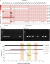
Fig. 1
Fig. 1
Characterization of PR-1 gene families in M. perniciosa and M. roreri genomes. a Heatmap of the number of gene copies per family of PR-1-like candidates per Moniliophthora isolate. Identification of genomes are in columns and PR-1 family names are in rows. b Amplification by PCR of MpPR-1c and MpPR-1d genes in the genomic DNA of eight M. perniciosa isolates. 1 Kb Ld = 1 Kb Plus DNA Ladder (Invitrogen), Neg = PCR negative control (no DNA). Expected fragment sizes were 687 bp for MpPR-1c and 902 bp for MpPR-1d. c Synteny analysis of a 10 Kb portion of the genome where the MpPR-1j, c, d genes are found in the three biotypes of M. perniciosa and M. roreri. The genomes analyzed were C-BA3, S-MG2, R-CO2, L-EC1 and L-EC2. Only identity above 75% to the C-biotype reference is shown
Fig. 2
Fig. 2
Phylogenetic cladogram of PR-1 proteins in Agaricales (Basidiomycota). a Phylogenetic relationships were inferred by maximum likelihood and branch support was obtained using 1000 bootstraps. Only branch support values greater than 70 are shown. PR-1c is indicated as a yellow branch inside the PR-1j clade, and PR-1n is indicated with a dark green dot and branch. Proteins with ancestral divergence to more than one family were named with the letters of the derived families. Full species names are in Additional file 1. The branch lengths were dimensioned for easy visualization. b The same phylogeny shown in “a” is presented with the respective branch lengths augmented for the Moniliophthora specific PR-1 genes
Fig. 3
Fig. 3
Phylogenetic reconstruction of PR-1 proteins in Moniliophthora and heatmap of expression Z-scores of PR-1. Phylogenetic relationships of PR-1 proteins from 18 M. perniciosa and 4 M. roreri isolates were inferred by maximum likelihood and branch support was obtained using 1000 bootstraps. The PRY1 protein of Saccharomyces cerevisiae was used as an outgroup. Clades filled with pink color indicate PR-1 families with evidence of positive selection detected with the site model test of CodeML. A version of this tree with non-collapsed branches can be found in Additional file 4. For each PR-1 family, the Z-score of log transformed expression levels of MpPR-1 and MrPR-1 from transcriptomic data was calculated for conditions (columns) and plotted as a heatmap. The heatmap includes MpPR-1 data from seven conditions of the C-biotype of M. perniciosa from the Witches’ Broom Transcriptomic Atlas, seven different time points of S-biotype infection in MicroTom tomato plants, and two conditions of M. roreri infection in cacao pods (frosty pod rot). Conditions highlighted with a grey background and asterisk indicate the biotrophic stage of the plant-pathogen interaction
Fig. 4
Fig. 4
Sequence alignment and phylogeny of PR-1i proteins in Moniliophthora isolates. Only a slice of the middle portion of the alignment is shown to highlight the sites with positive selection signs, indicated by red (p-value ≤ 0.01) or orange (p-value ≤ 0.05) bars in the bar chart of omega values below the alignment. Below the bar chart, annotations indicate the locations along the sequence of the CAP domains, caveolin-binding motif (CBM) and alpha-helices (α). On the 3D crystal structure of MpPR-1i protein (PBD:5V50) and on the bar chart, “A” indicates the site under positive selection detected in the CBM and “B” indicates the site under positive selection in alpha-helix 1. For figure of the whole sequence alignment and omega value bar chart of PR-1i and the other PR-1 families, see Additional file 5
Fig. 5
Fig. 5
Expression profile of MpPR-1 genes in response to two plant antimicrobial compounds. The necrotrophic mycelium of M. perniciosa C-biotype (C-BA1a) was grown in liquid media in the presence of eugenol, α-tomatin or DMSO (mock condition) for 7 days. The expression values (log2 transformed) for each MpPR-1 were obtained by RNA-Seq and subsequent quantification of read counts and between-sample normalization using size factors. Red bars indicate the mean of expression values within a group of replicates. Asterisks indicate that MpPR-1e, k, d, f are differentially expressed (s-value < 0.005) when compared to the mock condition, with blue asterisk indicating up-regulation and red indicating down-regulation
Fig. 6
Fig. 6
Proposed model for the adaptive evolution of PR-1 proteins in Moniliophthora towards the pathogenic lifestyle. All Moniliophthora PR-1 proteins derived independently from two ancient clades (PR-1b-like and PR-1d-like) within the Agaricales order, as indicated in PR-1 phylogeny. The subsequent diversification of PR-1a and PR-1e from PR-1b, and PR-1j from PR-1d, occured in putative saprotroph lineages before the divergence of Moniliophthora genus, suggesting a diversification not related to pathogenicity. Within Moniliophthora hypothetical pathogenic ancestors, five other PR-1 proteins were derived (c from j, h from b, f-g-i from e) and most of these new lineages showed evidence of positive selection in M. perniciosa samples (indicated by pink circles). New PR-1 copies (n and i2 in M. roreri, l and k in M. perniciosa) diverged within M. species. Recently diversified PR-1 genes in Moniliophthora, not only show an elevated rate of evolution and positive selection evidence but are also predominantly expressed during the biotrophic interaction (indicated by green highlights). This supports the hypothesis that these proteins accumulated adaptive changes related to pathogen lifestyle that might also contribute to the host specialization observed in Moniliophthora species and biotypes

Similar articles

Cited by

References

    1. Cantacessi C, Campbell BE, Visser A, Geldhof P, Nolan MJ, Nisbet AJ, et al. A portrait of the “SCP/TAPS” proteins of eukaryotes—developing a framework for fundamental research and biotechnological outcomes. Biotechnol Adv. 2009;27(4):376–388. doi: 10.1016/j.biotechadv.2009.02.005. - DOI - PubMed
    1. Gibbs GM, Roelants K, O’Bryan MK. The CAP superfamily: cysteine-rich secretory proteins, antigen 5, and pathogenesis-related 1 proteins—roles in reproduction, cancer, and immune defense. Endocr Rev. 2008;29(7):865–897. doi: 10.1210/er.2008-0032. - DOI - PubMed
    1. van Loon LC, Rep M, Pieterse CMJ. Significance of inducible defense-related proteins in infected plants. Annu Rev Phytopathol. 2006;44:135–162. doi: 10.1146/annurev.phyto.44.070505.143425. - DOI - PubMed
    1. Asojo OA, Goud G, Dhar K, Loukas A, Zhan B, Deumic V, et al. X-ray structure of Na-ASP-2, a pathogenesis-related-1 protein from the nematode parasite, Necator americanus, and a vaccine antigen for human hookworm infection. J Mol Biol. 2005;346(3):801–814. doi: 10.1016/j.jmb.2004.12.023. - DOI - PubMed
    1. Chalmers IW, McArdle AJ, Coulson RM, Wagner MA, Schmid R, Hirai H, et al. Developmentally regulated expression, alternative splicing and distinct sub-groupings in members of the Schistosomamansoni venom allergen-like (SmVAL) gene family. BMC Genomics. 2008;9(1):89. doi: 10.1186/1471-2164-9-89. - DOI - PMC - PubMed

Publication types

Supplementary concepts