. 2021 Jul 13;87(15):e0036721.
doi: 10.1128/AEM.00367-21. Epub 2021 Jul 13.

An Exposed Outer Membrane Hemin-Binding Protein Facilitates Hemin Transport by a TonB-Dependent Receptor in Riemerella anatipestifer

Affiliations

An Exposed Outer Membrane Hemin-Binding Protein Facilitates Hemin Transport by a TonB-Dependent Receptor in Riemerella anatipestifer

Mafeng Liu et al. Appl Environ Microbiol. .

Abstract

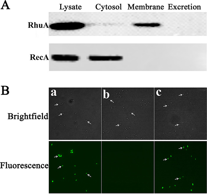
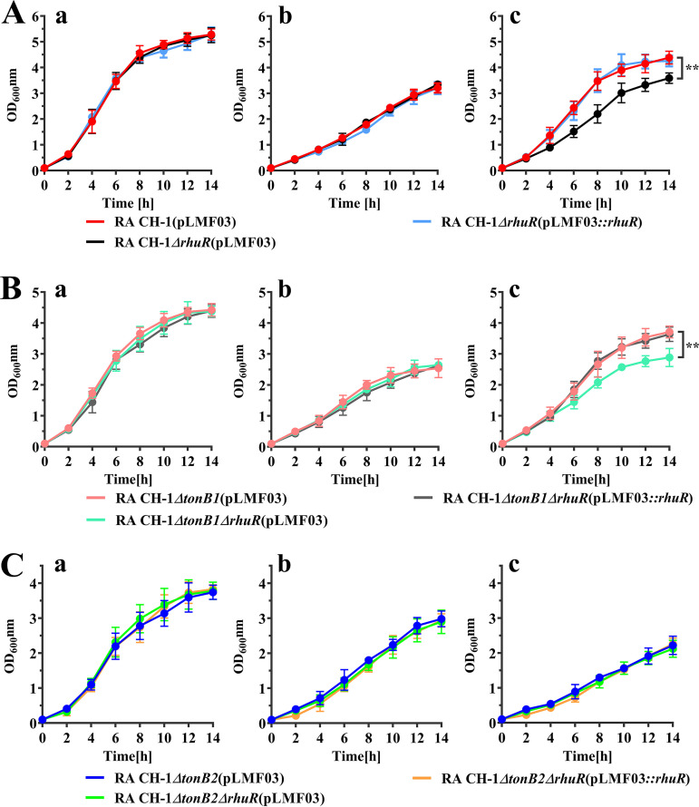
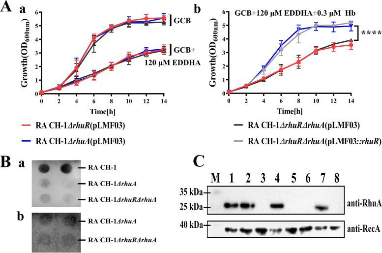
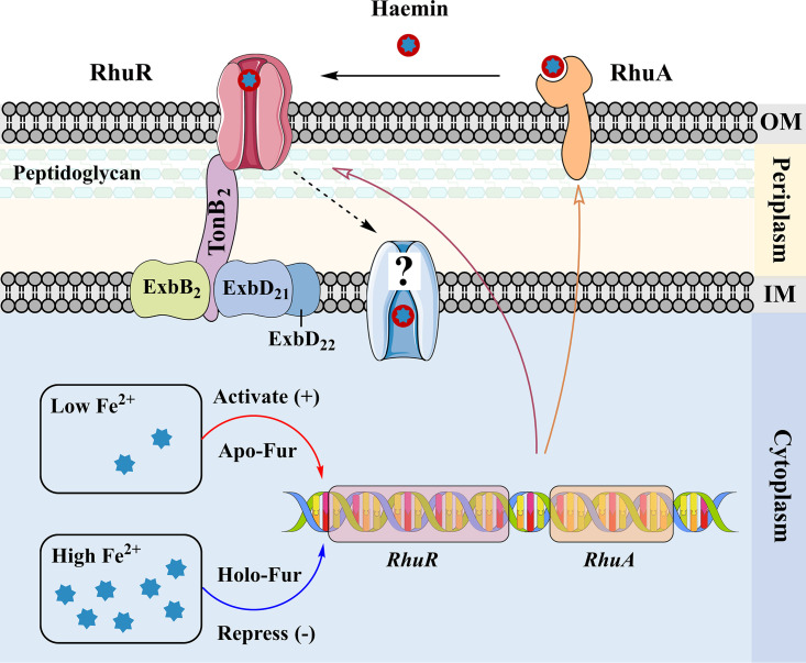
Iron is an essential element for the replication of most bacteria, including Riemerella anatipestifer, a Gram-negative bacterial pathogen of ducks and other birds. R. anatipestifer utilizes hemoglobin-derived hemin as an iron source; however, the mechanism by which this bacterium acquires hemin from hemoglobin is largely unknown. Here, rhuA disruption was shown to impair iron utilization from duck hemoglobin in R. anatipestifer CH-1. Moreover, the putative lipoprotein RhuA was identified as a surface-exposed, outer membrane hemin-binding protein, but it could not extract hemin from duck hemoglobin. Mutagenesis studies showed that recombinant RhuAY144A, RhuAY177A, and RhuAH149A lost hemin-binding ability, suggesting that amino acid sites at tyrosine 144 (Y144), Y177, and histidine 149 (H149) are crucial for hemin binding. Furthermore, rhuR, the gene adjacent to rhuA, encodes a TonB2-dependent hemin transporter. The function of rhuA in duck hemoglobin utilization was abolished in the rhuR mutant strain, and recombinant RhuA was able to bind the cell surface of R. anatipestifer CH-1 ΔrhuA rather than R. anatipestifer CH-1 ΔrhuR ΔrhuA, indicating that RhuA associates with RhuR to function. The sequence of the RhuR-RhuA hemin utilization locus exhibits no similarity to those of characterized hemin transport systems. Thus, this locus is a novel hemin uptake locus with homologues distributed mainly in the Bacteroidetes phylum. IMPORTANCE In vertebrates, hemin from hemoglobin is an important iron source for infectious bacteria. Many bacteria can obtain hemin from hemoglobin, but the mechanisms of hemin acquisition from hemoglobin differ among bacteria. Moreover, most studies have focused on the mechanism of hemin acquisition from mammalian hemoglobin. In this study, we found that the RhuR-RhuA locus of R. anatipestifer CH-1, a duck pathogen, is involved in hemin acquisition from duck hemoglobin via a unique pathway. RhuA was identified as an exposed outer membrane hemin-binding protein, and RhuR was identified as a TonB2-dependent hemin transporter. Moreover, the function of RhuA in hemoglobin utilization is RhuR dependent and not vice versa. The homologues of RhuR and RhuA are widely distributed in bacteria in marine environments, animals, and plants, representing a novel hemin transportation system of Gram-negative bacteria. This study not only was important for understanding hemin uptake in R. anatipestifer but also enriched the knowledge about the hemin transportation pathway in Gram-negative bacteria.

Keywords: Riemerella anatipestifer; hemin-binding protein.


Figures

FIG 1
FIG 1
Growth curves of R. anatipestifer (RA) CH-1(pLMF03) and its derivatives in different GCB media. The strains were grown in GCB overnight. Next, bacterial cells were inoculated into GCB, GCB containing 120 μM EDDHA, and GCB containing 120 μM EDDHA supplemented with 120 μM Fe(NO3)3 or 0.3 μM duck hemoglobin, separately, at an OD600 of 0.1. The OD600 value was measured at 2-h intervals for 14 h. The experiment was repeated at least three times, and the data were analyzed using Student’s t test. **, P < 0.01.
FIG 2
FIG 2
Subcellular localization and surface exposure of RhuA in R. anatipestifer CH-1. (A) Subcellular fractions of R. anatipestifer CH-1 were prepared and subjected to immunoblot analysis using antibodies against rRhuA. The localization of the cytoplasmic protein RecA was used as the control. Lysate, whole-cell lysate of R. anatipestifer CH-1; Cytosol, cytoplasmic protein fraction of R. anatipestifer CH-1; Membrane, membrane protein fraction of R. anatipestifer CH-1; Excretion, secreted proteins isolated from the R. anatipestifer CH-1 culture. (B) Immunofluorescence micrographs of bacteria labeled with anti-rRhuA serum as described in Materials and Methods. Micrographs were acquired with a Nikon Eclipse 80i microscope. (a) Strain R. anatipestifer CH-1(pLMF03); (b) strain R. anatipestifer CH-1 ΔrhuA(pLMF03); (c) strain R. anatipestifer CH-1 ΔrhuA(pLMF03::rhuA).
FIG 3
FIG 3
Detection of hemin binding to the rRhuA protein. (A) Equal amounts of recombinant protein or mixtures of recombinant protein and hemin were loaded into native polyacrylamide gels. (a) After electrophoresis, one gel was stained with Coomassie brilliant blue. (b) The proteins on the other gel were transferred to a nitrocellulose membrane and detected by ECL. Lane 1, mixture of rHasA and hemin; lane 2, rHasA; lane 3, mixture of rRhuA and hemin; lane 4, rRhuA. (B) Absorption spectra of hemin binding to rRhuA. Increasing concentrations of hemin (1 to 40 μM) were added to 20 μM rRhuA protein. The inset shows the absorbance values of hemin-rRhuA minus those of hemin alone at 397 nm at increasing hemin concentrations. Experiments were performed in triplicate, and the results of a single representative experiment are presented.
FIG 4
FIG 4
Detection of hemin extraction by rRhuA from duck hemoglobin. C600 ΔhemA(pAM238::hemR) was mixed with 4 ml of soft LB agar and poured onto LB plates. Holes were cut into the LB plates, and 20 μM hemin, a mixture of 20 μM hemin with 20 μM recombinant protein, 5 μM duck Hb, and a mixture of 5 μM duck Hb with 5 μM recombinant protein were added to the wells. rHasA, recombinant HasA of S. marcescens; rRhuA, recombinant RhuA of R. anatipestifer CH-1; rRecA, recombinant RecA of R. anatipestifer CH-1. The plates were incubated at 37°C overnight. The experiment was repeated three times, and a representative result is presented. The lucid zone represents the zone of C600 ΔhemA(pAM238::hemR) growth.
FIG 5
FIG 5
Affinity of rRhuA for hemin. rRhuA was immobilized on an NTA sensor chip (Nicoya). The kinetic parameters and concentrations used for the analysis are indicated. Protein-hemin interactions were monitored over a 5-min time period and reported as signal (picomole [pm]) values.
FIG 6
FIG 6
3D model of RhuA and sequence alignment of putative hemin-binding domains. (A) 3D model of RhuA built with Phyre2. The putative hemin-binding domain is shown in red. The potential crucial amino acid sites for hemin binding are indicated. (B) 3D model of RhuA built with I-TASSER. The putative hemin-binding domains are shown in red. The potential crucial amino acid sites for hemin binding are indicated. (C) Sequence alignment of putative hemin-binding domains (HBD) in RhuA and its orthologues. Sequence alignment was carried out using Clustal Omega, and figures were generated using Jalview 2. Universally conserved residues are highlighted in black, while putative conserved hemin-binding sites are highlighted in red. C. kapabacteria, “Candidatus Kapabacteria”; E. sp., Elizabethkingia sp.
FIG 7
FIG 7
Detection of hemin binding by rRhuA mutants. (A) Absorption spectra of 20 μM hemin binding to rRhuA or its mutants. (B) Mixtures of recombinant mutant proteins and hemin were loaded separately into native polyacrylamide gels. (a) After electrophoresis, one gel was stained with Coomassie brilliant blue. (b) The proteins on the other gel were transferred to a nitrocellulose membrane and detected by ECL (b). Mutant 1, RhuA142Y-A-144Y-A; mutant 2, RhuA177Y-A; mutant 3, RhuA144Y-A; mutant 4, RhuA176Y-A; mutant 5, RhuA144Y-A-176Y-A; mutant 6, RhuA149H-A.
FIG 8
FIG 8
Growth curves of R. anatipestifer CH-1, CH-1 ΔrhuR, and its derivative strains in different GCB media. (A) R. anatipestifer CH-1(pLMF03), CH-1 ΔrhuR(pLMF03), and CH-1 ΔrhuR(pLMF03)::rhuR bacterial cells in the exponential growth phase were inoculated into fresh GCB (a), GCB containing 120 μM EDDHA (b), and GCB containing 120 μM EDDHA supplemented with 0.3 μM duck hemoglobin (c). Bacteria were cultured at 37°C with shaking at 180 rpm, and the OD600 was measured every 2 h for 14 h. (B) R. anatipestifer CH-1 ΔtonB1(pLMF03), CH-1 ΔtonB1 ΔrhuR(pLMF03), and CH-1 ΔtonB1 ΔrhuR(pLMF03::rhuR) bacterial cells in the exponential growth phase were inoculated into fresh GCB (a), GCB containing 120 μM EDDHA (b), and GCB containing 120 μM EDDHA supplemented with 0.3 μM duck hemoglobin (c). Bacteria were cultured at 37°C with shaking at 180 rpm, and the OD600 was measured every 2 h for 14 h. (C) R. anatipestifer CH-1 ΔtonB2(pLMF03), CH-1 ΔtonB2 ΔrhuR(pLMF03), and CH-1 ΔtonB2 ΔrhuR(pLMF03::rhuR) bacterial cells in the exponential growth phase were inoculated into fresh GCB (a), GCB containing 120 μM EDDHA (b), and GCB containing 120 μM EDDHA supplemented with 0.3 μM duck hemoglobin (c). Bacteria were cultured at 37°C with shaking at 180 rpm, and the OD600 was measured every 2 h for 14 h. The error bars indicate the standard deviations from three repeated experiments. The data were analyzed using two-way analysis of variance (ANOVA). A P value of <0.05 indicates a statistically significant difference. **, P < 0.01.
FIG 9
FIG 9
Hemin binding assay of R. anatipestifer CH-1 and its derivative strains. Cells were incubated under iron starvation conditions, and a hemin-binding assay was performed. The mixture of 10 μg of hemin and 1 ml of bacteria (OD600 = 1) was incubated for 2 h and 4 h, respectively. The binding reactions were terminated by centrifugation at 10,000 × g for 5 min. The amount of unbound hemin was determined by measuring the OD of hemin at 405 nm with a NanoDrop 2000 instrument (Thermo Scientific). The amount of bound hemin on the cells was calculated by subtraction. Each value indicates the mean of the results from three experiments, and the vertical bars denote the standard deviations. ****, P < 0.0001; ***, P < 0.001; **, P < 0.01; *, P < 0.05.
FIG 10
FIG 10
The function of RhuA is dependent on RhuR. (Aa) Growth curves of R. anatipestifer CH-1 ΔrhuA(pLMF03), CH-1 ΔrhuR(pLMF03), and CH-1 ΔrhuR ΔrhuA(pLMF03) in GCB and GCB containing 120 μM EDDHA. (b) Growth curves of R. anatipestifer CH-1 ΔrhuA(pLMF03), CH-1 ΔrhuR(pLMF03), CH-1 ΔrhuR ΔrhuA(pLMF03), and CH-1 ΔrhuR ΔrhuA(pLMF03::rhuR) in GCB containing 120 μM EDDHA supplemented with 0.3 μM duck hemoglobin. Bacteria were cultured at 37°C with shaking at 180 rpm, and the OD600 was measured every 2 h for 14 h. (Ba) A total of 108 CFU of whole cells of R. anatipestifer CH-1 and its derivative strains were dropped on the filter as indicated (double spots for each strain). The filter was dried and blocked with a blocking solution. The dot blots were probed with mouse polyclonal anti-RhuA serum at a 1:400 dilution. (b) A total of 108 CFU of whole cells of R. anatipestifer CH-1 ΔrhuA(pLMF03) and CH-1 ΔrhuR ΔrhuA were dropped on the filter as indicated. The filter was dried and blocked with a blocking solution. The filter was incubated with rRhuA at a 4 μM concentration at room temperature for 30 min. After washing three times, the dot blots were probed with mouse polyclonal anti-RhuA serum at a 1:400 dilution. (C) rRhuA binds to the cell surface of R. anatipestifer CH-1 ΔrhuA but not the cell surface of R. anatipestifer CH-1 ΔrhuR. The binding of rRhuA to the cell surface of R. anatipestifer CH-1 ΔrhuA and its derivative strains was assessed as described in Materials and Methods. M, molecular weight; lane 1, mixture of rRhuA and rRecA; lane 2, R. anatipestifer CH-1; lane 3, R. anatipestifer CH-1 ΔrhuA; lane 4, sample of R. anatipestifer CH-1 ΔrhuA incubated with rRhuA and hemin; lane 5, R. anatipestifer CH-1 ΔrhuR ΔrhuA; lane 6, sample of R. anatipestifer CH-1 ΔrhuR ΔrhuA incubated with rRhuA and hemin; lane 7, sample of R. anatipestifer CH-1 ΔrhuA incubated with rRhuA; lane 8, sample of R. anatipestifer CH-1 ΔrhuR ΔrhuA incubated with rRhuA. ****, P < 0.0001.
FIG 11
FIG 11
Regulation of the RhuR-RhuA locus in R. anatipestifer CH-1. (A) R. anatipestifer CH-1 bacterial cells were cultured in GCB, GCB containing 120 μM EDDHA, and GCB containing 120 μM EDDHA supplemented with 240 μM Fe(NO3)3. Bacterial cells in the exponential growth phase were harvested, and the relative transcription of rhuR-rhuA was measured as described in Materials and Methods. (B) R. anatipestifer CH-1, CH-1 Δfur, and CH-1 Δfur(pLMF03::fur) bacterial cells were cultured in GCB. In parallel, R. anatipestifer CH-1 Δfur bacterial cells were cultured in GCB containing 120 μM EDDHA. Bacterial cells in the exponential growth phase were harvested, and the relative transcription of rhuR-rhuA was measured as described in Materials and Methods. (C) R. anatipestifer CH-1 bacterial cells were cultured in GCB, GCB containing 120 μM EDDHA, and GCB containing 120 μM EDDHA supplemented with 240 μM Fe(NO3)3. In parallel, R. anatipestifer CH-1 Δfur(pLMF03) and CH-1 Δfur(pLMF03::fur) bacterial cells were cultured in GCB. The bacteria were harvested and subjected to SDS-PAGE, and RhuA and RecA were then detected by Western blotting as described in Materials and Methods. (D) R. anatipestifer CH-1 bacterial cells were cultured in GCB and GCB containing 20 μM hemin. In parallel, R. anatipestifer CH-1 ΔhemA bacterial cells were cultured in GCB containing 20% δ-ala to an OD600 of 1 to 1.5, harvested, and inoculated into GCB and GCB containing 20 μM hemin. ****, P < 0.0001; ns, not significant.
FIG 12
FIG 12
Model of hemin acquisition from hemoglobin mediated by the RhuR-RhuA locus in R. anatipestifer. RhuA is a membrane-bound, surface-accessible putative lipoprotein. Hemin from hemoglobin is transferred to RhuA via an unidentified pathway. RhuA can transfer hemin to the TonB2-dependent hemin receptor RhuR to facilitate hemin utilization. Alternatively, RhuR can transport hemin from hemoglobin directly. In the cytoplasm, the transcription of rhuR-rhuA is increased when iron is limited, and this event is regulated by Fur. OM, outer membrane; IM, inner membrane.

Similar articles

Cited by

References

    1. Palmer LD, Skaar EP. 2016. Transition metals and virulence in bacteria. Annu Rev Genet 50:67–91. 10.1146/annurev-genet-120215-035146. - DOI - PMC - PubMed
    1. Choby JE, Skaar EP. 2016. Heme synthesis and acquisition in bacterial pathogens. J Mol Biol 428:3408–3428. 10.1016/j.jmb.2016.03.018. - DOI - PMC - PubMed
    1. Wandersman C, Delepelaire P. 2012. Haemophore functions revisited. Mol Microbiol 85:618–631. 10.1111/j.1365-2958.2012.08136.x. - DOI - PubMed
    1. Liao H, Cheng X, Zhu D, Wang M, Jia R, Chen S, Chen X, Biville F, Liu M, Cheng A. 2015. TonB energy transduction systems of Riemerella anatipestifer are required for iron and hemin utilization. PLoS One 10:e0127506. 10.1371/journal.pone.0127506. - DOI - PMC - PubMed
    1. Giardina BJ, Shahzad S, Huang W, Wilks A. 2019. Heme uptake and utilization by hypervirulent Acinetobacter baumannii LAC-4 is dependent on a canonical heme oxygenase (abHemO). Arch Biochem Biophys 672:108066. 10.1016/j.abb.2019.108066. - DOI - PMC - PubMed

Publication types

Supplementary concepts

LinkOut - more resources