. 2021 May 14;7(20):eabe4507.
doi: 10.1126/sciadv.abe4507. Print 2021 May.

The structure of an infectious immature flavivirus redefines viral architecture and maturation

Affiliations

The structure of an infectious immature flavivirus redefines viral architecture and maturation

Natalee D Newton et al. Sci Adv. .

Abstract

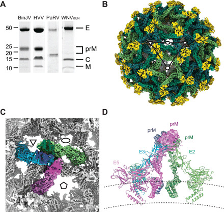
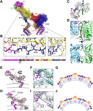
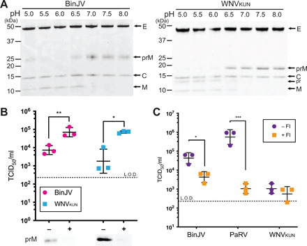
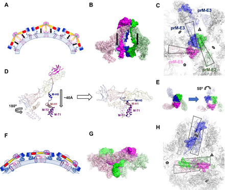
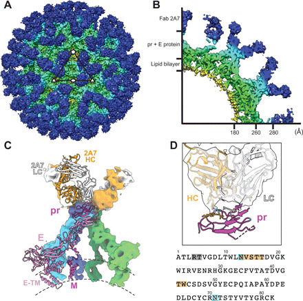
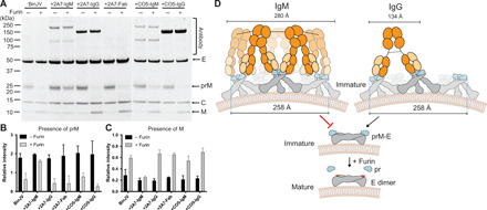
Flaviviruses are the cause of severe human diseases transmitted by mosquitoes and ticks. These viruses use a potent fusion machinery to enter target cells that needs to be restrained during viral assembly and egress. A molecular chaperone, premembrane (prM) maintains the virus particles in an immature, fusion-incompetent state until they exit the cell. Taking advantage of an insect virus that produces particles that are both immature and infectious, we determined the structure of the first immature flavivirus with a complete spike by cryo-electron microscopy. Unexpectedly, the prM chaperone forms a supporting pillar that maintains the immature spike in an asymmetric and upright state, primed for large rearrangements upon acidification. The collapse of the spike along a path defined by the prM chaperone is required, and its inhibition by a multivalent immunoglobulin M blocks infection. The revised architecture and collapse model are likely to be conserved across flaviviruses.


Figures

Fig. 1
Fig. 1. Cryo-EM reconstruction of BinJV.
(A) SDS-PAGE analysis of gradient purified BinJV, HVV, PaRV, and WNVKUN virions. Virus preparations are nonreduced and stained with SYPRO Ruby stain. E, prM, capsid (C), and M proteins indicated by arrows. (B) Surface cryo-EM reconstruction of BinJV. Density attributed to the pr proteins within the immature form is shown in yellow, and the three E proteins that make up the asymmetric unit were shown in shades of green. Ellipses, triangles, and pentagons represent the two-, three-, and fivefold icosahedral axes, respectively. (C) The asymmetric unit of BinJV is composed of three copies of E (green, pink, and cyan) and prM (dark green, magenta, and blue). (D) Side view of the asymmetric unit displayed as a cartoon and colored by symmetry position. Density is shown for prM. Lines illustrate the extent of the lipid bilayer.
Fig. 2
Fig. 2. The structure of the prM-E heterodimer reveals a furin site buried within the spike and a central pillar supporting the spike rather than the proposed drawstring.
(A) The electron density and structure of the fivefold prM-E heterodimer. The color scheme matches the domain diagram (bottom), which also indicates glycans (cyan), furin cleavage site (scissors), and unmodeled region (dashed line). (B) Zoom of the prM linker. (C) Section of the trimeric spike near the furin site viewed from virion exterior. Molecular surface and stick representation are colored according to the prM-E proximity to the twofold (green/lime), threefold (blue/cyan), and fivefold (magenta/pink) symmetry axes. (D) Zooms of the prM/E interfaces as in (C) (bottom) or in orthogonal orientation (top). (E and F) Structure of BinJV (E) compared to a main-chain model of immature DENV-1 [(H), PDB: 4B03]. E and prM is in pink and magenta, respectively. Arrows indicate that movements proposed in the drawstring model (E) are incompatible with the BinJV topology, supporting a prM-guided collapse (H). (F and I) Linker region electron density for BinJV (F) and DENV-1 [(I) EMD-2141]. Density is only visible for the BinJV model (magenta). None of the reconstructions show density for the proposed DENV-1 prM linker (blue; PDB: 4B03). (G and J) Schematics of the immature virion colored as (A) with membrane in blue. Instead of the previously proposed domain swap (G), prM forms a supporting pillar at the center of each BinJV spike (J).
Fig. 3
Fig. 3. Maturation of BinJV and WNVKUN particles by in vitro furin cleavage.
(A) Purified BinJV (left) and immature WNVKUN (right) were incubated with furin in buffer ranging from pH 5 to 8 and analyzed by nonreducing SDS-PAGE and visualized by SYPRO Ruby stain. Viral proteins indicated by arrows. (B) Infectivity of BinJV and immature WNVKUN virions in C6/36 cells with or without furin treatment. Cleavage was analyzed by SDS-PAGE, and virus titers were determined by TCID50. n = 3 biological replicates, and statistical analysis was performed by two-stage multiple t tests against nontreated (furin negative). *P ≤ 0.05 and **P ≤ 0.01. Means ± SD. Limit of detection (L.O.D.) for TCID50 is 2.3 log10 TCID50 ml−1. (C) Infectivity of BinJV, PaRV, and WNVKUN in C6/36 cells pretreated with or without 25 μM furin inhibitor (FI). Virus titers were determined by TCID50; n = 3 biological replicates, and statistical analysis was performed by two-stage multiple t tests against nontreated (FI negative). *P ≤ 0.05 and ***P ≤ 0.001. Means ± SD. Limit of detection for TCID50 is 2.3 log10 TCID50 ml−1.
Fig. 4
Fig. 4. Proposed model of maturation.
(A and F) A cartoon schematic showing several copies of prM-E arranged on a membrane (light blue), illustrating the collapse maturation model for the immature (A) and mature structure (F). Domains are colored as in Fig. 2. (B) The immature trimer of BinJV colored according to symmetry position with prM and E represented as a solid and semitransparent surfaces, respectively. The collapse model involves a refolding of the pr linker, which guides the E ectodomain toward M (see movie S1). (C and H) The transition from immature (C) to mature (H) structure involves minimal displacement of M and a simple collapse of E (see movie S2). Molecules are allowed to migrate from on icosahedral symmetry position to another (see movie S3).Views from the exterior of the virion. Four prM-E are outlined in wedged boxes and displayed as in (B). For the mature structure, subunits are colored according to their original icosahedral symmetry position as defined in (B) and (C). (D) Comparison of a single prM-E subunit between the immature form (left) to the mature form (right). The translation of pr and the ectodomain of E and the rotation of the E stem/TM are indicated. Domains are colored as in Fig. 2. (E) The pr and E components of the trimeric spike are asymmetric in the immature virion (left). By comparison, the spike would follow a pseudo-threefold symmetry if superposed to the M components of the immature spike (right). The arrow represents the twist of the pr and E component of prM-E3 in the actual immature spike. (G) Side view of a dimer of M-E in the mature structure. Mature structures are modeled from DENV-1 in (D), (G), and (H).
Fig. 5
Fig. 5. The pr-specific antibody 2A7 inhibits BinJV in an isotype-dependent manner.
(A) Purified 2A7 in Fab, IgG, and IgM form analyzed by reduced 4 to 12% SDS-PAGE. Heavy (HC) and light chains (LC) indicated by arrows. (B) Western blot probing with recombinant 2A7-Fab (before HRV3C digestion), IgG, and IgM. Purified BinJV from infected C6/36 cells was resolved by nonreducing 4 to 12% SDS-PAGE. (C) Neutralization of BinJV by 2A7-Fab, IgG, and IgM antibodies or anti-influenza CO5 antibody as isotype controls. Neutralization curves were determined by TCID50 on C6/36 cells. Means ± range. OD, optical density. (D) Neutralization by 2A7-IgM or control CO5-IgM of BinJV produced in C6/36 (A. albopictus), RML12 (A. albopictus), and Aag2 (A. aegypti) mosquito cells with 2A7-IgM or control CO5-IgM. Neutralization was determined by TCID50 on C6/36 cells. Means ± range. (E) Viral titers in A. albopictus mosquitoes injected intrathoracically with BinJV:2A7-IgM or BinJV:CO5-IgM. BinJV supernatant was incubated with either antibody (2A7-IgM or CO5-IgM) at 28°C for 1 hour before injection. Each point represents a single infected mosquito; means ± SD. Statistical analysis performed by unpaired t test with Welch’s correction. ***P ≤ 0.001.
Fig. 6
Fig. 6. Cryo-EM reconstruction of BinJV:2A7.
(A) Surface and (B) wedge cross-section of the cryo-EM reconstruction of BinJV:2A7-Fab. Map was colored according to radius (red, 0 to 30 Å; yellow, 31 to 140 Å; green, 141 to 180 Å; cyan, 181 to 280 Å; and blue, >280 Å). (C) Fit of a homology model of the 2A7-Fab and the structure of BinJV within the reconstruction of the complex. Dimers of prM-E are colored according to symmetry position: twofold with green/lime for prM/E, threefold with blue/cyan for prM/E, and fivefold with magenta/pink for prM/E. The HC and LC of the 2A7-Fab is represented in orange and white, respectively. Surfaces for one member of the asymmetric unit are omitted to show a cartoon representation. (D) Zoom of the pr:2A7 interface with the sequence of the N-terminal domain of pr displayed below. Residues of pr that are within 7 Å of the Fab are indicated by dark orange (HC) and gray (LC). N-linked glycan sites of pr are highlighted in light blue.
Fig. 7
Fig. 7. 2A7 blocks in vitro maturation in an isotype-dependent manner.
(A) Purified BinJV was incubated with anti-prM 2A7 antibodies (IgM, IgG, and Fab) or the control antibody CO5 (IgM and IgG) for 1 hour before incubating with furin and analyzing by SYPRO Ruby–stained SDS-PAGE. Virus and antibodies were mixed at equimolar concentrations. Viral proteins are indicated by arrows, and antibody fragments are in brackets. The relative intensity of the prM (B) and M (C) bands following incubation with and without furin was quantified. n = 2 independent experiments; means ± SD. (D) Model of neutralization of immature BinJV by the 2A7-IgM. Schematic representation of five prM-E trimers that are presented in gray (E) and blue (pr) surrounding the fivefold axis. Multimeric binding of IgM antibody are proposed to cross-link the prM-E spikes inhibiting low pH–mediated release of the furin site during endocytosis. Distance between antigen binding sites of IgM (51) and IgG (52) antibody isotypes and the span across the fivefold axis is annotated.

Similar articles

Cited by

References

    1. Pierson T. C., Diamond M. S., The continued threat of emerging flaviviruses. Nat. Microbiol. 5, 796–812 (2020). - PMC - PubMed
    1. Weaver S. C., Barrett A. D. T., Transmission cycles, host range, evolution and emergence of arboviral disease. Nat. Rev. Microbiol. 2, 789–801 (2004). - PMC - PubMed
    1. Blitvich B. J., Firth A. E., Insect-specific flaviviruses: A systematic review of their discovery, host range, mode of transmission, superinfection exclusion potential and genomic organization. Viruses 7, 1927–1959 (2015). - PMC - PubMed
    1. Cammisa-Parks H., Cisar L. A., Kane A., Stollar V., The complete nucleotide sequence of cell fusing agent (CFA): Homology between the nonstructural proteins encoded by CFA and the nonstructural proteins encoded by arthropod-borne flaviviruses. Virology 189, 511–524 (1992). - PubMed
    1. Hobson-Peters J., Harrison J. J., Watterson D., Hazlewood J. E., Vet L. J., Newton N. D., Warrilow D., Colmant A. M. G., Taylor C., Huang B., Piyasena T. B. H., Chow W. K., Setoh Y. X., Tang B., Nakayama E., Yan K., Amarilla A. A., Wheatley S., Moore P. R., Finger M., Kurucz N., Modhiran N., Young P. R., Khromykh A. A., Bielefeldt-Ohmann H., Suhrbier A., Hall R. A., A recombinant platform for flavivirus vaccines and diagnostics using chimeras of a new insect-specific virus. Sci. Transl. Med. 11, eaax7888 (2019). - PubMed

Publication types