. 2021 May 14;7(20):eabf5835.
doi: 10.1126/sciadv.abf5835. Print 2021 May.

A framework for highly multiplexed dextramer mapping and prediction of T cell receptor sequences to antigen specificity

Affiliations

A framework for highly multiplexed dextramer mapping and prediction of T cell receptor sequences to antigen specificity

Wen Zhang et al. Sci Adv. .

Abstract

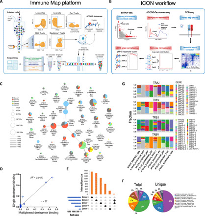
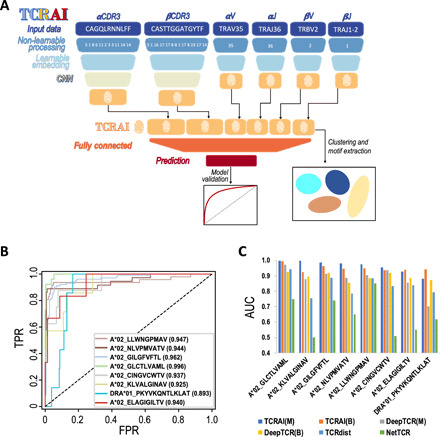
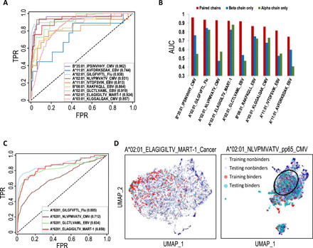
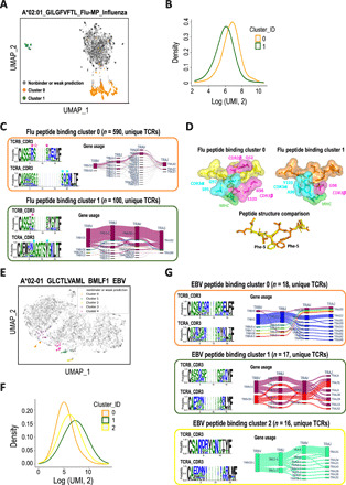
T cell receptor (TCR) antigen-specific recognition is essential for the adaptive immune system. However, building a TCR-antigen interaction map has been challenging due to the staggering diversity of TCRs and antigens. Accordingly, highly multiplexed dextramer-TCR binding assays have been recently developed, but the utility of the ensuing large datasets is limited by the lack of robust computational methods for normalization and interpretation. Here, we present a computational framework comprising a novel method, ICON (Integrative COntext-specific Normalization), for identifying reliable TCR-pMHC (peptide-major histocompatibility complex) interactions and a neural network-based classifier TCRAI that outperforms other state-of-the-art methods for TCR-antigen specificity prediction. We further demonstrated that by combining ICON and TCRAI, we are able to discover novel subgroups of TCRs that bind to a given pMHC via different mechanisms. Our framework facilitates the identification and understanding of TCR-antigen-specific interactions for basic immunological research and clinical immune monitoring.


Figures

Fig. 1
Fig. 1. Identification of pMHC binding T cells from the high-throughput dextramer binding data.
(A) Schema of the Immune Map platform. PBMC CD8+ T cells were enriched and stained with a pool of 50 dCODE dextramer antibodies. Dextramer-positive CD8+ T cells were sorted and then captured individually as inputs for 10x Genomics single-cell sequencing. (B) ICON workflow. Please see Materials and Methods for details. (C) Network of ICON identified pMHC binding unique TCRs. Each node represents a pMHC repertoire and is displayed as a pie chart of pMHC binding TCRs for each donor. The node size denotes the total number of unique TCRs. The thickness of an edge represents the number of shared unique TCR(s). (D) Correlation of the fraction of T cells binding to a given dextramer between the result from flow sorting on single dextramer binding and the relative abundance of pMHC binding T cells identified by ICON from the multiplexed dextramer binding data. (E) Uniqueness and overlap of pMHC binding unique TCRs among the five donors. (F) Pie charts of ICON identified pMHC binding TCRs. (G) V and J gene usage of the nine most abundant pMHC repertoires. The gene usage with less than 5% was combined and indicated in gray.
Fig. 2
Fig. 2. The framework and performance of TCRAI.
(A) Schematic of the TCRAI framework for a model receiving input of CDR3 and VJ genes of both the α and β chains. A trained TCRAI model creates a numerical fingerprint and prediction for a given TCR. CNN, convolutional neural network. Please see Materials and Methods for more details. (B) ROC curves for TCRAI (in binomial mode) classification performance using the eight curated public TCR-pMHC binding repertoires. Binders are unique TCRs that bind to a particular pMHC, and nonbinders are unique TCRs that bind to other pMHCs. Paired αβ TCR sequences were used as input data. FPR, false positive rate; TPR, true positive rate. (C) Comparison of classification performance. TCRAI was compared with predictive classifiers NetTCR, TCRdist, and DeepTCR. The AUC for NetTCR and TCRdist was generated using the original classifiers with default parameters. To compare with these two binomial classifiers (NetTCR and TCRdist), the AUC for DeepTCR (originally designed as a multinomial classifier) was derived from a slightly modified and hyperparameter optimized version of DeepTCR (Materials and Methods). TCRAI(M), TCRAI in multinomial mode; TCRAI(B), TCRAI in binary mode; DeepTCR(M), DeepTCR in multinomial mode; DeepTCR(B), DeepTCR in binary mode.
Fig. 3
Fig. 3. TCRAI prediction on the high-throughput dataset.
(A) ROC curves for TCRAI prediction on the nine most abundant pMHC binding repertoires. Binders are unique TCRs that bind to a particular pMHC, and nonbinders are unique TCRs that bind to other pMHCs. Paired αβ TCR sequences were used as input data. (B) Comparisons of TCRAI prediction on TCRα only, TCRβ only, and paired αβ chains as input data. (C) ROC curves for the independent tests of four overlapping pMHC repertoires between the curated public dataset and the high-throughput dataset. TCRAI was trained using pMHC repertoires identified from the high-throughput dataset and was tested on the curated public dataset. (D) UMAPs of both the training (high-throughput data) and testing (the gold-standard data) TCRAI fingerprints extracted from the models trained by the high-throughput data. The left panel shows the strong overlap between MART-1_cancer training and testing sets, while the poor overlap of NLVPMVATV_pp65_CMV training and testing datasets is shown in the right panel. The black circle highlights the region with almost no overlapping fingerprints of training and testing binders. UMAP, Uniform Manifold Approximation and Projection.
Fig. 4
Fig. 4. Characterization of pMHC binding TCRs.
(A) Clustering TCRAI fingerprints of high-confidence TCRs from a model trained by A*02:01_GILGFVFTL_Flu binders identified from the high-throughput dataset. (B) Dextramer signal (in UMI) distributions of the flu peptide binding clusters 0 and 1. (C) Conserved CDR3 motifs and gene usage in flu peptide binding TCR clusters. Structurally important residues are highlighted by filled stars and also shown in (D). The residue with an unfilled star missed the cutoff for inclusion in (D) but is nevertheless in close proximity (4.18 Å to the Phe-5 ring) and strongly conserved. Only the 30 most common unique quadruplets of gene usage are shown for cluster 0 to highlight the key variabilities. For motif construction, please see Materials and Methods for details. (D) 3D structures of flu peptide binding TCR-pMHC complexes for cluster 0 TCR (PDB 2VLJ) and cluster 1 TCR (PDB 5JHD). In the top panels, only nonpeptide residues within 4 Å of the Phe-5 ring are shown. (E) Clustering TCRAI fingerprints of high-confidence TCRs from a model trained by A*02-01_GLCTLVAML_BMLF1_EBV binders identified from the high-throughput dataset. (F) Dextramer signal distributions of the EBV peptide binding clusters. (G) Conserved CDR3 motifs and gene usage in the three EBV peptide binding clusters.
Fig. 5
Fig. 5. Immune phenotypes of pMHC binding CD8+ T cells.
(A) Classification of pMHC binding cells. (B) Heatmap of the expression of CD8+ T cell subtype marker genes and proteins. *: protein expression measured by CITE-seq. (C) pMHC binding landscape by CD8+ T cell immune subtypes. Bars indicate the number of pMHC binding T cells in log2 scale. (D) Expanded clonotypes are enriched in the non-naïve compartment. Each dot represents a unique TCR clone. (E) Pie chart of subpopulations of pMHC binding CD8+ T cells. (F) Fraction of HLA type matched and mismatched binding naïve and non-naïve T cells. Tpm, peripheral memory cells; Tcm, central memory cells; Tem, effector memory cells; Temra, terminally differentiated effector memory cells; Others, other memory cells with the marker gene expression pattern CD43loCD127loKLRG1high.

Similar articles

Cited by

References

    1. Krangel M. S., Mechanics of T cell receptor gene rearrangement. Curr. Opin. Immunol. 21, 133–139 (2009). - PMC - PubMed
    1. Mahe E., Pugh T., Kamel-Reid S., T cell clonality assessment: Past, present and future. J. Clin. Pathol. 71, 195–200 (2018). - PMC - PubMed
    1. Gascoigne N. R. J., Rybakin V., Acuto O., Brzostek J., TCR signal strength and T cell development. Annu. Rev. Cell Dev. Biol. 32, 327–348 (2016). - PubMed
    1. Jung D., Alt F. W., Unraveling V(D)J recombination; insights into gene regulation. Cell 116, 299–311 (2004). - PubMed
    1. Jackson K. J. L., Kidd M. J., Wang Y., Collins A. M., The shape of the lymphocyte receptor repertoire: Lessons from the B cell receptor. Front. Immunol. 4, 263 (2013). - PMC - PubMed

MeSH terms