. 2021 May 18;118(20):e2022357118.
doi: 10.1073/pnas.2022357118.

Knl1 participates in spindle assembly checkpoint signaling in maize

Affiliations

Knl1 participates in spindle assembly checkpoint signaling in maize

Handong Su et al. Proc Natl Acad Sci U S A. .

Abstract

The Knl1-Mis12-Ndc80 (KMN) network is an essential component of the kinetochore-microtubule attachment interface, which is required for genomic stability in eukaryotes. However, little is known about plant Knl1 proteins because of their complex evolutionary history. Here, we cloned the Knl1 homolog from maize (Zea mays) and confirmed it as a constitutive central kinetochore component. Functional assays demonstrated their conserved role in chromosomal congression and segregation during nuclear division, thus causing defective cell division during kernel development when Knl1 transcript was depleted. A 145 aa region in the middle of maize Knl1, that did not involve the MELT repeats, was associated with the interaction of spindle assembly checkpoint (SAC) components Bub1/Mad3 family proteins 1 and 2 (Bmf1/2) but not with the Bmf3 protein. They may form a helical conformation with a hydrophobic interface with the TPR domain of Bmf1/2, which is similar to that of vertebrates. However, this region detected in monocots shows extensive divergence in eudicots, suggesting that distinct modes of the SAC to kinetochore connection are present within plant lineages. These findings elucidate the conserved role of the KMN network in cell division and a striking dynamic of evolutionary patterns in the SAC signaling and kinetochore network.

Keywords: Knl1; SAC; cell division; kinetochore; maize.


Conflict of interest statement

The authors declare no competing interest.

Figures

Fig. 1.
Fig. 1.
Identification and annotation of the Knl1 homolog in maize. (A) Schematic diagram of Knl1 and SAC components in humans (H. sapiens), Arabidopsis (A. thaliana), and maize (Z. mays). Different-colored rectangles on human Knl1 represent the following: SILK and RVSF motif for PP1 binding domain; MELT repeats and KI1 motif for Bub3–Bub1 complex binding; KI2 motif for Bub3–BubR1 complex binding; coiled-coil domain for Zwint1 binding; and RWD domain for Mis12 complex binding. Ovals on Bub1 and BubR1 represent the TPR domains. Rectangles on Bub1 and BubR1 represent the Bub3-binding domain. Only RVSF, coiled-coil, and RWD domains are present in Arabidopsis and maize Knl1. The dotted lines and virtual boxes indicate there are no KI motifs in plant Knl1. (B) Model for evolution of the Bub/Mad family protein in humans, Arabidopsis, and maize. Some functional domains were shown with different-colored rectangles in the presumed ancestral Bub1/Mad3 protein. TPR domain is for Knl1 binding, GLEBS motif is for Bub3 binding, and a kinase domain is for histone H2A phosphorylation. A pseudokinase domain with a gray rectangle was shown in human BubR1. (CE) Colocalization of KMN network components on maize leptotene and pachytene chromosomes: ZmMis12 (red) and ZmKnl1 (green) (C); ZmNdc80 (red) and ZmKnl1 (green) (D); and ZmMis12 (red) and ZmNdc80 (green) (E). (F) Colocalization signals of ZmKnl1 (green) and ZmBmf1 (red) in maize pachytene chromosomes. The Insets indicate a higher-magnification view of colocalization signals of different proteins. Chromosomes stained with DAPI are shown in blue. (Scale bar, 10 μm.)
Fig. 2.
Fig. 2.
Identification of the BMF binding domain in ZmKnl1. (A) Schematic representation of the ZmKnl1 truncations used to define the ZmBmf1/2 binding domain. Eight truncated constructs of ZmKnl1 (M1 to M8) were produced. Blue indicates full-length ZmKnl1 proteins. Green indicates no Y2H interaction. Purple indicates Y2H interaction. (B) Y2H interactions between truncated ZmKnl1 variants and ZmBmf1 (Left three) or ZmBmf2 (Right three). (C) Luciferase complementation imaging assay of the interaction between ZmKnl1-M8 and ZmBmf1/2/3. Fluorescence signal intensities represent their interaction activities. (D and E) Schematic representation of ZmKnl1 substitution constructs used to define the ZmBmf1/2-binding domain. Y2H interaction was conducted between substituted ZmKnl1 variants and ZmBmf1/2. The sequential residues in this region were replaced with As.
Fig. 3.
Fig. 3.
The TPR domains of ZmBmf1 and ZmBmf2 are essential for binding with ZmKnl1. (AC) Schematic representations of the truncations and mutations of BMF proteins used to define the Knl1 binding domain (Left). Y2H system of ZmKnl1 with different BMF constructs (Right): ZmBmf1 (A), ZmBmf2 (B), and ZmBmf3 (C). The TPR motif is marked in green. Red indicates residues altered in the substitution mutants, as depicted in D. FL is full length; N is N terminus containing the TPR domain; C is C terminus without the intact TPR domain; C1 is C terminus without one TPR repeat unit; and C2 is C terminus without two TPR repeat units. Yeast cells at various dilutions were grown on selective (SD/−Trp/−Leu/−Ade/−His) media. (D) Multiple sequence alignment of the TPR domains of Bub1/Mad3 homologs from human (h), mouse (m), Saccharomyces cerevisiae (sc), Schizosaccharomyces pombe (sp), Arabidopsis (at), and maize (zm). The asterisks at the top of the alignment indicate completely conserved residues, and the colons and single dots indicate highly and moderately conserved residues, respectively. The red line indicates the alpha helix within the TPR domain. X indicates residues altered in the substitution mutants.
Fig. 4.
Fig. 4.
Distribution of ZmKnl1 signals in WT maize meiocytes: leptonema (A), pachynema (B), diakinesis (C), metaphase I (D), Telophase I (E), prometaphase II (F), metaphase II (G), and quartets (H). ZmKnl1 signals are shown in red. DAPI-stained chromosomes are shown in blue. (Scale bar, 10 μm.)
Fig. 5.
Fig. 5.
Knockout of ZmKnl1 by CRISPR/Cas9 leads to defects in chromosome congression during mitosis. (A and B) Prophase cells from the root tips of the control (A) (the transgene-negative lines were treated as the control) and a zmknl1 knockout mutant (B). (C and D) Representative chromosome congression in root-tip meristem cells during early growth in the control (C) and zmknl1 knockout mutant (D). Arrows indicate unaligned chromosomes. Tubulin signals are shown in green. DAPI-stained chromosomes are shown in blue. (Scale bar, 10 μm.)
Fig. 6.
Fig. 6.
ZmKNL1 Mu–insertion mutants display defective kernel development. (A) The Mu insertion in UFMu-06469 is located in the first exon of zmknl1. (B) Mature ear of a heterozygous zmknl1 mutant. Arrow indicates a homozygous UFMu-06469 (zmknl1) kernel. Red arrowheads indicate homozygous zmknl1 kernels. (Scale bar, 1 cm.) (C) WT (Left three) and zmknl1 (Right three) kernels at 25 DAP. (D) Comparison of germination in three WT (Left) and three zmknl1 (Right) seeds at 5 d after germination. (Scale bar, 1 cm.) (E) The chromosome behavior of endosperm cells in the WT and zmknl1 at 14 DAP at anaphase. The lagging chromosomes in the mutant are indicated by arrows. (Scale bar, 10 μm.)
Fig. 7.
Fig. 7.
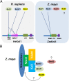
Models of the architecture of Knl1 with SAC signaling in humans and plants. (A) Comparison of the domains of Knl1 proteins and the architecture with SAC signaling between humans (H. sapiens) and maize (Z. mays). In humans, Knl1 MELT repeats cooperate with KI motifs to recruit SAC signals (Bub1, BubR1, and Bub3). In maize, the MELT repeats of Knl1 are lost, and a 145 aa region in the middle of Knl1 participates in interaction with Bmf1 and Bmf2 in maize. However, no interaction was detected between Knl1 and Bmf3 or Bub3. Different-colored rectangles on Knl1 represent the MELT, KI1, KI2, BMF (BMF-binding domain), coiled-coil (CC), and RWD domain. The solid lines represent detected interaction with the Y2H system. (B) Cell-cycle–dependent kinetochore localizations of Bmf1 and Bub3 were detected in maize, a typical feature of SAC signaling. The TPR domains of Bmf1 and Bmf2 are required for their interaction with Knl1. How Bub3 is recruited to the kinetochore and the role of Bmf3 in maize remains unknown.

Similar articles

Cited by

References

    1. Przewloka M. R., Glover D. M., The kinetochore and the centromere: A working long distance relationship. Annu. Rev. Genet. 43, 439–465 (2009). - PubMed
    1. Godek K. M., Kabeche L., Compton D. A., Regulation of kinetochore-microtubule attachments through homeostatic control during mitosis. Nat. Rev. Mol. Cell Biol. 16, 57–64 (2015). - PMC - PubMed
    1. Cheeseman I. M., The kinetochore. Cold Spring Harb. Perspect. Biol. 6, a015826 (2014). - PMC - PubMed
    1. Petrovic A., et al. ., Structure of the MIS12 complex and molecular basis of its interaction with CENP-C at human kinetochores. Cell 167, 1028–1040.e15 (2016). - PMC - PubMed
    1. Ladurner R., Straight A. F., MIS12/MIND control at the kinetochore. Cell 167, 889–891 (2016). - PubMed

Publication types

MeSH terms

Substances

LinkOut - more resources