

Assessment of Myofascial Trigger Points via Imaging: A Systematic Review
- PMID: 33990485
- PMCID: PMC8448923
- DOI: 10.1097/PHM.0000000000001789
Assessment of Myofascial Trigger Points via Imaging: A Systematic Review
Abstract
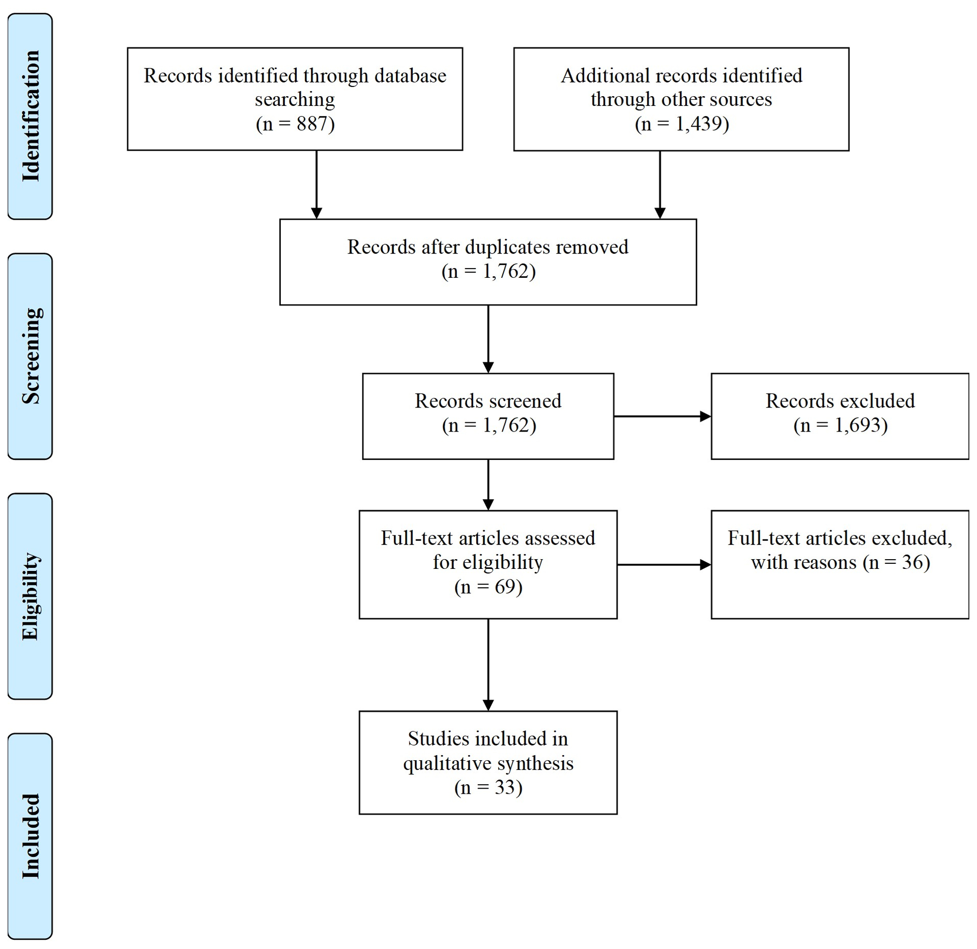
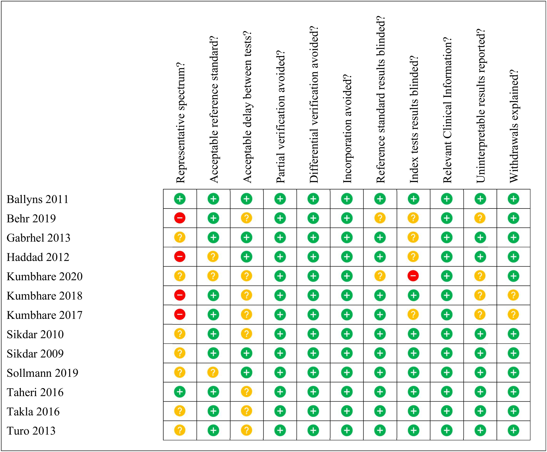
This study systematically reviewed the published literature on the objective characterization of myofascial pain syndrome and myofascial trigger points using imaging methods. PubMed, Embase, Ovid, and the Cochrane Library databases were used, whereas citation searching was conducted in Scopus. Citations were restricted to those published in English and in peer-reviewed journals between 2000 and 2021. Of 1762 abstracts screened, 69 articles underwent full-text review, and 33 were included. Imaging data assessing myofascial trigger points or myofascial pain syndrome were extracted, and important qualitative and quantitative information on general study methodologies, study populations, sample sizes, and myofascial trigger point/myofascial pain syndrome evaluation were tabulated. Methodological quality of eligible studies was assessed based on the Quality Assessment of Diagnostic Accuracy Studies criteria. Biomechanical properties and blood flow of active and latent myofascial trigger points assessed via imaging were found to be quantifiably distinct from those of healthy tissue. Although these studies show promise, more studies are needed. Future studies should focus on assessing diagnostic test accuracy and testing the reproducibility of results to establish the best performing methods. Increasing methodological consistency would further motivate implementing imaging methods in larger clinical studies. Considering the evidence on efficacy, cost, ease of use and time constraints, ultrasound-based methods are currently the imaging modalities of choice for myofascial pain syndrome/myofascial trigger point assessment.
Copyright © 2021 Wolters Kluwer Health, Inc. All rights reserved.
Conflict of interest statement
Financial disclosure statements have been obtained, and no conflicts of interest have been reported by the authors or by any individuals in control of the content of this article.
Figures

References
-
- Gerwin RD Classification, epidemiology, and natural history of myofascial pain syndrome. Curr Pain Headache Rep 5, 412–420 (2001). - PubMed
-
- Fishbain DA, Goldberg M, Meagher BR, Steele R & Rosomoff H Male and female chronic pain patients categorized by DSM-III psychiatric diagnostic criteria. Pain 26, 181–197 (1986). - PubMed
-
- Travell JG & Simons DG Myofascial pain and dysfunction: the trigger point manual. (Williams & Wilkins, 1983).
-
- Gerwin RD, Dommerholt J & Shah JP An expansion of Simons’ integrated hypothesis of trigger point formation. Current Science Inc 8, 468–475 (2004). - PubMed
-
- Yap EC Myofascial pain - An overview. Annals of the Academy of Medicine Singapore 36, 43–48 (2007). - PubMed
Publication types
MeSH terms
Grants and funding
LinkOut - more resources
Full Text Sources
Other Literature Sources
