. 2021 May 15;12(5):494.
doi: 10.1038/s41419-021-03786-6.

Loss of the WNT9a ligand aggravates the rheumatoid arthritis-like symptoms in hTNF transgenic mice

Affiliations

Loss of the WNT9a ligand aggravates the rheumatoid arthritis-like symptoms in hTNF transgenic mice

Stefan Teufel et al. Cell Death Dis. .

Abstract

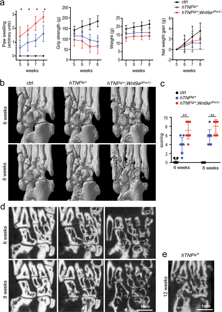
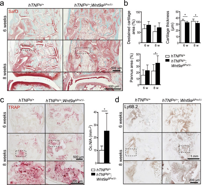
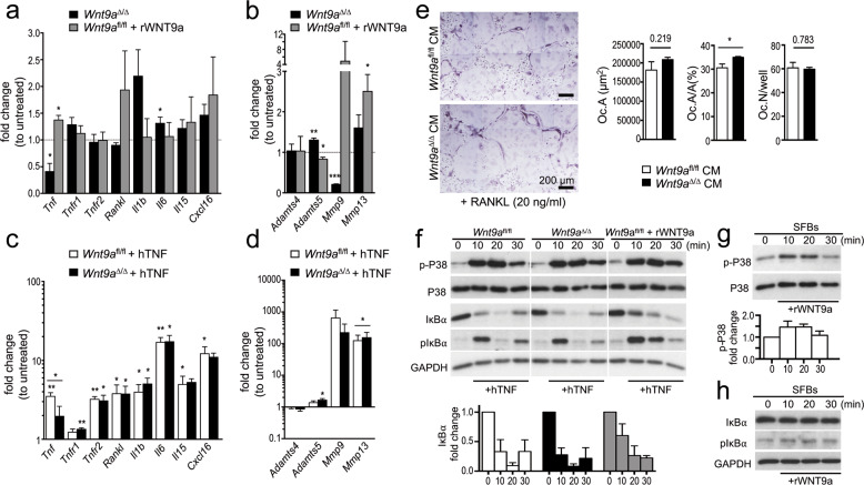
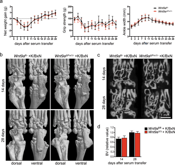
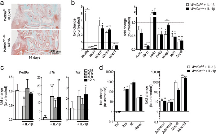
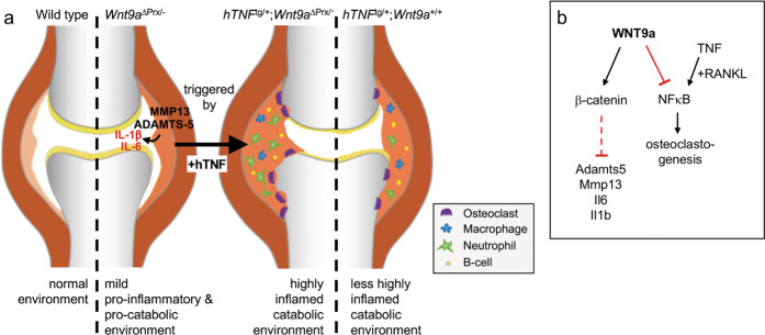
Agonists and antagonists of the canonical Wnt signaling pathway are modulators of pathological aspects of rheumatoid arthritis (RA). Their activity is primarily modifying bone loss and bone formation, as shown in animal models of RA. More recently, modulation of Wnt signaling by the antagonist Sclerostin has also been shown to influence soft-tissue-associated inflammatory aspects of the disease pointing towards a role of Wnt signaling in soft-tissue inflammation as well. Yet, nothing is known experimentally about the role of Wnt ligands in RA. Here we provide evidence that altering Wnt signaling at the level of a ligand affects all aspects of the rheumatoid arthritic disease. WNT9a levels are increased in the pannus tissue of RA patients, and stimulation of synovial fibroblasts (SFB) with tumor necrosis factor (TNF) leads to increased transcription of Wnt9a. Loss of Wnt9a in a chronic TNF-dependent RA mouse model results in an aggravation of disease progression with enhanced pannus formation and joint destruction. Yet, loss of its activity in the acute K/BxN serum-transfer induced arthritis (STIA) mouse model, which is independent of TNF signaling, has no effect on disease severity or progression. Thus, suggesting a specific role for WNT9a in TNF-triggered RA. In synovial fibroblasts, WNT9a can activate the canonical Wnt/β-catenin pathway, but it can also activate P38- and downregulate NFκB signaling. Based on in vitro data, we propose that loss of Wnt9a creates a slight proinflammatory and procatabolic environment that boosts the TNF-mediated inflammatory response.


Conflict of interest statement

The authors declare no competing interests.

Figures

Fig. 1
Fig. 1. Expression of Wnt genes under inflammatory conditions.
a Immunohistological analysis of WNT9a in human OA and RA tissue samples (n = 4). b qPCR analysis of WNT pathway-related genes in RA relative to OA SFB samples displayed as a dot plot with P-values noted above (n = 6). c qPCR analysis of murine Wnt9a, Wnt4, and Wnt16 in cultured deep layered chondrocytes (DLC; n = 3) and synovial fibroblasts (SFB; n = 6) after treatment with murine TNF for 48 h, displayed as relative fold change to untreated cells. d qPCR analysis of Wnt and Wnt pathway-related genes in SFBs treated with recombinant murine TNF (mTNF) or human TNF (hTNF) for 48 h, displayed as relative fold change to untreated cells (n ≥ 3). P-values: *<0.05, **<0.01, and ***<0.001.
Fig. 2
Fig. 2. Wnt9a-deficiency worsens clinical parameters of experimental arthritis and promotes bone destruction in hTNF transgenic mice.
a Assessment of disease progression by measurement of paw swelling, grip strength, and weight in control (ctrl), hTNFtg/+, and hTNFtg/+;Wnt9a∆Prx/− mice over 5–8 weeks. The net weight gain in the hTNFtg/+and hTNFtg/+;Wnt9a∆Prx/− mice was similar. Data are presented as the mean ± SD (ctrl: n = 19; hTNFtg/+: n = 34; hTNFtg/+;Wnt9a∆Prx/−: n = 21 for the 5- and 6-week timepoints, and ctrl: n = 9; hTNFtg/+: n = 25; hTNFtg/+;Wnt9a∆Prx/−: n = 12, for the 7- and 8-week timepoints). b Representative three-dimensional microCT images of ctrl, hTNFtg/+, and hTNFtg/+;Wnt9a∆Prx/− hind paw regions at the 6- and 8-week timepoint (n = 9). c Scoring of the three-dimensional microCT images with regard to the extent of bone destruction (arbitrary units) (n = 9). d Two-dimensional representative microCT images of ctrl, hTNFtg/+, and hTNFtg/+;Wnt9a∆Prx/− hind paw regions at the 6- and 8-week timepoint (n = 9). e For comparison, a two-dimensional representative microCT image of the hTNFtg/+ hind paw region at a 12-week timepoint (n = 3). P-values: *<0.05, **<0.01.
Fig. 3
Fig. 3. Wnt9a-deficiency promotes pannus formation and joint destruction in hTNF transgenic mice.
a Representative images of Safranin O stained tarsal joint sections from 6- and 8-week-old hTNFtg/+ and hTNFtg/+;Wnt9a∆Prx/−, showing increased pannus formation in the hTNFtg/+;Wnt9a∆Prx/− hind paw regions (n = 9). b Quantitative histomorphometric analysis of destaining of cartilage, cartilage thickness, and pannus area in 6- and 8-week-old hTNFtg/+ and hTNFtg/+;Wnt9a∆Prx/− hind paws (n = 9). c Representative images of tartrate-resistant acid phosphatase (TRAP) staining (red color) of tarsal joint sections from 6- and 8-week-old hTNFtg/+and hTNFtg/+;Wnt9a∆Prx/− mice (n ≥ 4). Quantification of TRAP-positive osteoclasts on sections of 8-week-old hTNFtg/+ (n = 7) and hTNFtg/+;Wnt9a∆Prx/− (n = 4) within the tarsal region (as in a). d Representative images of immunohistochemically stained sections of 6- and 8-week-old hTNFtg/+ and hTNFtg/+;Wnt9a∆Prx/− hind paws, showing the increased presence of Ly6B.2+ neutrophils in the Wnt9a-deficient background (n = 9). Data in b and c are presented as mean ± SD, P-values: *<0.05.
Fig. 4
Fig. 4. Molecular consequences in synovial fibroblasts after Wnt9a deletion or exogenous stimulation with WNT9a or TNF.
ad qPCR analysis of proinflammatory cytokines, TNF receptors 1 and 2, and Rankl in Wnt9a-deficient and wild-type (Wnt9afl/fl) SFBs (a, c), and of catabolic enzymes involved in cartilage degradation (b, d) stimulated with rWNT9a for 5 days (a, b) or treated with hTNF for 48 h (c, d). Data are displayed as fold expression changes relative to untreated SFBs (n = 3). e Images of osteoclast differentiation cultures treated for 4 days with conditioned medium (CM) from control (Wnt9afl/fl) or Wnt9a-deficient SFBs in the presence of 20 ng/ml RANKL; quantified for OC.A (average area covered by a single osteoclast), OC.A/A in % (osteoclast area per total area), and OC.N (osteoclast number per well). f Immunoblot for p-P38, P38, IκBα, and GAPDH using extracts from Wnt9afl/fl and Wnt9a∆/∆ hTNF-treated SFBs, and Wnt9afl/fl SFBs rWNT9a pretreated (2 h) followed by hTNF treatment for the indicated time; below the quantification of IκBα relative to GAPDH levels. g Immunoblot for p-P38 and P38 using extracts from rWNT9a-treated SFBs; below the quantification of the p-P38 relative to P38 levels. h Immunoblot for IκBα, pIκBα, and GAPDH (loading control) using extracts from rWNT9a-treated SFBs. (ah) n = 3. P-values: *<0.05, **<0.01.
Fig. 5
Fig. 5. Wnt9a-deficiency has no effect on disease parameters or bone and joint destruction in the acute K/BxN STIA model.
a Assessment of net weight gain, grip strength, and ankle width in K/BxN-treated control (Wnt9afl/−) and Wnt9a-deficient mice over 28 days. b Three- and c two-dimensional microCT images of Wnt9afl/− (control) and Wnt9a-deficient hind paws, 14 (n = 19) and 28 days (n = 9) after K/BxN serum transfer. d Quantification of the bone volume in the area outlined by the white dashed line in c, encompassing the navicular, intermediate, and lateral cuneiform bones, at the 14- (n = 10) and 28-day (n = 9) timepoint.
Fig. 6
Fig. 6. Histological assessment of K/BxN-treated mice and alterations in gene expression levels in Wnt9afl/fl and Wnt9a-deficient cells in response to IL-1β treatment.
a Representative images of Safranin O stained tarsal joint sections of the 14-day timepoint (n = 3). b qPCR analysis of Wnt genes and Wnt pathway-related genes in Wnt9afl/fl and Wnt9a-deficient SFBs after treatment with recombinant murine IL-1β. c qPCR analysis of short-term effects on the expression of Wnt9a, Il1b, and Tnf in response to stimulation with recombinant IL-1β. d qPCR analysis of long-term effects on the expression of cytokines and catabolic enzymes in Wnt9afl/fl (wild-type) and Wnt9a-deficient SFBs in response to stimulation with recombinant IL-1β for 48 h.
Fig. 7
Fig. 7. Model comparing the alterations in joints of wild-type versus Wnt9a-deficient, hTNFtg/+;Wnt9a∆Prx/−, and hTNFtg/+ animals and pathways utilized by WNT9a to control environmental changes in the joint.
a Compared to the situation in the wild-type, Wnt9a-deficient joints display a mild proinflammatory (slightly increased transcriptional levels of Il1b and Il6) and procatabolic (slightly increased transcriptional levels of Mmp13 and Adamts5) environment. In the hTNFtg/+;Wnt9a∆Prx/− joints, this is converted into a highly inflamed catabolic environment through the activity of TNF. Due to the predispositioning in the Wnt9a-deficient joints, the effects of the TNF transgene are more intense compared to the alteration in hTNFtg/+ joints. b WNT9a acts on the one hand through the Wnt/β-catenin pathway to maintain low transcriptional levels of the proinflammatory factors Il1b and Il6, and of the procatabolic factors Adamts5 and Mmp13 in synovial fibroblasts. On the other hand, WNT9a dampens the activity of the transcription factor NFκB, which is required for osteoclastogenesis downstream of TNF and RANKL.

Similar articles

Cited by

References

    1. Walsh NC, Crotti TN, Goldring SR, Gravallese EM. Rheumatic diseases: the effects of inflammation on bone. Immunol. Rev. 2005;208:228–251. doi: 10.1111/j.0105-2896.2005.00338.x. - DOI - PubMed
    1. Lam J, et al. TNF-alpha induces osteoclastogenesis by direct stimulation of macrophages exposed to permissive levels of RANK ligand. J. Clin. Invest. 2000;106:1481–1488. doi: 10.1172/JCI11176. - DOI - PMC - PubMed
    1. Redlich K, et al. Osteoclasts are essential for TNF-alpha-mediated joint destruction. J. Clin. Invest. 2002;110:1419–1427. doi: 10.1172/JCI0215582. - DOI - PMC - PubMed
    1. Pettit AR, et al. TRANCE/RANKL knockout mice are protected from bone erosion in a serum transfer model of arthritis. Am. J. Pathol. 2001;159:1689–1699. doi: 10.1016/S0002-9440(10)63016-7. - DOI - PMC - PubMed
    1. Keffer J, et al. Transgenic mice expressing human tumour necrosis factor: a predictive genetic model of arthritis. EMBO J. 1991;10:4025–4031. doi: 10.1002/j.1460-2075.1991.tb04978.x. - DOI - PMC - PubMed

Publication types