. 2021 May 14;12(1):2791.
doi: 10.1038/s41467-021-23146-4.

Cryo-EM structures of an insecticidal Bt toxin reveal its mechanism of action on the membrane

Affiliations

Cryo-EM structures of an insecticidal Bt toxin reveal its mechanism of action on the membrane

Matthew J Byrne et al. Nat Commun. .

Abstract

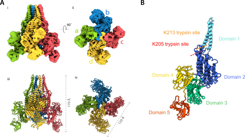
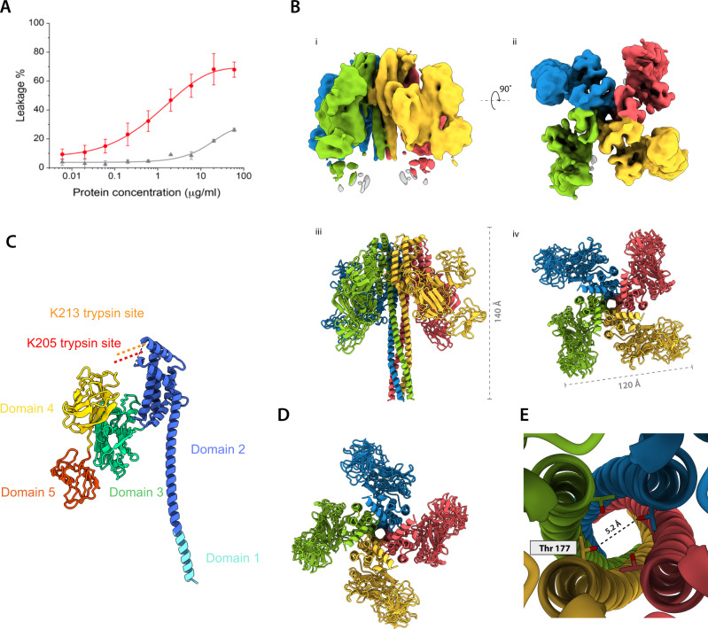
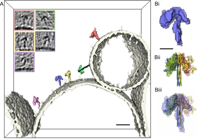
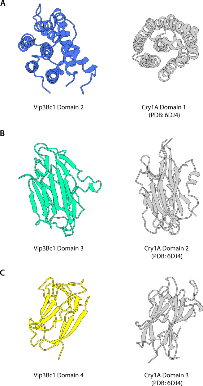
Insect pests are a major cause of crop losses worldwide, with an estimated economic cost of $470 billion annually. Biotechnological tools have been introduced to control such insects without the need for chemical pesticides; for instance, the development of transgenic plants harbouring genes encoding insecticidal proteins. The Vip3 (vegetative insecticidal protein 3) family proteins from Bacillus thuringiensis convey toxicity to species within the Lepidoptera, and have wide potential applications in commercial agriculture. Vip3 proteins are proposed to exert their insecticidal activity through pore formation, though to date there is no mechanistic description of how this occurs on the membrane. Here we present cryo-EM structures of a Vip3 family toxin in both inactive and activated forms in conjunction with structural and functional data on toxin-membrane interactions. Together these data demonstrate that activated Vip3Bc1 complex is able to insert into membranes in a highly efficient manner, indicating that receptor binding is the likely driver of Vip3 specificity.


Conflict of interest statement

The authors declare no competing interests.

Figures

Fig. 1
Fig. 1. Structure of Vip3Bc1.
A Quaternary structure of Vip3Bc1 tetramer as shown from two views of the complex related by 90° for the EM density (i, ii) and model (iii, iv). B Model of Vip3Bc1 illustrating domain structure as a monomer. The model shown corresponds to subunit A. Primary trypsin processing site K205 is indicated. Domain structure with respect to the primary sequence can be seen in Supplementary Fig. 4.
Fig. 2
Fig. 2. Vip3Bc1act has a greater propensity to perturb liposome membranes compared with Vip3Bc1.
A Digestion of the complex increases activity according to CF dye release from LUVs upon addition of Vip3Bc1act (red) and Vip3Bc1 (grey). Plotted data represents the mean and the error bars the standard deviation of the measurements from three independent replicates (Full data shown Supplementary Table 3 and 4). B Quaternary structure of Vip3Bc1act tetramer as shown from two views of the complex related by 90° for the EM density (I, ii) and model (iii, iv). C Model of Vip3Bc1act asymmetric unit, illustrating domain structure as a monomer. D Vip3Bc1act tetrameric assembly as shown from the top conformation is consistent with a pore. E Pore structure of Vip3Bc1act.
Fig. 3
Fig. 3. Vip3act directly visualised on liposome membranes.
Segmented density from cryo-ET shows interaction of Vip3Bc1act (coloured) with the LUV membrane (white). Images inset show section through the tomogram of the matching particle in segmentation. Full tomogram can be visualised in Supplementary Movie 1. Scale bar 20 nm. B Scaled comparison between segmented density for a single particle in the tomogram, with 5 nm scale bar (i) compared to Vip3Bc1act model (ii) with an overlay showing the stalk density fits the 4-helix bundle well, and the density which is distal to the membrane is consistent with single particle Vip3Bc1act model. The helical stalk modelled in Vip3Bc1act does not account for all of the stalk density observed in the sub-volume.
Fig. 4
Fig. 4. Structural homology of Vip3 domains to counterpart 3D-cry domain.
Structural homology of Vip3 domains to counterpart 3D-cry domain. A Vip3Bc1 domain 2 and 3D-Cry domain 1 (Cry1A domain 1, PDB 6DJ4 shown) share a predominantly hydrophobic alpha helix surrounded by other helices in a bundle. B Vip3Bc1 domain 3 and 3D-Cry domain 2 (Cry1A domain 2 PDB 6DJ4) share a beta prism fold with three sides made up of antiparallel beta sheets. C Domains 4 Vip3Bc1 and 3D-Cry domain 3 (Cry1A domain 3, PDB 6DJ4 shown) share a twisted beta-sheet ‘jelly roll’ topology.
Fig. 5
Fig. 5. Proposed model of Vip3Bc1 conformational rearrangement upon processing.
Cleavage at K205 liberates helices α1-α4 allowing dissociation of inter-subunit interactions at the tip of the complex (i), the helices of domains 2 that are C-terminal to the cut site (from α5 onwards), remain in essentially their original position stabilising the central interface of the tetramer. The α4 helices form a central helical bundle while the other helices N-terminal to the cut site (α1-3) are able to unfurl and move down between the propeller domains to form a new helical bundle (ii–v), which interacts with the membrane via the ends of the helical bundle (vi).

References

    1. Sharma, S., Kooner, R. & Arora, R. Insect Pests and Crop Losses. in Breeding Insect Resistant Crops for Sustainable Agriculture 45–66 (Springer, 2017).
    1. Gan Y, et al. Improving farming practices reduces the carbon footprint of spring wheat production. Nat. Commun. 2014;5:5012. doi: 10.1038/ncomms6012. - DOI - PMC - PubMed
    1. Moar WJ, Berry C, Narva KE. The structure/function of new insecticidal proteins and regulatory challenges for commercialization. J. Invertebr. Pathol. 2017;142:1–4. doi: 10.1016/j.jip.2017.02.001. - DOI - PubMed
    1. Carriere Y, Fabrick JA, Tabashnik BE. Can Pyramids and Seed Mixtures Delay Resistance to Bt Crops? Trends Biotechnol. 2016;34:291–302. doi: 10.1016/j.tibtech.2015.12.011. - DOI - PubMed
    1. Chakroun M, Banyuls N, Bel Y, Escriche B, Ferre J. Bacterial Vegetative Insecticidal Proteins (Vip) from Entomopathogenic Bacteria (vol 80, pg 329, 2016) Microbiol. Mol. Biol. Rev. 2016;80:III–III. doi: 10.1128/MMBR.00039-16. - DOI - PMC - PubMed

Publication types

MeSH terms