. 2021 May 14;11(1):10331.
doi: 10.1038/s41598-021-89542-4.

Chorioamnionitis induces hepatic inflammation and time-dependent changes of the enterohepatic circulation in the ovine fetus

Affiliations

Chorioamnionitis induces hepatic inflammation and time-dependent changes of the enterohepatic circulation in the ovine fetus

Cathelijne Heymans et al. Sci Rep. .

Abstract

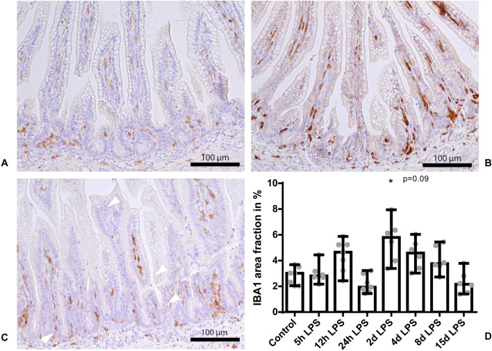
Chorioamnionitis, inflammation of fetal membranes, is an important cause of preterm birth and a risk factor for the development of adverse neonatal outcomes including sepsis and intestinal pathologies. Intestinal bile acids (BAs) accumulation and hepatic cytokine production are involved in adverse intestinal outcomes. These findings triggered us to study the liver and enterohepatic circulation (EHC) following intra-amniotic (IA) lipopolysaccharide (LPS) exposure. An ovine chorioamnionitis model was used in which circulatory cytokines and outcomes of the liver and EHC of preterm lambs were longitudinally assessed following IA administration of 10 mg LPS at 5, 12 or 24h or 2, 4, 8 or 15d before preterm birth. Hepatic inflammation was observed, characterized by increased hepatic cytokine mRNA levels (5h - 2d post IA LPS exposure) and increased erythropoietic clusters (at 8 and 15 days post IA LPS exposure). Besides, 12h after IA LPS exposure, plasma BA levels were increased, whereas gene expression levels of several hepatic BA transporters were decreased. Initial EHC alterations normalized over time. Concluding, IA LPS exposure induces significant time-dependent changes in the fetal liver and EHC. These chorioamnionitis induced changes have potential postnatal consequences and the duration of IA LPS exposure might be essential herein.


Conflict of interest statement

The authors declare no competing interests.

Figures

Figure 1
Figure 1
Study design. At 5, 12, or 24 hours or 2, 4, 8 or 15 days (black arrows) pregnant ewes received an IA injection with 10 mg LPS before preterm delivery at 125 days of gestation (term ~ 150 days). An IA saline injection at comparable time points to LPS injections was given to control animals. Timing shown in gestational days.
Figure 2
Figure 2
Scoring of H&E slides on a 0 to 4 scale for degree of sinusoidal dilatation, shape and size of central veins and number and location of extramedullary hematopoietic clusters. Representative images of control (A), at day 8 after IA LPS administration (B) and at day 15 after LPS administration (C). Data are presented as median with IQR (D). D: Pathologic score with increased number of extramedullary hematopoietic clusters in the animals at day 8 and day 15 after IA LPS administration. Specifically, the increased extramedullary hematopoiesis, which manifests as clustering and conflation of hepatic erythropoiesis in the parenchyma, is indicated by white triangles (b + c). *P < 0.05 and **P < 0.01 compared to control.
Figure 3
Figure 3
Relative mRNA expression of IL-8 (A), TNF-α (B) and IL-18 (C) in AU in the liver. Data are presented as median with IQR. A: IL-8 tended to increase 5 hours after IA LPS administration. P = 0.09 compared to control. B: TNF-α mRNA levels tended to be increased 12 hours after IA LPS administration. P = 0.07 compared to control. C: IL-18 mRNA levels were increased at 24 hours and 2 days after IA LPS administration. *P = 0.05 and **P < 0.005 compared to control.
Figure 4
Figure 4
Relative hepatic NTCP and BSEP gene expression in AU. Data are presented as median with IQR. NTCP (a) and BSEP (b) were decreased 12 hours after IA LPS administration. *P < 0.05 compared to control.
Figure 5
Figure 5
tBAs concentrations in plasma in μmol/L. Data are presented as median with IQR. Increased tBAs concentration in animals 12 hours after IA LPS administration. *P < 0.05 compared to control.
Figure 6
Figure 6
Relative gene expression of ASBT in AU in the terminal ileum. Data are presented as median with IQR. Increased ASBT expression in animals 24 hours and 8 days after IA LPS administration. *P < 0.05 compared to control. P = 0.07 compared to control.
Figure 7
Figure 7
An overview of the enterohepatic circulation (EHC) of bile acids (BAs) and changes herein related to FIRS and liver inflammation. CYP7A1 and CYP27A1 play a role in BAs production, which can be inhibited by FGF19. Physiologically, BAs are transported from hepatocytes via BSEP, via the gallbladder subsequently into the intestinal lumen. Most BAs are reabsorbed in the terminal ileum by ASBT. Following enterocyte uptake, BAs bind to IBABP to traverse the cytoplasm of epithelial cells. BAs exit the basolateral site of the enterocyte via OSTα-OSTβ. Via the portal vein, BAs are transported back to the liver. NTCP takes up the BAs into the hepatocyte to be recycled. Decreased expression of NTCP and BSEP and resultant increased plasma BA levels are associated with FIRS and liver inflammation. Increased ASBT expression may also be inflammation dependent, or a compensatory mechanism to the postulated lower intraluminal BAs supply to maintain a constant BA pool in the EHC.
Figure 8
Figure 8
IBA1 immunoreactivity in the terminal ileum. Representative images of control (A), at day 2 after IA LPS administration (B) and at day 4 after LPS administration (C). Data are presented as medium with IQR (D). D: Increased IBA1 positive surface area in animals 2 days and 4 days after IA LPS exposure. *P < 0.05 compared to control. P = 0.09 compared to control.
Figure 9
Figure 9
Relative gene expression of IL-18 in AU in the terminal ileum. Data are presented as median with IQR. IL-18 mRNA levels tended to be increased 24 hours after IA LPS administration. P = 0.07 compared to control.

Similar articles

Cited by

References

    1. Murphy SL, Mathews TJ, Martin JA, Minkovitz CS, Strobino DM. Annual summary of vital statistics: 2013–2014. Pediatrics. 2017 doi: 10.1542/peds.2016-3239. - DOI - PubMed
    1. Goldenberg RL, Hauth JC, Andrews WW. Intrauterine infection and preterm delivery. N. Engl. J. Med. 2000;342(20):1500–1507. doi: 10.1056/NEJM200005183422007. - DOI - PubMed
    1. Galinsky R, Polglase GR, Hooper SB, Black MJ, Moss TJ. The consequences of chorioamnionitis: preterm birth and effects on development. J. Pregnancy. 2013;2013:412831. doi: 10.1155/2013/412831. - DOI - PMC - PubMed
    1. Kim CJ, et al. Acute chorioamnionitis and funisitis: definition, pathologic features, and clinical significance. Am. J. Obstet. Gynecol. 2015;213(4 Suppl):S29–52. doi: 10.1016/j.ajog.2015.08.040. - DOI - PMC - PubMed
    1. Gussenhoven R, et al. Chorioamnionitis, neuroinflammation, and injury: timing is key in the preterm ovine fetus. J. Neuroinflammation. 2018;15(1):113. doi: 10.1186/s12974-018-1149-x. - DOI - PMC - PubMed

Publication types

MeSH terms