Review
. 2021 Aug;22(8):529-547.
doi: 10.1038/s41580-021-00366-6. Epub 2021 May 14.

The principles of directed cell migration

Affiliations
Review

The principles of directed cell migration

Shuvasree SenGupta et al. Nat Rev Mol Cell Biol. 2021 Aug.

Abstract

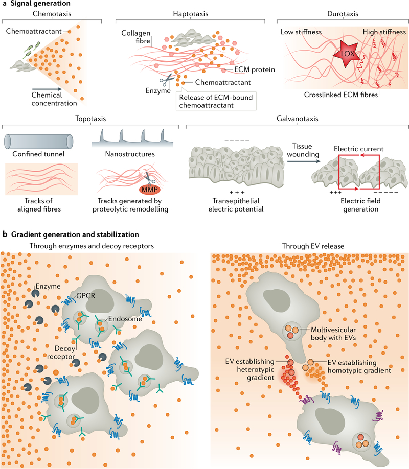
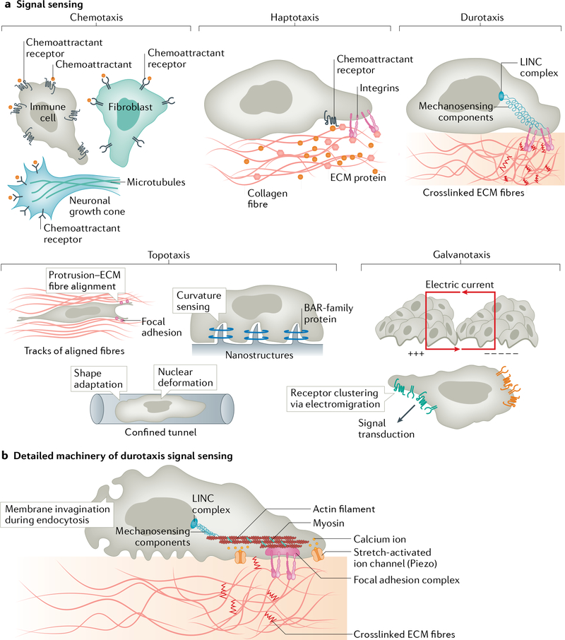
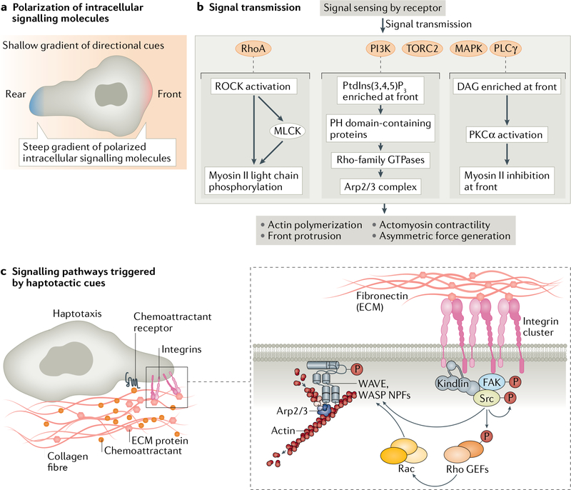
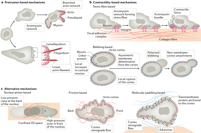
Cells have the ability to respond to various types of environmental cues, and in many cases these cues induce directed cell migration towards or away from these signals. How cells sense these cues and how they transmit that information to the cytoskeletal machinery governing cell translocation is one of the oldest and most challenging problems in biology. Chemotaxis, or migration towards diffusible chemical cues, has been studied for more than a century, but information is just now beginning to emerge about how cells respond to other cues, such as substrate-associated cues during haptotaxis (chemical cues on the surface), durotaxis (mechanical substrate compliance) and topotaxis (geometric features of substrate). Here we propose four common principles, or pillars, that underlie all forms of directed migration. First, a signal must be generated, a process that in physiological environments is much more nuanced than early studies suggested. Second, the signal must be sensed, sometimes by cell surface receptors, but also in ways that are not entirely clear, such as in the case of mechanical cues. Third, the signal has to be transmitted from the sensing modules to the machinery that executes the actual movement, a step that often requires amplification. Fourth, the signal has to be converted into the application of asymmetric force relative to the substrate, which involves mostly the cytoskeleton, but perhaps other players as well. Use of these four pillars has allowed us to compare some of the similarities between different types of directed migration, but also to highlight the remarkable diversity in the mechanisms that cells use to respond to different cues provided by their environment.


Conflict of interest statement

Competing interests

The authors declare no competing interests.

Figures

Fig. 1 |
Fig. 1 |. Generating the signal.
a | The diverse ways by which cues for directional migration are generated. During chemotaxis, soluble chemoattractants released from bacteria or cellular sources diffuse to form chemical gradients. During haptotaxis, extracellular matrix (ECM) proteins and chemokines released from cellular sources are deposited onto the ECM and generate gradients of immobilized chemical cues. In some cases, ECM-bound chemokines are released from the matrix by cellular proteolytic activities (scissors) and provide soluble cues for chemotaxis. During durotaxis, gradients of stiffness can be generated by lysyl oxidase (LOX)-mediated ECM crosslinking. During topotaxis, the geometry of the existing tissue structures, aligned fibres or tracks generated by proteolytic remodelling (via matrix metalloproteinases (MMPs), scissors) or deformation provides directional signals. During galvanotaxis, electric fields generated at wounding sites as a result of the loss of transepithelial potential provide guidance cues for cells involved in damage repair. b | Cartoon explaining how stable gradients are generated and maintained during chemotaxis. Uniformly present soluble chemicals are either degraded by enzymes or scavenged (via endocytic internalization) by decoy receptors to establish a gradient (left). Cells release extracellular vesicles (EVs), such as exosomes carrying either identical (homotypic gradient) or distinct (heterotypic gradient) chemical cues, to generate a stable secondary gradient (right). GPCR, G protein-coupled receptor.
Fig. 2 |
Fig. 2 |. Sensing the signal.
a | Various ways cells sense directional cues. During chemotaxis, cells sense the signal through surface receptors (G protein-coupled receptor (GPCRs), receptor tyrosine kinases or other transmembrane receptors), which bind the soluble chemical cues. During haptotaxis, cells detect surface-bound cues through integrin receptors and GPCRs. During durotaxis, substrate stiffness is sensed by an array of mechanically coupled components located on the cell surface, in the cytosol or at the nuclear envelope. During topotaxis, cells detect the geometry of available space and adapt their shape by changing the orientation of membrane protrusions in parallel to the aligned extracellular matrix (ECM) fibres, sensing topology-induced membrane curvature by BAR-family proteins, or gauging nuclear deformation resulting from compression and shape change. During galvanotaxis, the electric field is sensed by electromigration of membrane components (including signalling receptors) towards the cathode (+++) or the anode (− − − − −). b | Molecular machinery for durotactic sensing. Cells sense gradients of stiffness using mechanosensors at the cell surface (including integrin receptors in focal adhesions, invaginated membranes and stress-activated ion channels), inside the cytoplasm (including components of focal adhesions, actin filaments, microtubules and other mechanosensitive proteins) or at the nucleus (LINC complex).
Fig. 3 |
Fig. 3 |. Transmitting the signal.
a | Cartoon depicting how shallow gradients of extracellular directional cues are transmitted into steep gradients of intracellular signalling molecules at the front and rear of cells. b | Flow chart describing how various signalling pathways activated by sensing of directional cues lead to changes in the cytoskeletal machinery. c | Cartoon highlighting the molecular machinery that transmits haptotactic signals. Sensing gradients of fibronectin through integrin engagement activates non-receptor tyrosine kinases: focal adhesion kinase (FAK) and Src-family kinases. These kinases phosphorylate a variety of substrates, triggering the formation of new protein complexes, including guanine nucleotide exchange factors (GEFs), and the activation of Rac, which leads to branched actin network formation through Arp2/3 complex activation via the nucleation-promoting factors (NPFs) WASP and WAVE, ultimately generating lamellipodial structures. DAG, diacylglycerol; PH, pleckstrin homology; PI3K, phosphoinositide 3-kinase; PtdIns(3,4,5)P3, phosphatidylinositol 3,4,5-trisphosphate.
Fig. 4 |
Fig. 4 |. Executing the signal.
Depiction of how asymmetric force is generated through protrusion-based, contractility-based or alternative mechanisms. a | Formation of protrusions such as pseudopodia, lamellipodia and/or filopodia is driven by branched and linearly polymerized actin networks. b | Contractility-based mechanisms of migration (characteristic of mesenchymal cells) depend on the establishment of stress fibres, where a contractile array of actomyosin networks is mechanically coupled to the substrate through integrin-based focal adhesions (top). Many cell types, in particular in the in vivo context, migrate via extension of blebs (bottom), which are generated through local increases in cortical tension on the non-blebbing side of the cell (marked with arrows) and asymmetric membrane tearing (delamination), or by local rupture of the cortex, or both. c | Cells moving through a confined space use the nuclei as a piston to generate zones of high cytosolic pressure at the leading edge. Cells moving in the absence of substrate adhesion depend on friction between cells and the environment, generated by retrograde flow of the cortical actin. Cells can also use molecular paddling to swim through the environment using horizontal rearward flow of transmembrane proteins anchored to the actin cortex (advection).

Similar articles

Cited by

References

    1. Borrell V Recent advances in understanding neocortical development. F1000Res 10.12688/f1000research.20332.1 (2019). - DOI - PMC - PubMed
    1. Pawar KB, Desai S, Bhonde RR, Bhole RP & Deshmukh AA Wound with diabetes: present scenario and future. Curr. Diabetes Rev. 10.2174/1573399816666200703180137 (2020). - DOI - PubMed
    1. Janssen E & Geha RS Primary immunodeficiencies caused by mutations in actin regulatory proteins. Immunol. Rev. 287, 121–134 (2019). - PubMed
    1. Novikov NM, Zolotaryova SY, Gautreau AM & Denisov EV Mutational drivers of cancer cell migration and invasion. Br. J. Cancer 10.1038/s41416-020-01149-0 (2020). - DOI - PMC - PubMed
    1. García-Cuesta EM et al. The role of the CXCL12/CXCR4/ACKR3 axis in autoimmune diseases. Front. Endocrinol. 10, 585 (2019). - PMC - PubMed

Publication types

LinkOut - more resources