Review
. 2021 Jun;11(6):e02176.
doi: 10.1002/brb3.2176. Epub 2021 May 14.

Cerebral blood flow impairment and cognitive decline in heart failure

Affiliations
Review

Cerebral blood flow impairment and cognitive decline in heart failure

Ana Ovsenik et al. Brain Behav. 2021 Jun.

Abstract

Background and purpose: Cognitive decline is an important contributor to disability in patients with chronic heart failure, affecting 25%-50% of patients. The aim of this review is to stress the importance of understanding pathophysiological mechanisms of heart failure involved in cognitive decline.

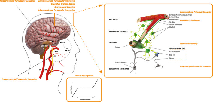
Methods: An extensive PubMed search was conducted for the literature on the basic mechanisms of cerebral blood flow regulation, the effect of cardiac dysfunction on cerebral blood flow, and possible mechanisms underlying the association between cardiac dysfunction and cognitive decline.

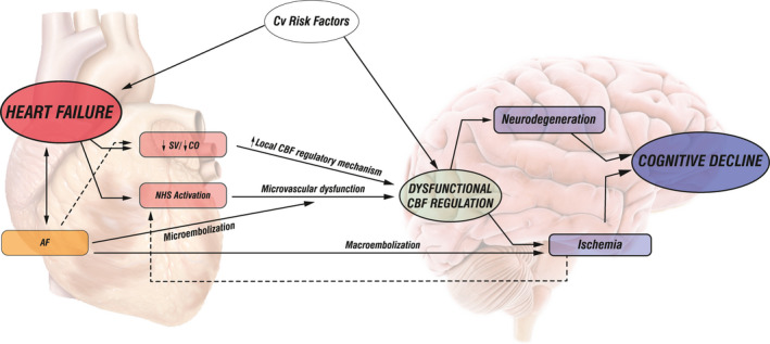
Results: Published literature supports the thesis that cardiac dysfunction leads to cerebral blood flow impairment and predisposes to cognitive decline. One of the postulated mechanisms underlying cognitive decline in chronic heart failure is chronic regional hypoperfusion of critical brain areas. Cognitive function may be further compromised by microvascular damage due to cardiovascular risk factors. Furthermore, it is implied that cerebral blood flow assessment could enable early recognition of patients at risk and help guide appropriate therapeutic strategies.

Conclusion: Interdisciplinary knowledge in the fields of neurology and cardiology is essential to clarify heart and brain interconnections in chronic heart failure. Understanding and identifying the basic neuropathophysiological changes in chronic heart failure could help with developing methods for early recognition of patients at risk, followed by institution of therapeutic actions to prevent or decrease cognitive decline.

Keywords: autonomic nervous system; cerebral autoregulation; cerebral blood flow; cognitive decline; heart failure; neurovascular coupling.


Conflict of interest statement

None to declare.

Figures

FIGURE 1
FIGURE 1
Schematic presentation of cerebrovascular tree structure and cerebral blood flow regulatory mechanisms. The brain receives arterial perfusion through two internal carotid arteries (ICA) and two vertebral arteries (VA) that interconnect intracranially through the circle of Willis, which gives rise to several vessels branching into progressively smaller arteries that run along the surface of the brain and then penetrate the brain tissue continuing deeper into the brain until they terminate in a network of capillaries. The main mechanisms regulating cerebral blood flow (CBF) include cerebral autoregulation (CA), activity of perivascular nerves, regulation by blood gasses, and neurovascular coupling. Cerebral autoregulation (CA) ensures CBF is maintained approximately constant across a wide range of mean arterial pressure (MAP), with neck vessels and large cerebral arteries serving as the first line of defense against hyperperfusion of the downstream microvasculature. CBF is highly dependent on extraparenchymal and intraparenchymal perivascular innervation. Extraparenchymal cerebral blood vessels are extensively innervated by autonomic and sensory nerve fibers deriving from the superior cervical ganglion (SCG), sphenopalatine ganglion (SPG), otic ganglion (OG), and the trigeminal ganglion (TG), while intraparenchymal vessels receive innervation through projections from subcortical neurons originating in locus coeruleus (LC), nucleus basalis (NB), and dorsal raphe nucleus (DRN). Pial arterioles are considered to be the main site of resistance regulation associated with changes in the partial pressure of blood gasses and of pH. Neurovascular coupling, which defines the association between neurons, microvascular endothelial cells, and glial cells, initiates local CBF changes on the level of capillaries. This signal is then transmitted upstream to remote vessels, including pial arterioles, which leads to broader blood flow changes. Additionally, neurovascular coupling may be modulated through subcortical neuronal activity. Internal carotid artery (ICA); vertebral artery (VA); superior cervical ganglion (SCG), otic ganglion (OT); sphenopalatine ganglion (SPG); trigeminal ganglion (TG); superior cervical ganglion (SCG); locus coeruleus (LC); dorsal raphe nucleus (DRN); nucleus basalis (NB)
FIGURE 2
FIGURE 2
Schematic presentation of the association among impaired cardiac function in chronic heart failure, cerebral blood flow, and cognitive decline. Left: The cerebral blood flow (CBF) changes in chronic heart failure (CHF) result from cardiac dysfunction (reduced stroke volume—SV/cardiac output—CO) and activation of the neurohumoral system (NHS). Reduced SV or CO induces local CBF regulatory mechanisms, leading to arteriolar vasodilatation, compromising further vasodilatation potential, and impairing CBF regulation. NHS activation provokes vasoconstriction of cerebral vascular bed, induces structural changes in cerebral resistance vessels, and causes microvascular dysfunction leading to impaired CBF regulation. Atrial fibrillation (AF) as a specific condition may lead to CBF impairment through mechanisms activated in CHF, through microembolic events causing microvascular dysfunction and impairing CBF regulation, or through macroembolic events leading to cerebral ischemia. Right: Cognitive decline (CD) in CHF results from dysfunctional CBF regulation causing hypoperfusion and ischemia of critical brain areas or from microvascular dysfunction due to cardiovascular risk factors associated with neurodegenerative disorders. Stroke volume (SV); cardiac output (cardiac output); neurohumoral System (NHS); atrial fibrillation (AF)

Similar articles

Cited by

References

    1. Aires, A. , Andrade, A. , Azevedo, E. , Gomes, F. , Araujo, J. P. , & Castro, P. (2020). Neurovascular coupling impairment in heart failure with reduction ejection fraction. Brain Sciences, 10, 714. 10.3390/brainsci10100714 - DOI - PMC - PubMed
    1. Azhan, A. , & Wong, F. Y. (2012). Challenges in understanding the impact of blood pressure management on cerebral oxygenation in the preterm brain. Frontiers in Physiology, 3, 471. - PMC - PubMed
    1. Bornstein, R. A. , Starling, R. C. , Myerowitz, P. D. , & Haas, G. J. (1995). Neuropsychological function in patients with end‐stage heart failure before and after cardiac transplantation. Acta Neurologica Scandinavica, 91, 260–265. - PubMed
    1. Brassard, P. , Tymko, M. M. , & Ainslie, P. N. (2017). Sympathetic control of the brain circulation: Appreciating the complexities to better understand the controversy. Autonomic Neuroscience: Basic & Clinical, 207, 37–47. - PubMed
    1. Caldas, J. R. , Panerai, R. B. , Haunton, V. J. , Almeida, J. P. , Ferreira, G. S. , Camara, L. , Nogueira, R. C. , Bor‐Seng‐Shu, E. , Oliveira, M. L. , Groehs, R. R. , Ferreira‐Santos, L. , Teixeira, M. J. , Galas, F. R. , Robinson, T. G. , Jatene, F. B. , & Hajjar, L. A. (2017). Cerebral blood flow autoregulation in ischemic heart failure. American Journal of Physiology Regulatory, Integrative and Comparative Physiology, 312, R108–R113. - PubMed

LinkOut - more resources