. 2021 Aug 15:707:108899.
doi: 10.1016/j.abb.2021.108899. Epub 2021 May 12.

The nucleotide binding affinities of two critical conformations of Escherichia coli ATP synthase

Affiliations

The nucleotide binding affinities of two critical conformations of Escherichia coli ATP synthase

Yunxiang Li et al. Arch Biochem Biophys. .

Abstract

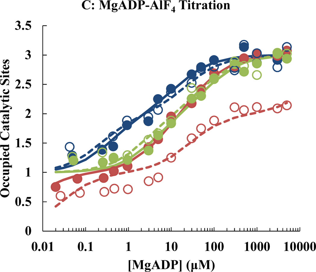
ATP synthase is essential in aerobic energy metabolism, and the rotary catalytic mechanism is one of the core concepts to understand the energetic functions of ATP synthase. Disulfide bonds formed by oxidizing a pair of cysteine mutations halted the rotation of the γ subunit in two critical conformations, the ATP-waiting dwell (αE284C/γQ274C) and the catalytic dwell (αE284C/γL276C). Tryptophan fluorescence was used to measure the nucleotide binding affinities for MgATP, MgADP and MgADP-AlF4 (a transition state analog) to wild-type and mutant F1 under reducing and oxidizing conditions. In the reduced state, αE284C/γL276C F1 showed a wild-type-like nucleotide binding pattern; after oxidation to lock the enzyme in the catalytic dwell state, the nucleotide binding parameters remained unchanged. In contrast, αE284C/γQ274C F1 showed significant differences in the affinities of the oxidized versus the reduced state. Locking the enzyme in the ATP-waiting dwell reduced nucleotide binding affinities of all three catalytic sites. Most importantly, the affinity of the low affinity site was reduced to such an extent that it could no longer be detected in the binding assay (Kd > 5 mM). The results of the present study allow to present a model for the catalytic mechanism of ATP synthase under consideration of the nucleotide affinity changes during a 360° cycle of the rotor.

Keywords: ATP synthase; Catalytic mechanism; Disulfide crosslink; Nucleotide binding; Rotational catalysis; Tryptophan fluorescence.


Conflict of interest statement

Conflict of interest

The authors declare that there is no conflict of interest.

Figures

Fig. 1.
Fig. 1.
Titration of WT and mutant F1 with nucleotides. Data for WT are shown in blue, those for the αE284C/γQ274C mutant in red, and those for the αE284C/γL276C mutant in green. Filled circles represent the experimental data of F1 in absence of α-γ crosslink; open circles represent the experimental data of F1 in presence of α-γ crosslink. Each data point represents the average of at least three independent assays. Solid lines are the least-squares best fit curves of the filled circle sets, whereas dashed lines are the least-squares best fit curves of the open circle sets. The curves were fitted as described in Materials and Methods, using a linear x-axis. Conversion to a log scale is only for illustration purposes, to allow presentation of binding events with vastly different affinities in a single figure.
Fig. 1.
Fig. 1.
Titration of WT and mutant F1 with nucleotides. Data for WT are shown in blue, those for the αE284C/γQ274C mutant in red, and those for the αE284C/γL276C mutant in green. Filled circles represent the experimental data of F1 in absence of α-γ crosslink; open circles represent the experimental data of F1 in presence of α-γ crosslink. Each data point represents the average of at least three independent assays. Solid lines are the least-squares best fit curves of the filled circle sets, whereas dashed lines are the least-squares best fit curves of the open circle sets. The curves were fitted as described in Materials and Methods, using a linear x-axis. Conversion to a log scale is only for illustration purposes, to allow presentation of binding events with vastly different affinities in a single figure.
Fig. 2.
Fig. 2.
Catalytic site affinity changes in the rotary mechanism of ATP synthase. (A) Revised catalytic mechanism [15] incorporating the results of the present study. For details, see part 4.2. of the Discussion. It should be noted that the red arrow symbolizes the changes in the rotational angle of γ; it is not associated with a specific structural feature of γ. The hypothetical intermediate conformation βIM during closing of β might or might not be the same as the half-closed conformation βHC seen in crystal structures [14,41]. (B) Catalytic site affinity as a function of the rotational angle of γ. For each specified angle, the approximate range of experimentally-determined affinities is given (for details, see part 4.2. of the Discussion). The letters indicate the symbols for the different affinities of the catalytic sites used in Fig. 2A (H, high; M, medium; L, low; O, open site; asterisk, ATP-waiting dwell; no asterisk, catalytic dwell). The red horizontal arrow and the red L** indicate that γ probably has to undergo thermal rotational fluctuations in the positive direction before MgATP can bind effectively (see part 4.4. of the Discussion; see also state B in Fig. 2A).
Fig. 2.
Fig. 2.
Catalytic site affinity changes in the rotary mechanism of ATP synthase. (A) Revised catalytic mechanism [15] incorporating the results of the present study. For details, see part 4.2. of the Discussion. It should be noted that the red arrow symbolizes the changes in the rotational angle of γ; it is not associated with a specific structural feature of γ. The hypothetical intermediate conformation βIM during closing of β might or might not be the same as the half-closed conformation βHC seen in crystal structures [14,41]. (B) Catalytic site affinity as a function of the rotational angle of γ. For each specified angle, the approximate range of experimentally-determined affinities is given (for details, see part 4.2. of the Discussion). The letters indicate the symbols for the different affinities of the catalytic sites used in Fig. 2A (H, high; M, medium; L, low; O, open site; asterisk, ATP-waiting dwell; no asterisk, catalytic dwell). The red horizontal arrow and the red L** indicate that γ probably has to undergo thermal rotational fluctuations in the positive direction before MgATP can bind effectively (see part 4.4. of the Discussion; see also state B in Fig. 2A).

References

    1. Weber J, Structural biology: Toward the ATP synthase mechanism, Nat. Chem. Biol 6 (2010) 794–795. 10.1038/nchembio.458. - DOI - PubMed
    1. Guo H, Suzuki T, Rubinstein JL, Structure of a bacterial ATP synthase, Elife. 8 (2019) e43128. 10.7554/eLife.43128. - DOI - PMC - PubMed
    1. Junge W, Sielaff H, Engelbrecht S, Torque generation and elastic power transmission in the rotary FoF1-ATPase, Nature. 459 (2009) 364–370. 10.1038/nature08145. - DOI - PubMed
    1. Nakamoto RK, Baylis Scanlon JA, Al-Shawi MK, The rotary mechanism of the ATP synthase, Arch. Biochem. Biophys 476 (2008) 43–50. 10.1016/j.abb.2008.05.004. - DOI - PMC - PubMed
    1. Yoshida M, Muneyuki E, Hisabori T, ATP synthase - A marvellous rotary engine of the cell, Nat. Rev. Mol. Cell Biol 2 (2001) 669–677. 10.1038/35089509. - DOI - PubMed

Publication types

MeSH terms

LinkOut - more resources