. 2021 Sep:343:113761.
doi: 10.1016/j.expneurol.2021.113761. Epub 2021 May 12.

Non-invasive, neurotoxic surgery reduces seizures in a rat model of temporal lobe epilepsy

Affiliations

Non-invasive, neurotoxic surgery reduces seizures in a rat model of temporal lobe epilepsy

Yanrong Zhang et al. Exp Neurol. 2021 Sep.

Abstract

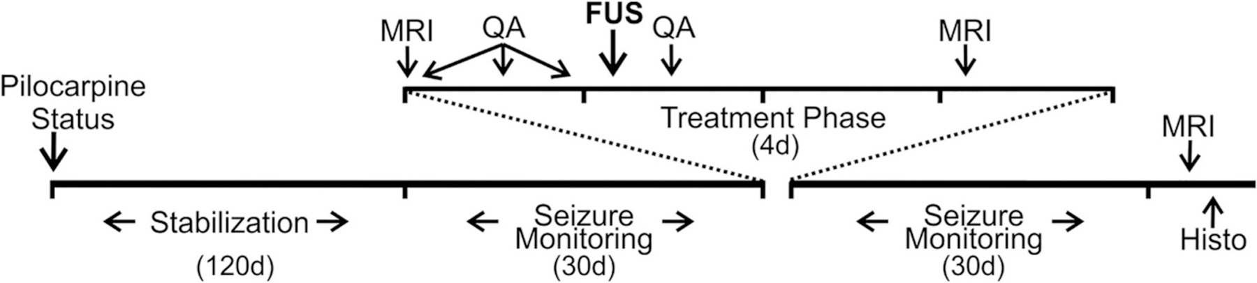
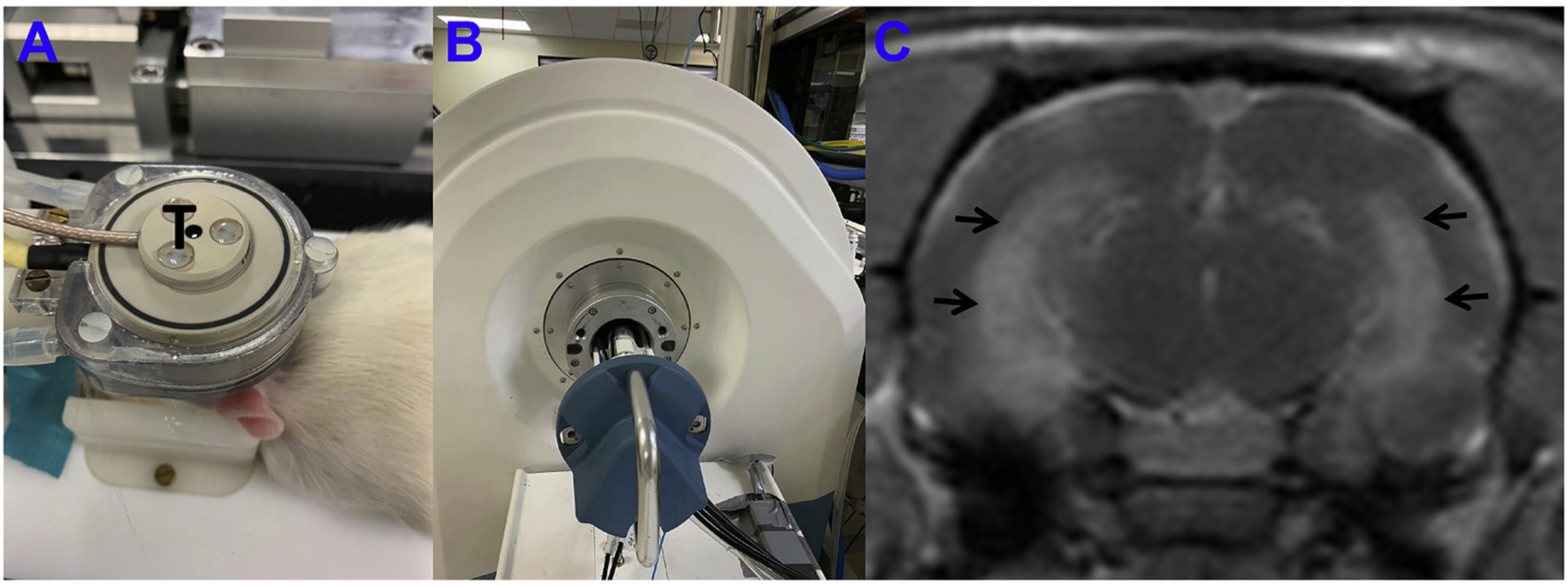
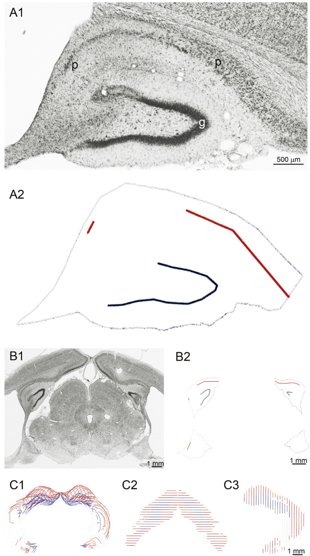
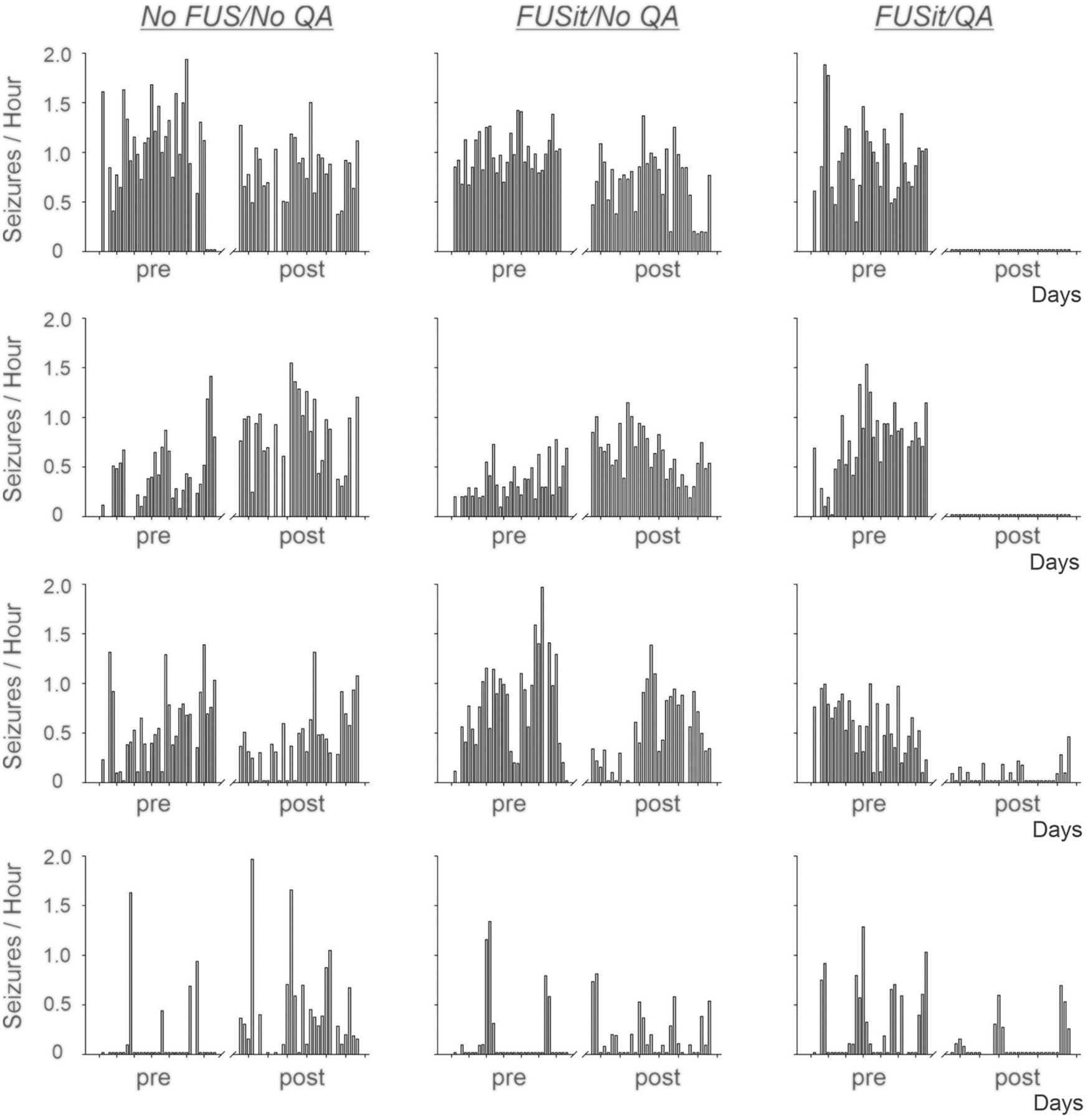
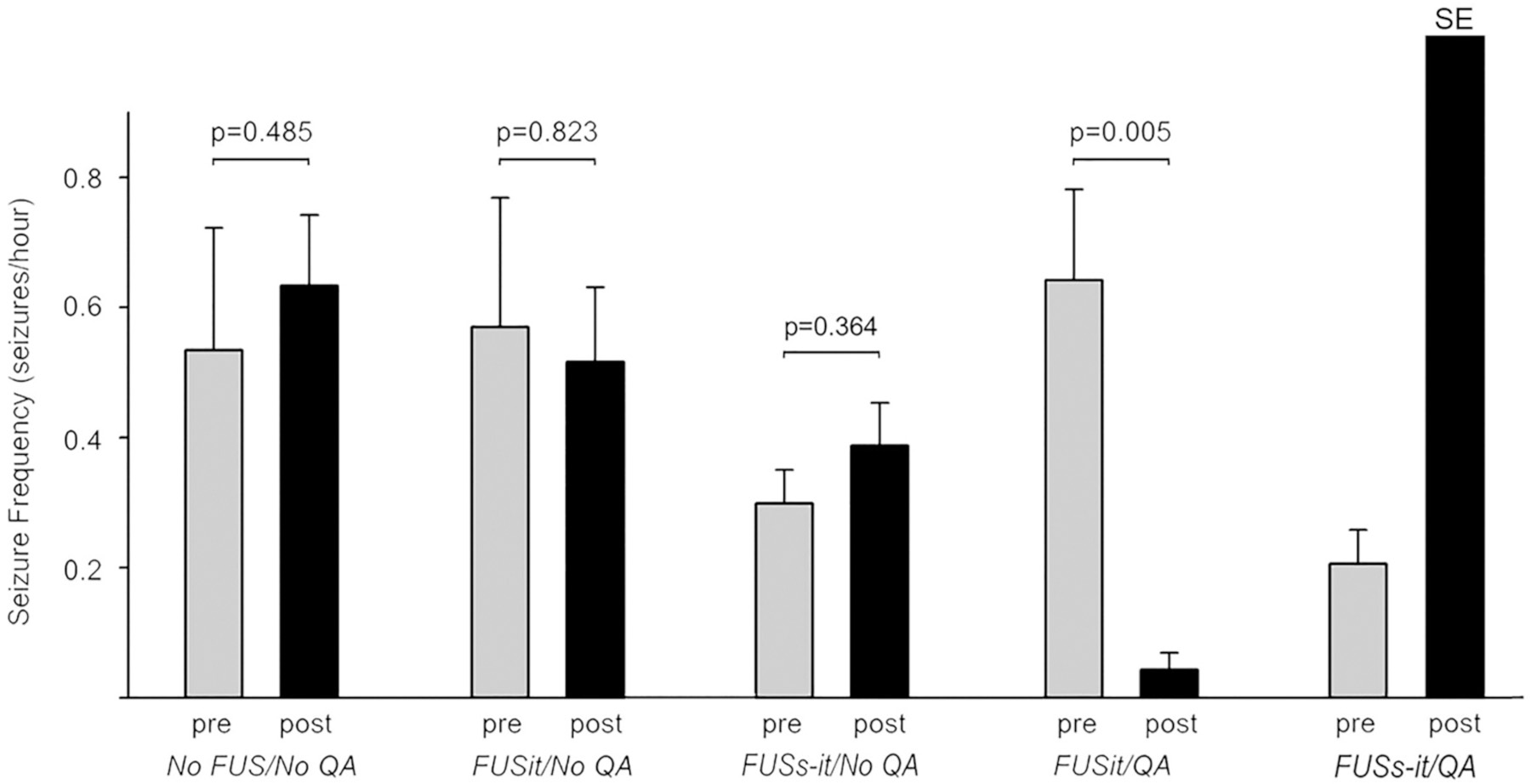
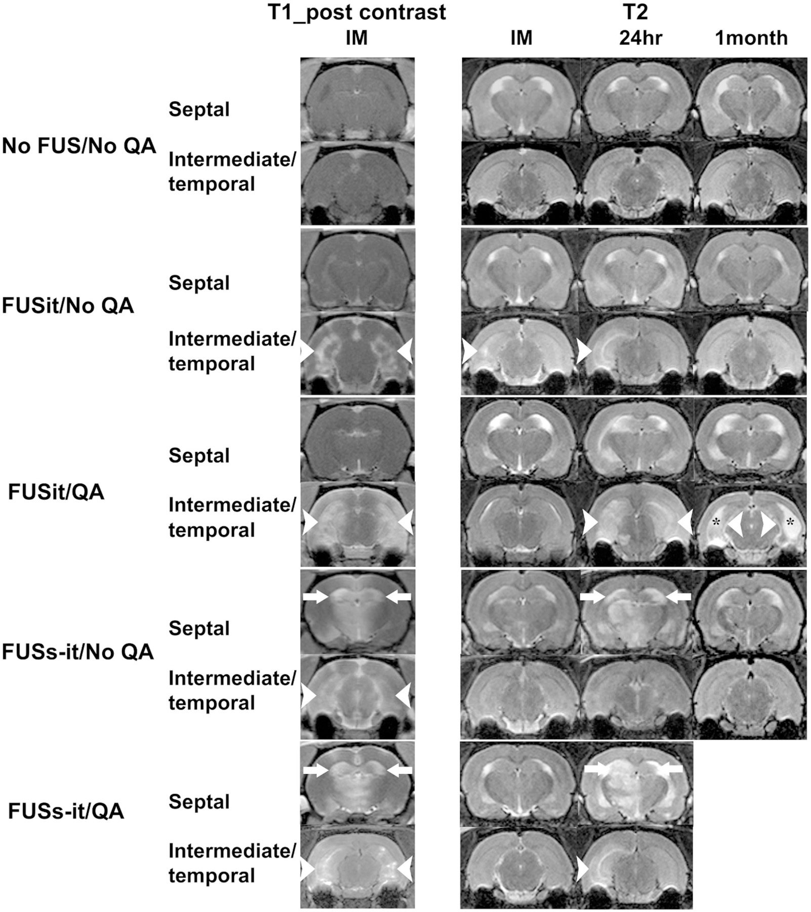
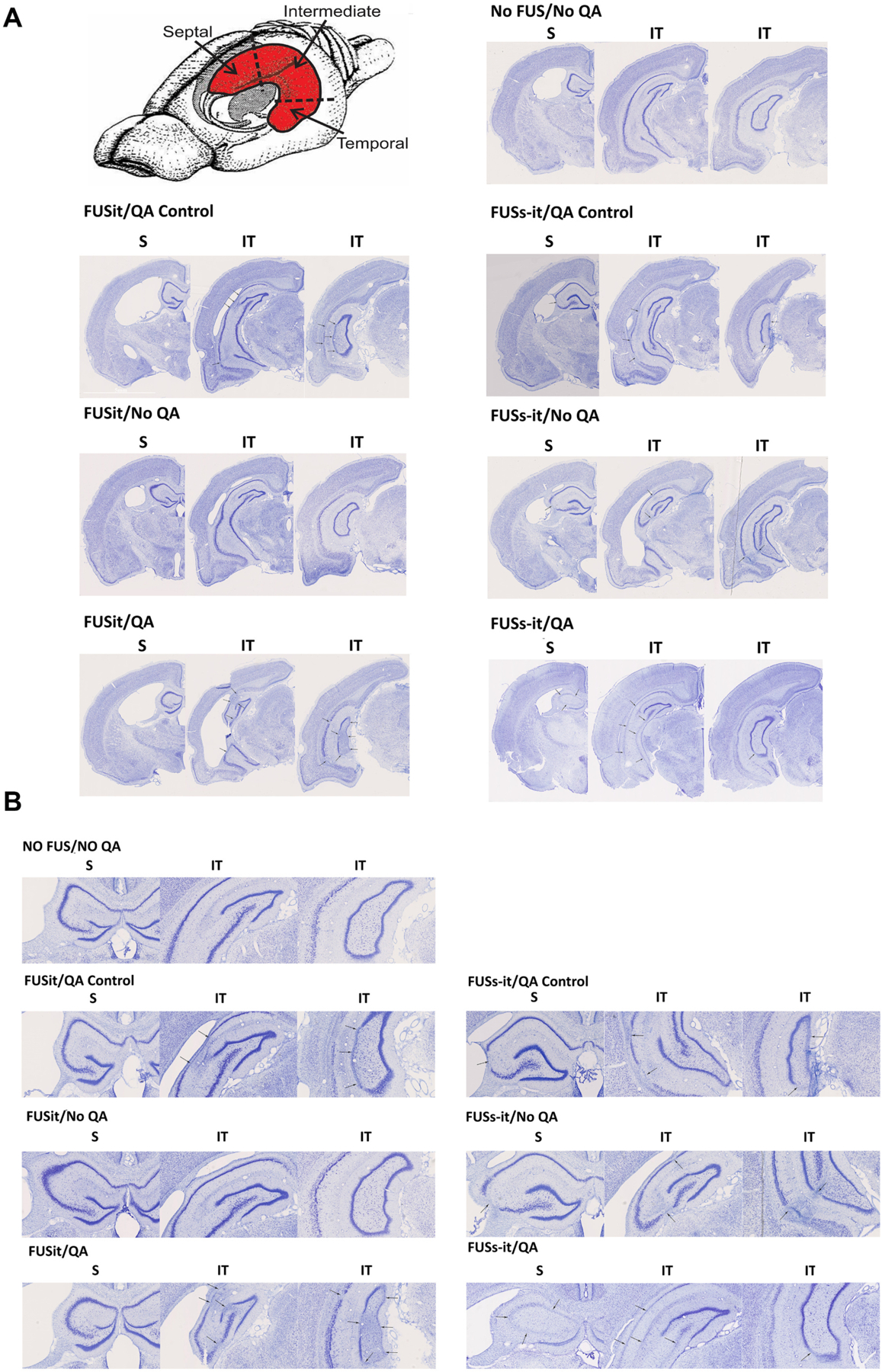
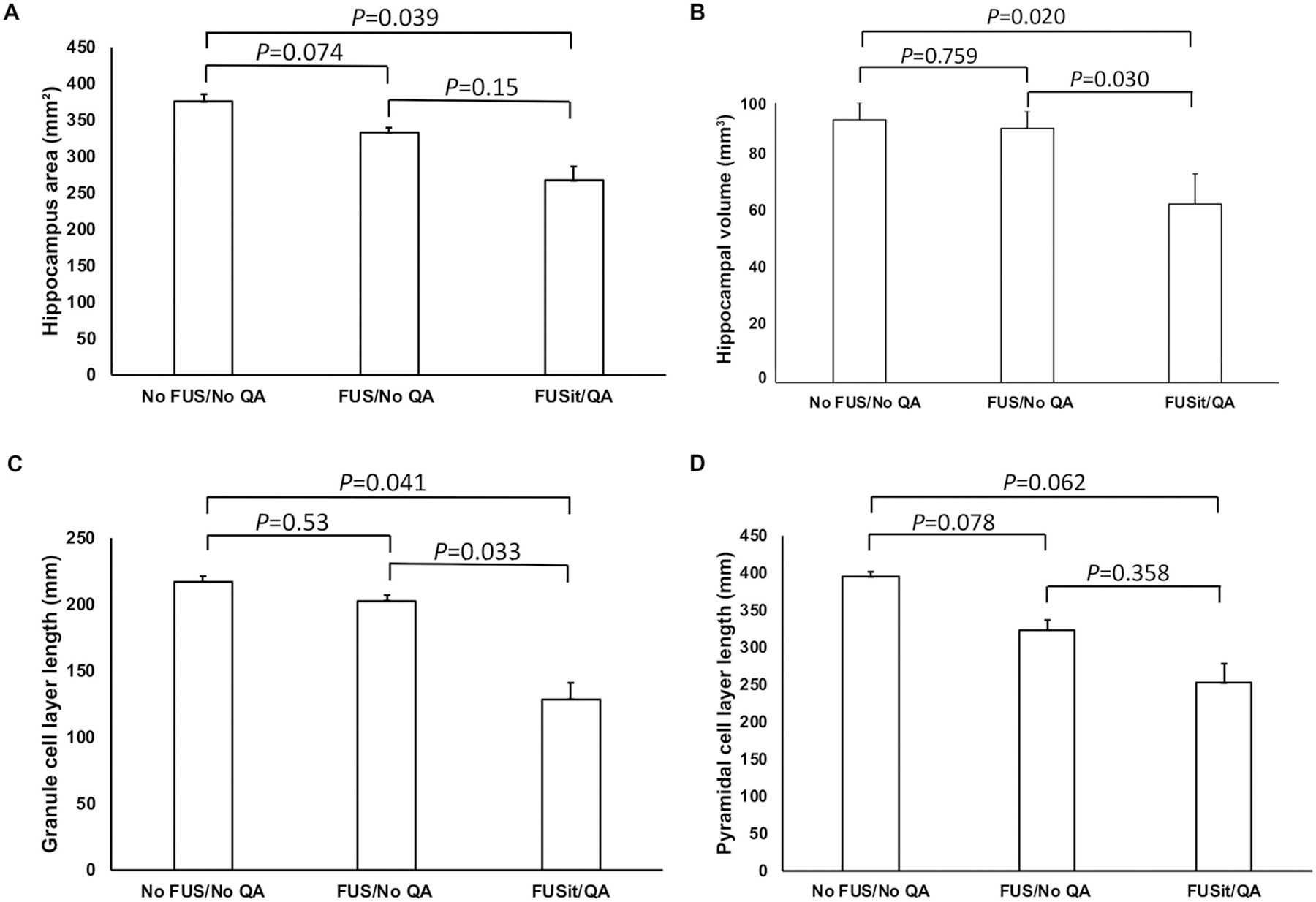
Surgery can be highly effective for treating certain cases of drug resistant epilepsy. The current study tested a novel, non-invasive, surgical strategy for treating seizures in a rat model of temporal lobe epilepsy. The surgical approach uses magnetic resonance-guided, low-intensity focused ultrasound (MRgFUS) in combination with intravenous microbubbles to open the blood-brain barrier (BBB) in a transient and focal manner. During the period of BBB opening, a systemically administered neurotoxin (Quinolinic Acid: QA) that is normally impermeable to the BBB gains access to a targeted area in the brain, destroying neurons where the BBB has been opened. This strategy is termed Precise Intracerebral Non-invasive Guided Surgery (PING). Spontaneous recurrent seizures induced by pilocarpine were monitored behaviorally prior to and after PING or under control conditions. Seizure frequency in untreated animals or animals treated with MRgFUS without QA exhibited expected seizure rate fluctuations frequencies between the monitoring periods. In contrast, animals treated with PING targeting the intermediate-temporal aspect of the hippocampus exhibited substantial reductions in seizure frequency, with convulsive seizures being eliminated entirely in two animals. These findings suggest that PING could provide a useful alternative to invasive surgical interventions for treating drug resistant epilepsy, and perhaps for treating other neurological disorders in which aberrant neural circuitries play a role.

Keywords: Epilepsy surgery; Focused ultrasound; Magnetic resonance-guided; Neuronal loss; Non-invasive; Quinolinic acid.


Conflict of interest statement

Declaration of Competing Interest

These are no financial and non-financial competing interests.

Figures

Fig. 1.
Fig. 1.
Timeline of the study. Pilocarpine-induced status epilepticus (Status) was followed by a stabilization period of 120 days. Behavioral seizures were then monitored for 30 days. A 60-day period then intervened (not shown) prior to initiating treatment. A 4-day treatment phase included two episodes of imaging (MRI), four intraperitoneal injections of Quinolinic Acid (QA), and the delivery of focused ultrasound (FUS). Behavioral seizures were then monitored for 30 days. At the end of seizure monitoring, imaging was again performed, after which animals were euthanized and prepared for histological analyses (Histo).
Fig. 2.
Fig. 2.
Experimental apparatus and post contrast T1-weighted image immediately after sonication. A: FUS system, a 650-kHz transducer (T) in brown rests upon the top of the rat head and can move in X-Y planes and be focused in the Z axis. B: A 3 T MRI scanner was used to detect the BBB-opening after sonication. C: Post contrast T1-weighted image immediately post sonication; enhancement of the bilateral intermediate-temporal hippocampus (black arrows) indicated BBB opening.
Fig. 3.
Fig. 3.
Method for measuring hippocampal cell layer loss and volume loss. A1: Nissl-stained hippocampus from a rat in the FUSit/QA group. Intact cell layers in the subiculum and Ammon’s horn (p) and in the granule cell layer (g) could be distinguished from gaps in those layers, where neuronal loss had occurred. A2: Reconstruction shows intact pyramidal cell layer (red), intact granule cell layer (blue), and outline of hippocampus (grey). B1: Nissl-stained coronal brain section from same rat. The left, dorsal hippocampus is same as that shown in panel A. B2: Reconstructed intact cell layers and hippocampal outline. Reconstruction of entire 1-in-6 series of sections shown from the coronal view (C1), dorsal view (C2), and side view (C3).
Fig. 4.
Fig. 4.
Daily convulsive seizure frequencies in individual animals before and after treatment. Daily convulsive seizure frequencies (seizures/Hour) recorded prior to (pre) and after (post) treatment are shown for animals in the No FUS/NoQA, FUSit/No QA, and FUSit/QA groups. Seizure frequencies were relatively stable in the pre- and post-treatment recording periods for animals in the No FUS/No QA and FUSit/No QA groups. In contrast, seizure frequencies were reduced substantially after treatment in the animals in the FUSit/QA group. In two of these animals, convulsive seizures were eliminated entirely.
Fig. 5.
Fig. 5.
Group seizure frequencies before (pre) and after (post) treatment. Average seizure frequencies (mean +/− SEM) are shown. Seizure frequencies did not differ significantly between pre- and post-treatment recording periods in either the No FUS/No QA or FUSit/NoQA groups. In contrast, seizure frequency was reduced substantially and significantly post-treatment in the FUSit/QA group. The animals from FUSs-it/QA group showed status epilepticus (SE) immediately after treatment. P values are from Students’ t-test comparing pre and post-treatment values for individual groups.
Fig. 6.
Fig. 6.
MRI evidence of BBB opening and tissue damage post-treatment. Post-contrast T1 images show areas of hyperintensity (BBB opening) in the regions targeted by FUS. BBB opening is indicated by white arrowheads in the intermediate-temporal hippocampus and by white arrows in the septal hippocampus. The untreated (No FUS/No QA) animal did not show signs of BBB opening. Examples of T2 images obtained immediately, 24 h, and 1 month post-FUS are shown for each of the five groups of animals. The untreated (No FUS/No QA) animal did not show signs of damage (abnormal bright or dark signal) at any of the post-treatment time points. Evidence of unilateral hyperintensity which indicates edema was seen immediately and at 24 h post-treatment (white arrowheads), but no abnormal signal was seen at the1 month time point in the FUSit/No QA example. Evidence of both edema (higher intensity signal indicated by white arrowheads and arrows) at the 24 h and atrophy (the thickness of hippocampus was significantly reduced compared to the ones on T2 images acquired immediately and 24 h post-FUS, the area that had used to be occupied by hippocampus was filled with cerebrospinal fluid indicated with black stars) was observed and 1 month time points for the FUSit/QA example. The one animal in the FUSs-it/QA group that survived the initial post-treatment period showed signs of edema in both the intermediate-temporal and septal hippocampus at 24 h. Part of the thalamus underlying the septal hippocampus in the FUSs-it/QA example also exhibited unilateral BBB opening, as well as evidence of edema at 24 h. No images are available at the 1-month time point for the FUSs-it/QA animals because they were euthanized prior to that time point.
Fig. 7.
Fig. 7.
Location of PING-induced neuronal loss. A drawing of the rodent brain with the hippocampus (in red) shows the aspects of the hippocampus that were assessed for neuronal loss. Examples of the types of neuronal loss that occurred in the different groups of animals are shown in Nissl-stained, coronal sections taken along the longitudinal axis of the hippocampus (arrows, 7A and 7B with different magnification). Obvious neuron loss was evident in the hilus of all rats including the untreated animals (No FUS/No QA) and the animal receiving FUS-only targeting the intermediate-temporal (it) aspect of the hippocampus (FUSit/No QA). In contrast, the animals induced epilepsy with pilocarpine injection and receiving QA together with FUS in the ‘it’ hippocampus (FUSit/QA), displayed substantial neuronal loss, involving CA1, CA3, and the dentate gyrus. The animals that did not receive pilocarpine injection, receiving QA together with FUS (FUSit/QA Control and FUSs-it/QA Control) all showed neuronal loss involving CA1 and CA3. The animal receiving FUS-only to both the it and septal aspects of the hippocampus without QA (FUSs-it/No QA) exhibited limited, sporadic cell loss. In contrast, the animal receiving FUS to both the it and septal aspects of the hippocampus together with QA (FUSs-it/QA) exhibited substantial neuronal loss in both the septal and it aspects of the hippocampus.
Fig. 8.
Fig. 8.
Quantification of hippocampal size and the length of neuronal cell body laminae. A. The area of the hippocampus was reduced significantly in the FUSit/QA group (all p values are from comparisons using Students’ t-test). B. The length of the granule cell layer was also reduced significantly in the FUSit/QA group. C. The length of the pyramidal cell layer also showed a strong trend toward reduction in the FUSit/QA group. D. The hippocampal volume decreased significantly in the FUSit/QA group.

Similar articles

Cited by

References

    1. Arida RM, Scorza FA, Peres CA, Cavalheiro EA, 1999. The course of untreated seizures in the pilocarpine model of epilepsy. Epilepsy Res 34, 99–107. - PubMed
    1. Baud MO, Kleen JK, Mirro EA, Andrechak JC, King-Stephens D, Chang EF, Rao VR, 2018a. Multi-day rhythms modulate seizure risk in epilepsy. Nat. Commun 9, 88. - PMC - PubMed
    1. Baud MO, Perneger T, Racz A, Pensel MC, Elger C, Rydenhag B, Malmgren K, Cross JH, McKenna G, Tisdall M, Lamberink HJ, Rheims S, Ryvlin P, Isnard J, Mauguiere F, Arzimanoglou A, Akkol S, Deniz K, Ozkara C, Lossius M, Rektor I, Kalviainen R, Vanhatalo LM, Dimova P, Minkin K, Staack AM, Steinhoff BJ, Kalina A, Krsek P, Marusic P, Jordan Z, Fabo D, Carrette E, Boon P, Rocka S, Mameniskiene R, Vulliemoz S, Pittau F, Braun KPJ, Seeck M, 2018b. European trends in epilepsy surgery. Neurology 91, e96–e106. - PubMed
    1. Baud MO, Ghestem A, Benoliel JJ, Becker C, Bernard C, 2019. Endogenous multidien rhythm of epilepsy in rats. Exp. Neurol 315, 82–87. - PubMed
    1. Beskid M, Rozycka Z, Taraszewska A, 1997. Quinolinic acid: effect on the nucleus arcuatus of the hypothalamus in the rat (ultrastructural evidence). Exp. Toxicol. Pathol 49, 477–481. - PubMed

Publication types

MeSH terms