. 2021 Sep:343:113757.
doi: 10.1016/j.expneurol.2021.113757. Epub 2021 May 12.

Cervical spinal cord injury-induced neuropathic pain in male mice is associated with a persistent pro-inflammatory macrophage/microglial response in the superficial dorsal horn

Affiliations

Cervical spinal cord injury-induced neuropathic pain in male mice is associated with a persistent pro-inflammatory macrophage/microglial response in the superficial dorsal horn

Eric V Brown et al. Exp Neurol. 2021 Sep.

Abstract

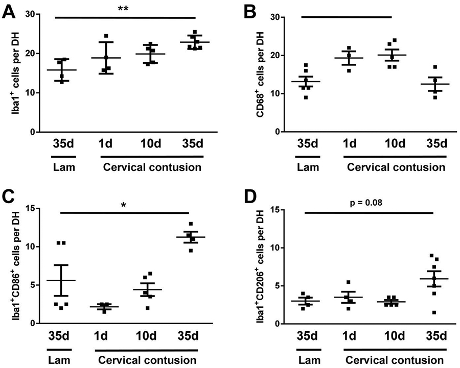
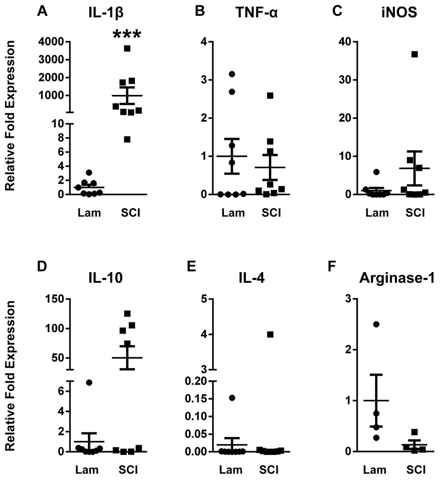
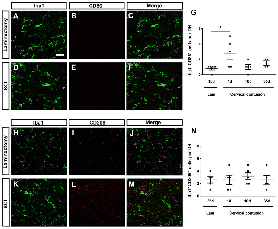
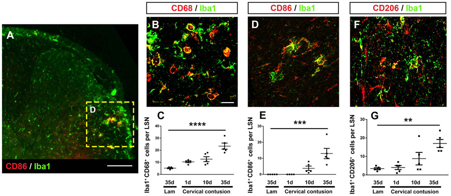
A significant portion of individuals living with traumatic spinal cord injury (SCI) experiences some degree of debilitating neuropathic pain (NP). This pain remains largely intractable in a majority of cases, due in part to an incomplete understanding of its underlying mechanisms. Central sensitization, an increase in excitability of pain transmission neurons located in superficial dorsal horn (sDH), plays a key role in development and maintenance of SCI-induced NP. Resident microglia and peripheral monocyte-derived macrophages (referred to collectively as MMΦ) are involved in promoting SCI-induced DH neuron hyperexcitability. Importantly, these MMΦ consist of populations of cells that can exert pro-inflammatory or anti-inflammatory signaling within injured spinal cord. It is critical to spatiotemporally characterize this heterogeneity to understand MMΦ contribution to NP after SCI. Given that a majority of SCI cases are cervical in nature, we used a model of unilateral C5/C6 contusion that results in persistent at-level thermal hyperalgesia and mechanical allodynia, two forms of NP-related behavior, in the forepaw. The aim of this study was to characterize the sDH MMΦ response within intact cervical spinal cord segments caudal to the lesion (i.e. the location of primary afferent nociceptive input from the forepaw plantar surface). Cervical SCI promoted a persistent MMΦ response in sDH that coincided with the chronic NP phenotype. Using markers of pro- and anti-inflammatory MMΦ, we found that the MMΦ population within sDH exhibited significant heterogeneity that evolved over time post-injury, including a robust and persistent increase in pro-inflammatory MMΦ that was especially pronounced at later times. C5/C6 contusion SCI also induced below-level thermal hyperalgesia and mechanical allodynia in the hindpaw; however, we did not observe a pronounced MMΦ response in sDH of L4/L5 spinal cord, suggesting that different inflammatory cell mechanisms occurring in sDH may be involved in at-level versus below-level NP following SCI. In conclusion, our findings reveal significant MMΦ heterogeneity both within and across pain transmission locations after SCI. These data also show a prominent and persistent pro-inflammatory MMΦ response, suggesting a possible role in DH neuron hyperexcitability and NP.

Keywords: Central sensitization; Contusion; Dorsal horn; Heterogeneity; Hyperexcitability; Macrophage; Neuropathic pain; SCI; Spinal cord injury.


Conflict of interest statement

Conflict of interest: The authors declare no competing financial interests.

Figures

Figure 1:
Figure 1:. Cervical contusion SCI promoted a persistent increase in MMΦ numbers within superficial dorsal horn.
SCI induced thermal hyperalgesia and mechanical allodynia in the ipsilateral (A, C) and contralateral (B, D) forepaws. Diagram showing location of contusion in relation to site of histological analysis (E). Diagram of spinal cord cross section highlighting region of analysis in ipsilateral sDH (E). Representative images of Iba1 immunohistochemistry in sDH of laminectomy-only (F), 1 day post-injury (G), 10 days post-injury (H) and 35 days post-injury (I). Laminae I and II are outlined with dotted red line. Compared to laminectomy-only (J), cervical contusion also induced marked changes in the morphology of Iba1+ cells at 35 days post-injury (K). Quantification of Iba1+ cell counts (L). Representative images showing Iba1 and CD68 co-labeling in the DH 10 days post injury (M-O). Cervical contusion resulted in increases in number of cells expressing the phagocytic marker CD68 (P). Data displayed as mean +/− SEM, * p<0.05, ** p<0.001, *** p<0.0001. (n = 5-7 animals per group for behavioral testing; n = 5-6 animals per group for histology analyses). F-G: 20x objective; JK, M-O: 40x objective. Scale bars: 200 μm (F-I), 50 μm (J-K), 25 μm (M-O.)
Figure 2:
Figure 2:. Cervical contusion SCI induced a pro-inflammatory MMΦ response in superficial dorsal horn.
Representative double-labeling for Iba1 and CD86 in laminectomy-only (A-C) and contusion SCI at 10 days (G) and 35 days (D-F) post-injury: Iba1 is shown in green; CD86 is shown in red; overlay shown in panels C, F and G. Per cell expression profile of CD86 intensity in Iba1+ cells (H). Graph of expression binning (I). Representative double-labeling for Iba1 and MARCO in laminectomy-only (J-L) and contusion SCI at 35 days post-injury (M-O). Iba1 is shown in green; MARCO in red; overlay in K and N. Quantification of double-labeled cells in (P). Data displayed as mean +/− SEM, *** p<0.0001. (n = 5-6 animals per group for histology analyses). All images: 40x objective. Scale bars: 25 μm (A-C, G), 25 μm (D-F), 50 μm (J-O).
Figure 3:
Figure 3:. Cervical contusion induced an anti-inflammatory MMΦ response in the superficial dorsal horn.
Representative double-labeling for Iba1 and CD206 in laminectomy-only (A-C) and contusion SCI at 10 days (G) 35 days (D-F) post-injury: Iba1 is shown in green; CD206 is shown in red; overlay shown in panels C, F and G. Per cell expression profile of CD206 intensity in Iba1+ cells (H). Graph of expression binning (I). Representative double-labeling for Iba1 and YM1 in laminectomy-only (J-L) and contusion SCI at 35 days post-injury (M-O): Iba1 is shown in green; YM1 is shown in red; overlay shown in panels K and N. Quantification of double-labeled cells in (P). Data displayed as mean +/− SEM, *p<0.05, *** p<0.0001. (n = 6 animals per group for histology analyses). All images: 40x objective. Scale bars: 25 μm (all panels).
Figure 4:
Figure 4:. Cervical contusion induced a persistent change in MMΦ inflammatory profile in superficial dorsal horn of contralateral cervical spinal cord.
Quantification in the contralateral cervical sDH of: number of Iba1+ cells (A), CD68+ cells (B), Iba1+CD206+ cells (C), and Iba1+CD86+ cells (D). (n=4-6 animals per group). Data displayed as mean +/− SEM. *p<0.05, **p<0.01.
Figure 5:
Figure 5:. Cervical contusion SCI alters expression of both pro- and anti-inflammatory genes in the ipsilateral dorsal cervical spinal cord.
At 10 days post-injury, there was increased expression of IL-1β (p=0.0002) (A), but no significant changes in TNF-α (B) or iNOS (C) expression. There was an increase in IL-10 expression in a subset of SCI animals, but this effect was not significant overall p>0.05) at 10 days post-injury compared to laminectomy-only control (D). IL-4 expression was unchanged by injury (E). There was a non-significant decrease in Arginase-1 expression after SCI (p = 0.057; F). Transcript expression was determined by qRT-PCR from sub-dissected ipsilateral dorsal quadrant obtained 1-2 segments caudal to the lesion site. (n = 8 animals per group for PCR analyses, except n = 4 animals per group for Arginase-1).
Figure 6:
Figure 6:. Cervical contusion SCI induced a below-level neuropathic pain-related behavioral phenotype, but did not alter MMΦ numbers in lumbar spinal cord dorsal horn.
Cervical contusion induced thermal hyperalgesia in ipsilateral (A) but not contralateral (B) hind paw, and induced mechanical allodynia in both ipsilateral (C) and contralateral (D) hindpaws. Representative double-labeling for Iba1 and CD68 in laminectomy-only (E-G) and contusion SCI 35 days post injury (H-J). Iba1 is shown in green; CD68 shown in red, overlay shown in G and J. Quantification of numbers of Iba1+ cells (K) and Iba1+ CD68+ co-labeled cells (L). *p<0.05, ***p<0.001, ****p<0.0001. (n = 5-7 animals per group for behavioral testing; n = 5 animals per group for histology analyses). All images: 40x objective. Scale bars: 30 μm (all panels).
Figure 7:
Figure 7:. Cervical contusion SCI did not alter the MMΦ inflammatory profile in lumbar dorsal horn.
Representative double-labeling for Iba1 and CD86 in laminectomy-only (A-C) and contusion SCI at 35 days post-injury (D-F): Iba1 is shown in green; CD86 is shown in red; overlay shown in panels C and F. Quantification of number of CD86+ Iba1+ co-labeled cells (G). Representative double-labeling for Iba1 and CD206 in laminectomy-only (H-J) and contusion SCI at 35 days post-injury (K-M). Iba1 is shown in green; CD206 in red; overlay in J and M. Quantification of CD206+ Iba1+ co-labeled cells (N). Data displayed as mean +/− SEM. (n = 5 animals per group for histology analyses). All images: 40x objective. Scale bars: 30 μm (all panels).
Figure 8:
Figure 8:. Cervical contusion SCI induced a MMΦ response in the lumbar lateral spinal nucleus.
Low magnification image of the LSN in the lumbar spinal cord (ipsilateral to contusion) labeled with Iba1 and CD86 (A). Higher-magnification representative image of CD68 and Iba1 co-labeling (B); quantification of numbers of Iba1+ CD68+ double-labeled cells (C). Representative image of CD86 and Iba1 co-labeling (D) and quantification of numbers of Iba1+ CD86+ double-labeled cells (E). Representative image of CD206 and Iba1 co-labeling (F) and quantification of numbers of Iba1+ CD206+ double-labeled cells (G). Data displayed as mean +/− SEM. *p<0.05. **p<0.01. ***p<0.001. ****p<0.0001. (n = 5 animals per group for histology analyses). A: Tile-scan 20x objective; B, D, F: 40x objective. Scale bars: 150 μm (A), 30 μm (B, D, F).

Similar articles

Cited by

References

    1. NB Finnerap IJ, sindrup SH Bach FW, Jensen TS, Pain and Dysesthesia in Patients with Spinal Cord Injury a Postal Survey. Spinal Cord, 2001. 39: p. 256–262. - PubMed
    1. Siddall PJ, et al., A longitudinal study of the prevalence and characteristics of pain in the first 5 years following spinal cord injury. Pain, 2003. 103(3): p. 249–257. - PubMed
    1. Diana D Cardenas MPJ, Treatments for Chronic Pain in Person with Spinal Cord Injury. J Spinal Cord Med, 2006. 29: p. 109–117. - PMC - PubMed
    1. Carlton SM, et al., Peripheral and central sensitization in remote spinal cord regions contribute to central neuropathic pain after spinal cord injury. Pain, 2009. 147(1-3): p. 265–76. - PMC - PubMed
    1. Detloff MR, et al., Remote activation of microglia and pro-inflammatory cytokines predict the onset and severity of below-level neuropathic pain after spinal cord injury in rats. Experimental neurology, 2008. 212(2): p. 337–47. - PMC - PubMed

Publication types

MeSH terms

Substances