. 2021 Sep;75(3):647-658.
doi: 10.1016/j.jhep.2021.04.050. Epub 2021 May 13.

Liver injury in COVID-19 and IL-6 trans-signaling-induced endotheliopathy

Affiliations

Liver injury in COVID-19 and IL-6 trans-signaling-induced endotheliopathy

Matthew J McConnell et al. J Hepatol. 2021 Sep.

Abstract

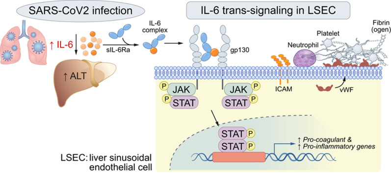
Background and aims: COVID-19 is associated with liver injury and elevated interleukin-6 (IL-6). We hypothesized that IL-6 trans-signaling in liver sinusoidal endothelial cells (LSECs) leads to endotheliopathy (a proinflammatory and procoagulant state) and liver injury in COVID-19.

Methods: Coagulopathy, endotheliopathy, and alanine aminotransferase (ALT) were retrospectively analyzed in a subset (n = 68), followed by a larger cohort (n = 3,780) of patients with COVID-19. Liver histology from 43 patients with COVID-19 was analyzed for endotheliopathy and its relationship to liver injury. Primary human LSECs were used to establish the IL-6 trans-signaling mechanism.

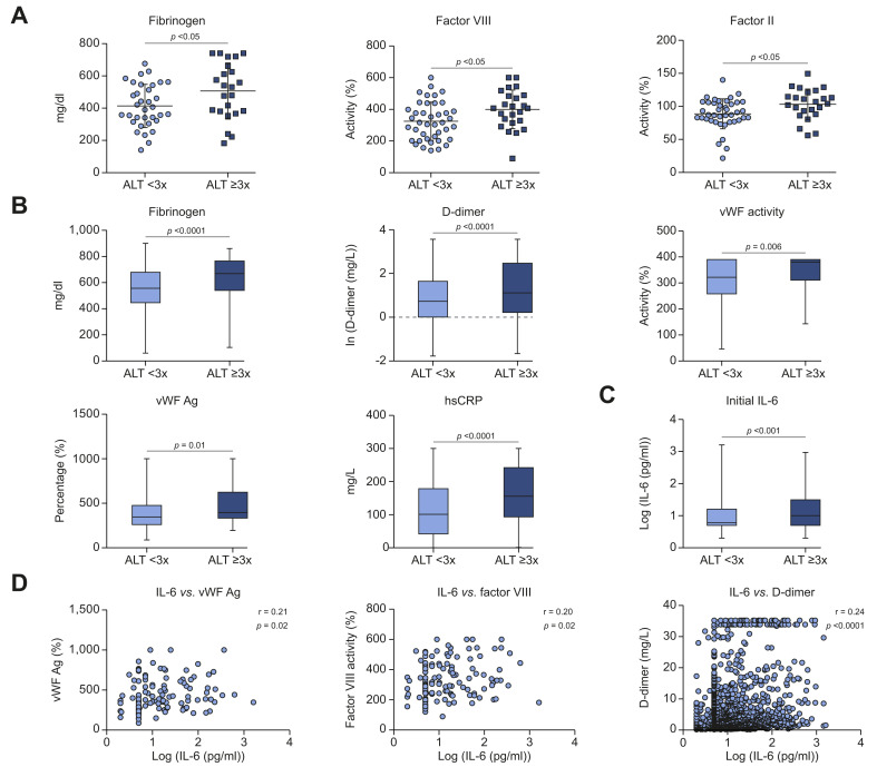
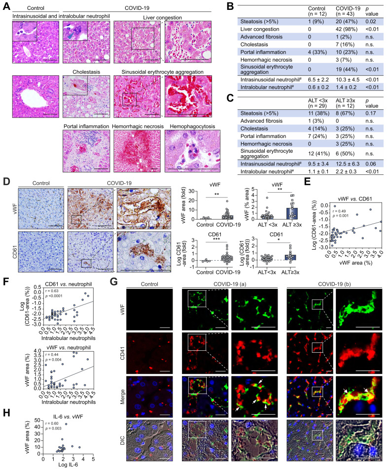
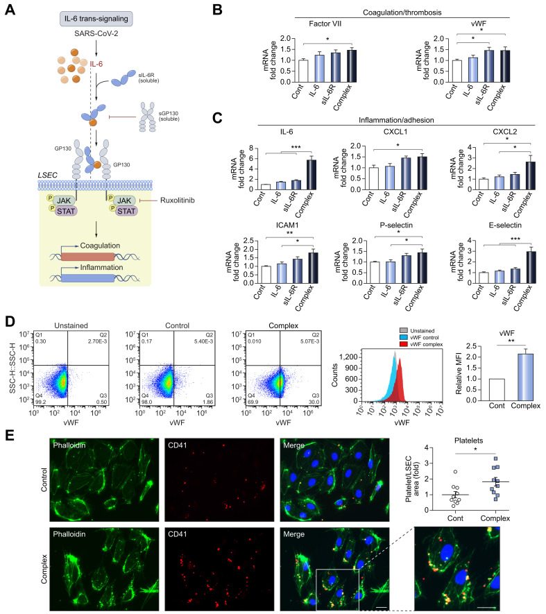
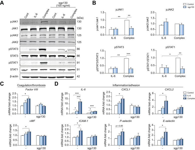
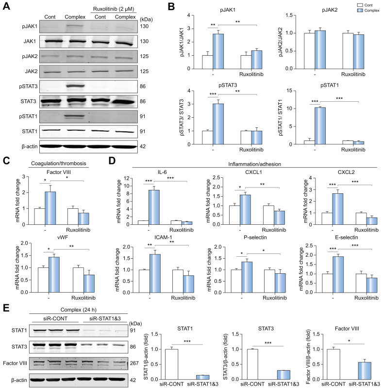
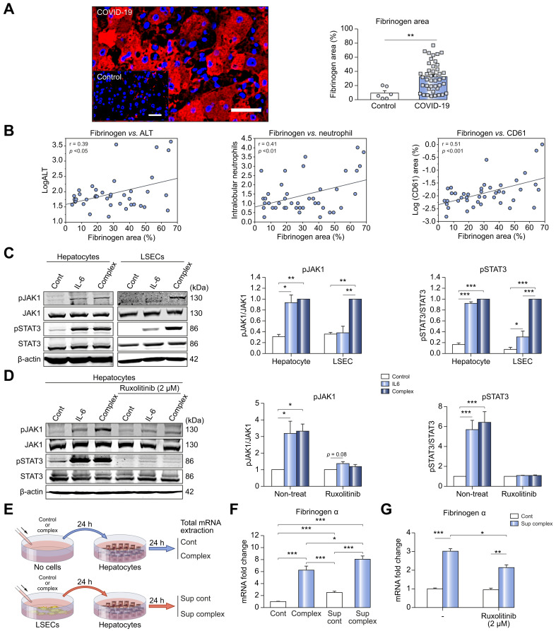
Results: Factor VIII, fibrinogen, D-dimer, von Willebrand factor (vWF) activity/antigen (biomarkers of coagulopathy/endotheliopathy) were significantly elevated in patients with COVID-19 and liver injury (elevated ALT). IL-6 positively correlated with vWF antigen (p = 0.02), factor VIII activity (p = 0.02), and D-dimer (p <0.0001). On liver histology, patients with COVID-19 and elevated ALT had significantly increased vWF and platelet staining, supporting a link between liver injury, coagulopathy, and endotheliopathy. Intralobular neutrophils positively correlated with platelet (p <0.0001) and vWF (p <0.01) staining, and IL-6 levels positively correlated with vWF staining (p <0.01). IL-6 trans-signaling leads to increased expression of procoagulant (factor VIII, vWF) and proinflammatory factors, increased cell surface vWF (p <0.01), and increased platelet attachment in LSECs. These effects were blocked by soluble glycoprotein 130 (IL-6 trans-signaling inhibitor), the JAK inhibitor ruxolitinib, and STAT1/3 small-interfering RNA knockdown. Hepatocyte fibrinogen expression was increased by the supernatant of LSECs subjected to IL-6 trans-signaling.

Conclusion: IL-6 trans-signaling drives the coagulopathy and hepatic endotheliopathy associated with COVID-19 and could be a possible mechanism behind liver injury in these patients.

Lay summary: Patients with SARS-CoV-2 (severe acute respiratory syndrome coronavirus 2) infection often have liver injury, but why this occurs remains unknown. High levels of interleukin-6 (IL-6) and its circulating receptor, which form a complex to induce inflammatory signals, have been observed in patients with COVID-19. This paper demonstrates that the IL-6 signaling complex causes harmful changes to liver sinusoidal endothelial cells and may promote blood clotting and contribute to liver injury.

Keywords: SARS-CoV-2; coagulopathy; endothelial cell dysfunction; thrombosis.


Conflict of interest statement

Conflict of interest The authors declare no conflicts of interest that pertain to this work. Please refer to the accompanying ICMJE disclosure forms for further details.

Figures

None
Graphical abstract
Fig. 1
Fig. 1
Procoagulant factors and IL-6 are significantly increased in COVID-19 patients with liver injury. (A) Serum levels of fibrinogen (n = 57), factor VIII activity (n = 65), and factor II activity (n = 65) in patients with liver injury in an initial sample of patients with COVID-19 (n = 68). (B,C) Retrospective analysis using a large database of COVID-19 inpatients from 3/10/2020 to 12/1/2020 (n = 3,780). (B) Serum levels of fibrinogen (n = 3,344), D-dimer (n = 3,478), vWF activity (n = 129), vWF Ag (n = 131), and hsCRP (n = 2,828) in patients with COVID-19. (C) Initial IL-6 levels in patients with COVID-19. ALT <3x (n = 1,110) and ≥3x (n = 481). (D) Correlation between serum IL-6 and vWF antigen (n = 119), factor VIII activity (n = 126), and D-dimer (n = 1,600). Means compared with 2-tailed Student’s t test for (A) and medians compared with 2-tailed Mann-Whitney U test for (B), (C). Correlation in (D) assessed with Pearson correlation. Patients excluded in each analysis if necessary data not available. ALT, alanine aminotransferase; hsCRP, high-sensitivity C-reactive protein; IL-6, interleukin-6; vWF, von Willebrand factor; vWF Ag, VWF antigen.
Fig. 2
Fig. 2
Expression of vWF in the liver is associated with increased platelet adhesion and liver injury in COVID-19. (A) Representative liver histological features in patients with COVID-19 and controls. Scale bars:100 μm. (B) Summary of histological features (H&E staining) of the livers from patients with COVID-19 (n = 43) and controls (n = 12). #: Neutrophils per high-power field (C) Summary of histological features associated with liver injury: ALT <3x (n = 29) and ≥3x (n = 12). (D) Immunohistochemistry for vWF and CD61 in the liver from patients with COVID-19 and controls. Scale bars:100 μm. Comparisons of vWF and CD61 positive areas in the liver from patients with COVID-19 (n = 43) and controls (n = 5) (Upper and lower left graphs), as well as those from patients with liver injury (n = 12) and those without (n = 29) (upper and lower right graphs). (E) Correlation between vWF and CD61 positive areas in the livers of patients with COVID-19 (n = 43). (F) Correlation between intralobular neutrophils and CD61-positive area (n = 43) and vWF-positive area (n = 43) in the livers of patients with COVID-19. (G) Immunofluorescence of vWF and CD41 (a platelet marker) in the livers from patients with COVID-19 and controls. Scale bars: 25 μm. In patients with COVID-19, vWF is expressed mainly on LSECs, and CD41-positive platelets attach to these LSECs (arrow head). A lower level of vWF is expressed on CD41-positive platelets (arrow). (a) and (b) are different fields from the same patient. (H) Correlation between IL-6 and vWF-positive area in COVID-19 patients. Comparisons between 2 groups were obtained using Welch’s t test for continuous variables, Chi-squared test was used for categorical variables. Pearson’s correlation co-efficients were calculated to examine the correlation among vWF area, CD61 area, and intralobular neutrophils. Spearman correlation was used to examine the correlation between serum IL-6 level and vWF area. Biochemical liver injury in 2 patients with COVID-19 could not be classified (no available ALT value). Data are mean ± SEM. ∗p <0.05, ∗∗p <0.01, ∗∗∗p <0.001. ALT, alanine aminotransferase; DIC, differential interference contrast; IL-6, interleukin-6; LSECs, liver sinusoidal endothelial cells; vWF, von Willebrand factor.
Fig. 3
Fig. 3
IL-6 trans-signaling induces endotheliopathy and increases cell surface levels of vWF and platelet attachment to LSECs. (A) Schema of IL-6 trans-signaling and blocking by soluble gp130 or ruxolitinib. (B,C) qPCR of markers for (B) procoagulant and (C) proinflammatory endotheliopathy. Human primary LSECs were incubated with control, human recombinant IL-6 (20 ng/ml), human recombinant sIL-6R (20 ng/ml) or IL-6/sIL-6R complex (20 ng/ml) for 1 hour. Graphs show the fold-change (control is set to 1). n = 6. (D) Flow cytometry for cell surface levels of vWF in LSECs treated with control or IL-6/sIL-6R complex (20 ng/ml) for 6 hours. n = 3. (E) Platelet attachment to LSECs. Phalloidin (Green, F-actin for cell structure), CD41 (Red, platelet). LSECs were treated with control or IL-6/sIL-6R complex (20 ng/ml) for 4 hours and incubated with platelets for 2 hours. Scale bar = 10 μm n = 10. Data are mean ± SEM of at least 3 experiments. One-way ANOVA with Tukey’s test for multiple groups, or 2-tailed unpaired t test for 2 groups was used. ∗p <0.05, ∗∗p <0.01, ∗∗∗p <0.001. CXCL, chemokine (C-X-C motif) ligand; ICAM1, intercellular adhesion molecule 1; IL-6, interleukin-6; LSECs, liver sinusoidal endothelial cells; qPCR, quantitative PCR; sIL-6R, soluble IL-6 receptor; vWF, von Willebrand factor.
Fig. 4
Fig. 4
Soluble gp130 blocks JAK1 and STAT3 phosphorylation induced by IL-6 trans-signaling in LSECs. Human primary LSECs were treated with control, IL-6 (20 ng/ml) or IL-6/sIL-6R complex (20 ng/ml) in the presence or absence of sgp130 Fc (100 ng/ml) for 15 minutes (western blot) and 1 hour (qPCR). (A) A representative blot. β-actin (loading control). n = 3. (B) Activation (phosphorylation) of JAK1, JAK2, STAT3 and STAT1. The fold-change for quantification of western blot. (complex without sgp130 is set to 1). n = 3. qPCR of markers for (C) procoagulant and (D) proinflammatory endotheliopathy in LSECs with or without sgp130 Fc (100 ng/ml). n = 6. Control is set to 1 for each experiment and data presented as fold-change vs. complex. Data are mean ± SEM of at least 3 experiments. One-way ANOVA with Tukey’s test for multiple groups, or 2-tailed unpaired t test for 2 groups was used. ∗p <0.05, ∗∗p <0.01, ∗∗∗p <0.001. CXCL, chemokine (C-X-C motif) ligand; gp130, glycoprotein 130; ICAM1, intercellular adhesion molecule 1; IL-6, interleukin-6; JAK, Janus kinase; LSECs, liver sinusoidal endothelial cells; qPCR, quantitative PCR; sgp130, soluble gp130; sIL-6R, soluble IL-6 receptor; STAT, signal transducer and activator of transcription.
Fig. 5
Fig. 5
JAK inhibitor ruxolitinib blocks JAK1/STAT3 induction by IL-6 trans-signaling and blocks procoagulant endotheliopathy in LSECs. Human primary LSECs were treated with control or IL-6/sIL-6R complex (20 ng/ml) in the presence or absence of ruxolitinib (2 μM) for 15 minutes (western blot) and 1 hour (qPCR). Cells were incubated with ruxolitinib for 20 minutes before treatment with complex. (A) Representative western blot. (B) Quantification of western blot. Control is set to 1 for each experiment and data presented as fold-change vs. complex. n = 3. qPCR of markers for (C) procoagulant and (D) proinflammatory endotheliopathy in LSECs with or without ruxolitinib. The graphs show the fold-change (control is set to 1). n = 6. (E) Representative western blot and its quantification. LSECs were treated with siRNA-STAT1 & 3 [STAT1 (10 nM), STAT3 (20 nM) for 64 hours] and then incubated with control or IL-6/sIL-6R complex for 24 hours. n = 3. Data are mean ± SEM of at least 3 experiments. Two-tailed unpaired t test was used. ∗p <0.05, ∗∗p <0.01, ∗∗∗p <0.001. IL-6, interleukin-6; JAK, Janus kinase; LSECs, liver sinusoidal endothelial cells; qPCR, quantitative PCR; sIL-6R, soluble IL-6 receptor; STAT, signal transducer and activator of transcription.
Fig. 6
Fig. 6
Fibrinogen levels are increased in livers of patients with COVID-19 by IL-6 signaling amplified by LSEC-hepatocyte crosstalk. (A) Representative immunolabeling of fibrinogen (Red) in livers from patients with COVID-19. Scale bar: 50 μm. Quantification of fibrinogen positive area (right panel) in patients with COVID-19 (n = 43) and controls (n = 6). (B) Correlation between fibrinogen area and ALT (n = 41), neutrophil infiltration (n = 43), and CD61 (n = 43) in livers of patients with COVID-19. (C) Western blot analyses in primary hepatocytes and primary LSECs treated with control, IL-6 alone (20 ng/ml) or IL-6/sIL-6R complex (20 ng/ml). Representative western blot (left) and its quantification (right panel). n = 3. Complex is set to 1 for each experiment and data presented as fold-change. (D) Western blot analyses in mouse primary hepatocytes treated with control, IL-6 alone (20 ng/ml) or IL-6/sIL-6R complex (20 ng/ml) in the presence or absence of ruxolitinib (2 μM) for 15 minutes. Ruxolitinib was added 20 minutes before treatment with IL-6 alone or complex. n = 3. The graphs show the fold-change (control is set to 1). (E) Experimental scheme for (F & G). (F) mRNA expression of fibrinogen. n = 9. The graphs show the fold-change (control is set to 1). (G) mRNA expression of fibrinogen in mouse primary hepatocytes treated with control medium or supernatant from LSECs treated with IL-6/sIL-6R complex for 24 hours in the presence or absence of ruxolitinib. n = 3. The graphs show the fold-change (control is set to 1). Data are the mean ± SEM of at least 3 experiments. Pearson’s correlation coefficients were calculated to examine the correlation among fibrinogen area, ALT, neutrophil infiltration and CD61 area. One-way ANOVA with Tukey’s test for multiple groups, or two-tailed unpaired t test for 2 groups was used. ∗p <0.05, ∗∗p <0.01, ∗∗∗p <0.001. ALT, alanine aminotransferase; IL-6, interleukin-6; LSECs, liver sinusoidal endothelial cells; sIL-6R, soluble IL-6 receptor.

Comment in

Similar articles

Cited by

References

    1. Woolf S.H., Chapman D.A., Lee J.H. COVID-19 as the leading cause of death in the United States. JAMA. 2021;325:123–124. - PMC - PubMed
    1. Saviano A., Wrensch F., Ghany M.G., Baumert T.F. Liver disease and COVID-19: from pathogenesis to clinical care. Hepatology. 2020 - PMC - PubMed
    1. Phipps M.M., Barraza L.H., LaSota E.D., Sobieszczyk M.E., Pereira M.R., Zheng E.X., et al. Acute liver injury in COVID-19: prevalence and association with clinical outcomes in a large U.S. Cohort. Hepatology. 2020;72:807–817. - PMC - PubMed
    1. Hundt M.A., Deng Y., Ciarleglio M.M., Nathanson M.H., Lim J.K. Abnormal liver tests in COVID-19: a retrospective observational cohort study of 1,827 patients in a major U.S. Hospital network. Hepatology. 2020;72:1169–1176. - PMC - PubMed
    1. Romana P.F., Antonio N., Fabio D.Z., Francesco S., Maurizio P., Antonio G. Correlation between liver function tests abnormalities and interleukin-6 serum levels in patients with SARS-CoV-2 infection. Gastroenterology. 2020 - PMC - PubMed

Publication types