Effects of tDCS dose and electrode montage on regional cerebral blood flow and motor behavior
- PMID: 33991697
- PMCID: PMC8653867
- DOI: 10.1016/j.neuroimage.2021.118144
Effects of tDCS dose and electrode montage on regional cerebral blood flow and motor behavior
Abstract
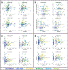
We used three dose levels (Sham, 2 mA, and 4 mA) and two different electrode montages (unihemispheric and bihemispheric) to examine DOSE and MONTAGE effects on regional cerebral blood flow (rCBF) as a surrogate marker of neural activity, and on a finger sequence task, as a surrogate behavioral measure drawing on brain regions targeted by transcranial direct current stimulation (tDCS). We placed the anodal electrode over the right motor region (C4) while the cathodal or return electrode was placed either over a left supraorbital region (unihemispheric montage) or over the left motor region (C3 in the bihemispheric montage). Performance changes in the finger sequence task for both hands (left hand: p = 0.0026, and right hand: p = 0.0002) showed a linear tDCS dose response but no montage effect. rCBF in the right hemispheric perirolandic area increased with dose under the anodal electrode (p = 0.027). In contrast, in the perirolandic ROI in the left hemisphere, rCBF showed a trend to increase with dose (p = 0.053) and a significant effect of montage (p = 0.00004). The bihemispheric montage showed additional rCBF increases in frontomesial regions in the 4mA condition but not in the 2 mA condition. Furthermore, we found strong correlations between simulated current density in the left and right perirolandic region and improvements in the finger sequence task performance (FSP) for the contralateral hand. Our data support not only a strong direct tDCS dose effect for rCBF and FSP as surrogate measures of targeted brain regions but also indirect effects on rCBF in functionally connected regions (e.g., frontomesial regions), particularly in the higher dose condition and on FSP of the ipsilateral hand (to the anodal electrode). At a higher dose and irrespective of polarity, a wider network of sensorimotor regions is positively affected by tDCS.
Keywords: Arterial spin labeling; Bihemispheric electrical stimulation; Motor learning; Neural excitability; Sensorimotor network; rCBF change; tDCS.
Copyright © 2021 The Author(s). Published by Elsevier Inc. All rights reserved.
Figures


Similar articles
-
Transcranial Direct Current Stimulation to Provide Neuroprotection and Enhance Cerebral Blood Flow in Stroke: A Comprehensive Review.Medicina (Kaunas). 2024 Dec 14;60(12):2061. doi: 10.3390/medicina60122061. Medicina (Kaunas). 2024. PMID: 39768940 Free PMC article. Review.
-
Short periods of bipolar anodal TDCS induce no instantaneous dose-dependent increase in cerebral blood flow in the targeted human motor cortex.Sci Rep. 2022 Jun 10;12(1):9580. doi: 10.1038/s41598-022-13091-7. Sci Rep. 2022. PMID: 35688875 Free PMC article.
-
Effects of transcranial direct current stimulation (tDCS) on human regional cerebral blood flow.Neuroimage. 2011 Sep 1;58(1):26-33. doi: 10.1016/j.neuroimage.2011.06.018. Epub 2011 Jun 16. Neuroimage. 2011. PMID: 21703350 Free PMC article.
-
Current intensity- and polarity-specific online and aftereffects of transcranial direct current stimulation: An fMRI study.Hum Brain Mapp. 2020 Apr 15;41(6):1644-1666. doi: 10.1002/hbm.24901. Epub 2019 Dec 20. Hum Brain Mapp. 2020. PMID: 31860160 Free PMC article.
-
A Literature Review on Optimal Stimulation Parameters of Transcranial Direct Current Stimulation for Motor Recovery After Stroke.Brain Neurorehabil. 2024 Nov 28;17(3):e24. doi: 10.12786/bn.2024.17.e24. eCollection 2024 Nov. Brain Neurorehabil. 2024. PMID: 39649716 Free PMC article. Review.
Cited by
-
Transcranial Direct Current Stimulation to Provide Neuroprotection and Enhance Cerebral Blood Flow in Stroke: A Comprehensive Review.Medicina (Kaunas). 2024 Dec 14;60(12):2061. doi: 10.3390/medicina60122061. Medicina (Kaunas). 2024. PMID: 39768940 Free PMC article. Review.
-
The Effect of Single-Session Transcranial Direct Current Stimulation on Cerebral Blood Flow: A Systematic Review and Meta-Analysis.Brain Topogr. 2025 Aug 28;38(5):60. doi: 10.1007/s10548-025-01140-z. Brain Topogr. 2025. PMID: 40875070
-
Effects of bihemispheric transcranial direct current stimulation on motor recovery in subacute stroke patients: a double-blind, randomized sham-controlled trial.J Neuroeng Rehabil. 2023 Feb 27;20(1):27. doi: 10.1186/s12984-023-01153-4. J Neuroeng Rehabil. 2023. PMID: 36849990 Free PMC article. Clinical Trial.
-
Left Prefrontal tDCS during Learning Does Not Enhance Subsequent Verbal Episodic Memory in Young Adults: Results from Two Double-Blind and Sham-Controlled Experiments.Brain Sci. 2023 Jan 31;13(2):241. doi: 10.3390/brainsci13020241. Brain Sci. 2023. PMID: 36831783 Free PMC article.
-
Short periods of bipolar anodal TDCS induce no instantaneous dose-dependent increase in cerebral blood flow in the targeted human motor cortex.Sci Rep. 2022 Jun 10;12(1):9580. doi: 10.1038/s41598-022-13091-7. Sci Rep. 2022. PMID: 35688875 Free PMC article.
References
-
- Agboada D, Mosayebi-Samani M, Kuo M-F, Nitsche MA, 2020. Induction of long-term potentiation-like plasticity in the primary motor cortex with repeated anodal transcranial direct current stimulation – Better effects with intensified protocols? Brain Stimul 13, 987–997. doi:10.1016/j.brs.2020.04.009. - DOI - PubMed
-
- Antal A, Polania R, Schmidt-Samoa C, Dechent P, Paulus W, 2011. Transcranial direct current stimulation over the primary motor cortex during fMRI. Neuroimage 55, 590–596. - PubMed
Publication types
MeSH terms
Substances
Grants and funding
LinkOut - more resources
Full Text Sources
Other Literature Sources
Miscellaneous

