

Age, Gender, and Laterality of Retinal Vascular Occlusion: A Retrospective Study from the IRIS® Registry
- PMID: 33991710
- PMCID: PMC9178780
- DOI: 10.1016/j.oret.2021.05.004
Age, Gender, and Laterality of Retinal Vascular Occlusion: A Retrospective Study from the IRIS® Registry
Abstract
Purpose: Retinal vascular occlusion is a leading cause of profound irreversible visual loss, but the understanding of the disease is insufficient. We systematically investigated the age, gender, and laterality at the onset of retinal artery occlusion (RAO) and retinal vein occlusion (RVO) in the Intelligent Research in Sight (IRIS®) Registry.
Design: Retrospective registry cohort.
Participants: Patients with retinal vascular occlusion participating in the IRIS® Registry.
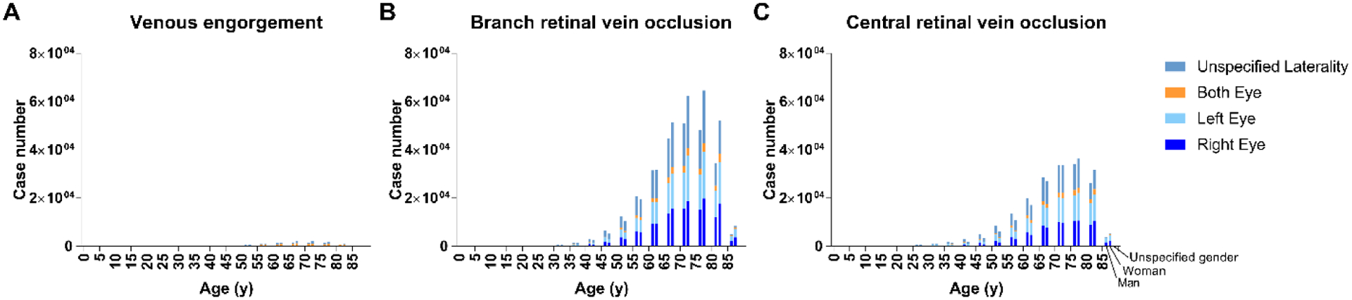
Methods: Patients who received a diagnosis of retinal vascular occlusion between 2013 and 2017 were included. Those with unspecified gender or laterality were excluded when conducting the relevant analyses. Patients were categorized into RAO, with subtypes transient retinal artery occlusion (TRAO), partial retinal artery occlusion (PRAO), branch retinal artery occlusion (BRAO), and central retinal artery occlusion (CRAO), and into RVO, with subtypes venous engorgement (VE), branch retinal vein occlusion (BRVO), and central retinal vein occlusion (CRVO). Age was evaluated as a categorical variable (5-year increments). We investigated the association of age, gender, and laterality with the onset frequency of retinal vascular occlusion subtypes.
Main outcome measures: The frequency of onset of RAO and RVO subtypes by age, gender and laterality.
Results: A total of 1 251 476 patients with retinal vascular occlusion were included, 23.8% of whom had RAO, whereas 76.2% had RVO. Of these, 1 248 656 and 798 089 patients were selected for analyses relevant to gender and laterality, respectively. The onset frequency of all subtypes increased with age. PRAO, BRAO, CRAO, and CRVO presented more frequently in men (53.5%, 51.3%, 52.6%, and 50.4%, respectively), whereas TRAO, VE, and BRVO presented more frequently in women (54.9%, 56.0%, and 54.5% respectively). All RAO subtypes and BRVO showed a right-eye onset preference (TRAO, 51.7%; PRAO, 54.4%; BRAO, 53.5%; CRAO, 53.4%; and BRVO, 51.0%), whereas VE and CRVO exhibited a left-eye onset preference (53.3% and 50.9%, respectively).
Conclusions: Although retinal vascular occlusion incidence increases with age regardless of subtypes, we found various subtype-specific disease-onset differences related to gender and, in particular, ocular laterality. These findings may improve understanding of the specific cause of retinal vascular occlusions of different subtypes and their relationships with structural and anatomic asymmetries of the vascular system.
Keywords: Big data; Demographics; Epidemiology; Retinal artery occlusion; Retinal vein occlusion.
Copyright © 2021 American Academy of Ophthalmology. Published by Elsevier Inc. All rights reserved.
Figures


References
-
- Atchison EA, Wood KM, Mattox CG et al. The Real-World Effect of Intravitreous Anti–Vascular Endothelial Growth Factor Drugs on Intraocular Pressure. Ophthalmology. 2018;125(5):676–682 - PubMed
-
- Chiang MF, Sommer A, Rich WL, Lum F, Parke DW. The 2016 American Academy of Ophthalmology IRIS® Registry (Intelligent Research in Sight) Database. Ophthalmology. 2018;125(8):1143–1148 - PubMed
Publication types
MeSH terms
Grants and funding
LinkOut - more resources
Full Text Sources
Other Literature Sources
