. 2021 Aug:342:113765.
doi: 10.1016/j.expneurol.2021.113765. Epub 2021 May 13.

Mild traumatic brain injury increases vulnerability to cerebral ischemia in mice

Affiliations

Mild traumatic brain injury increases vulnerability to cerebral ischemia in mice

Zachary M Weil et al. Exp Neurol. 2021 Aug.

Abstract

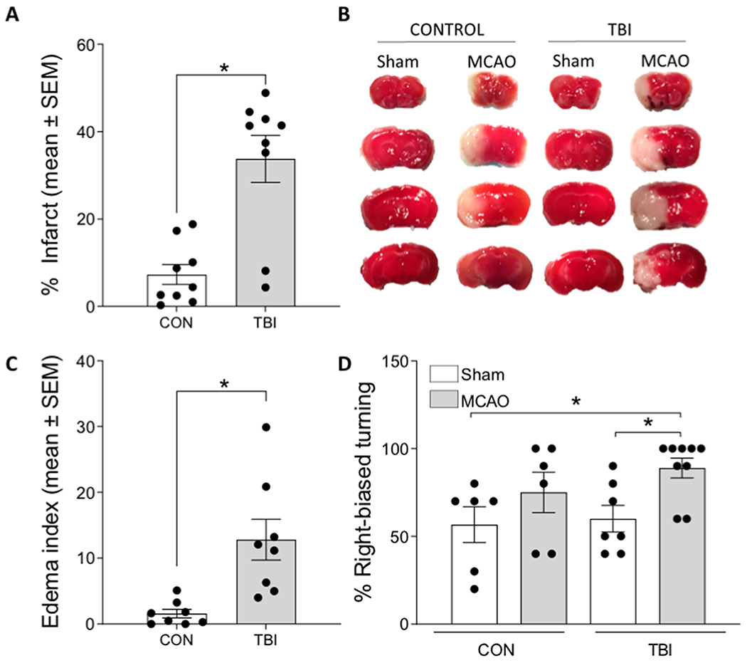
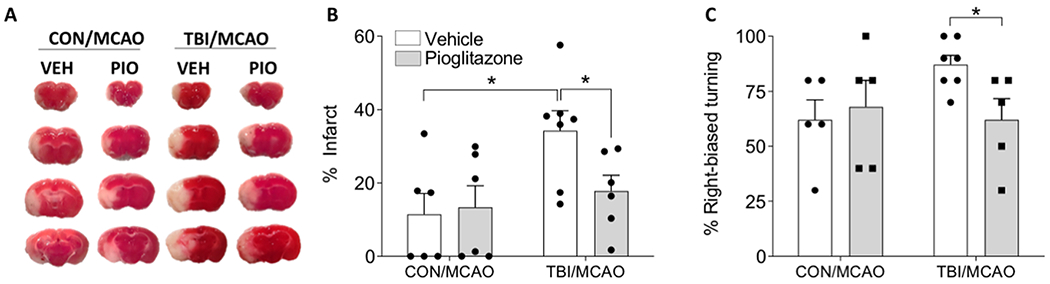
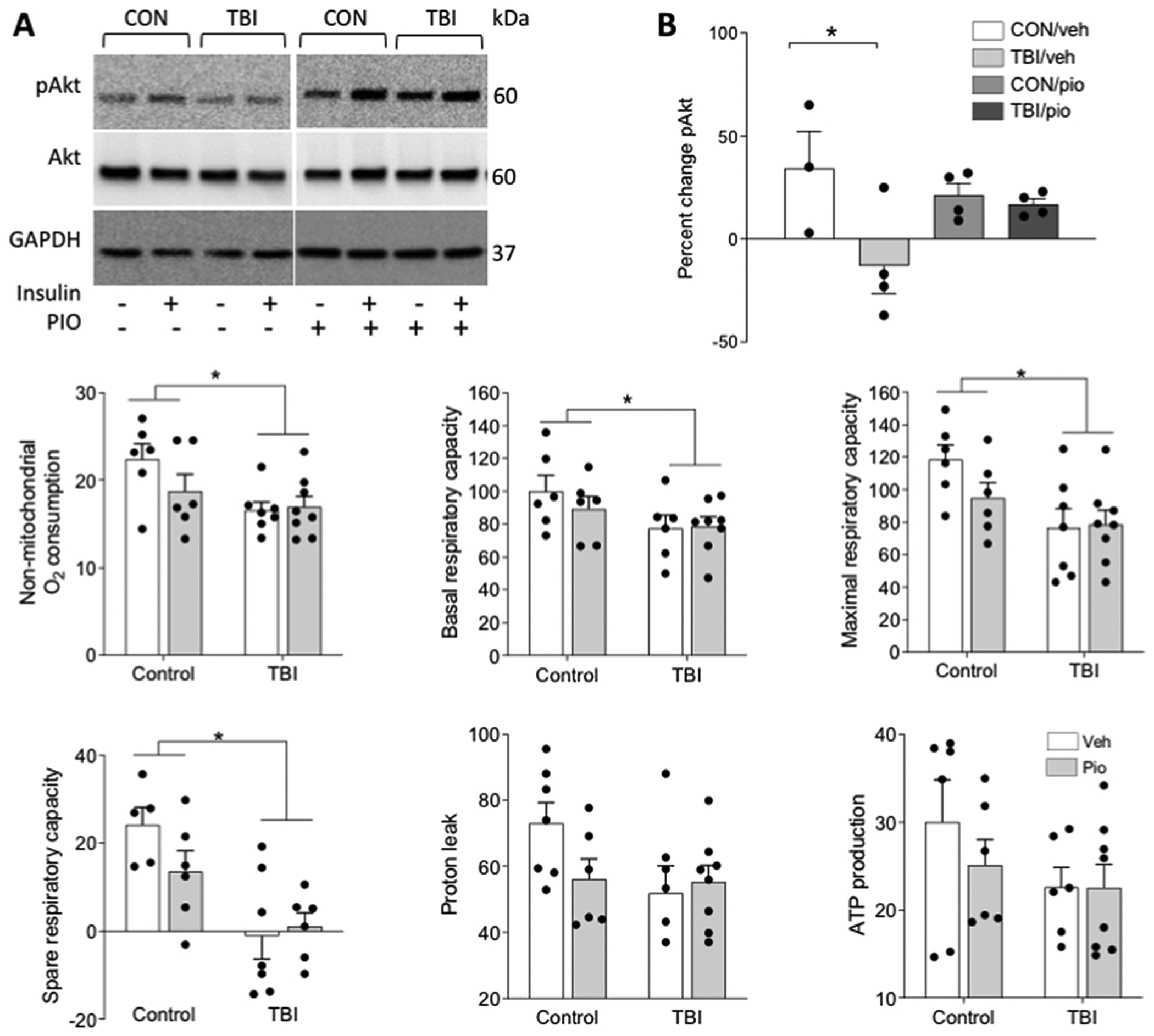
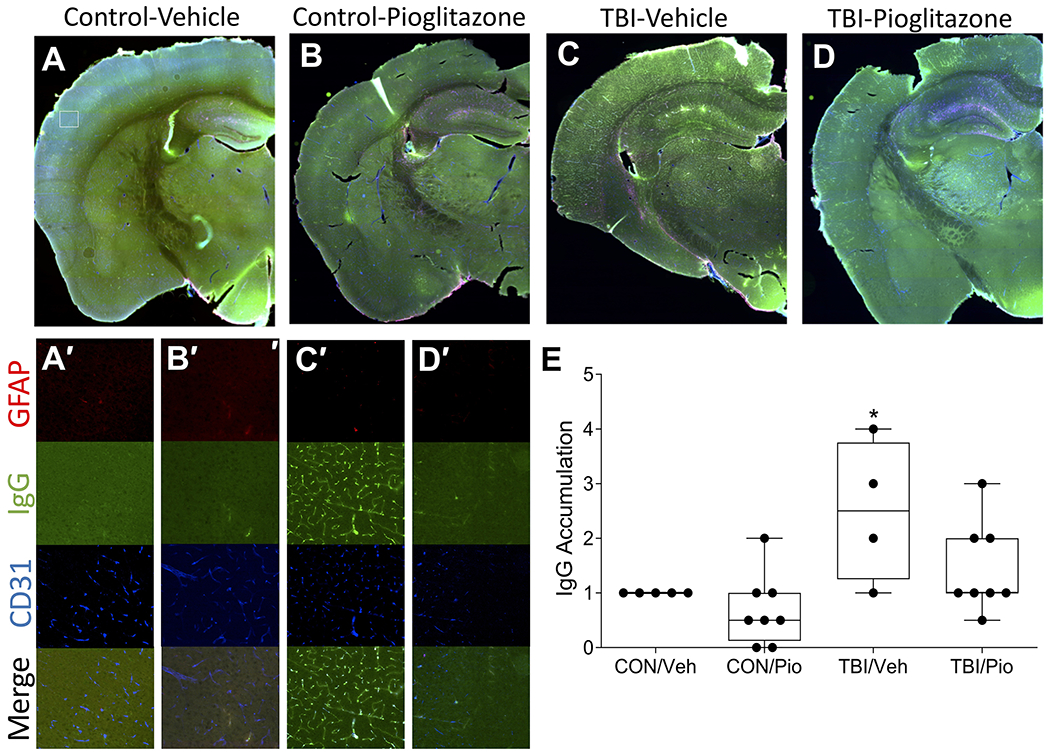
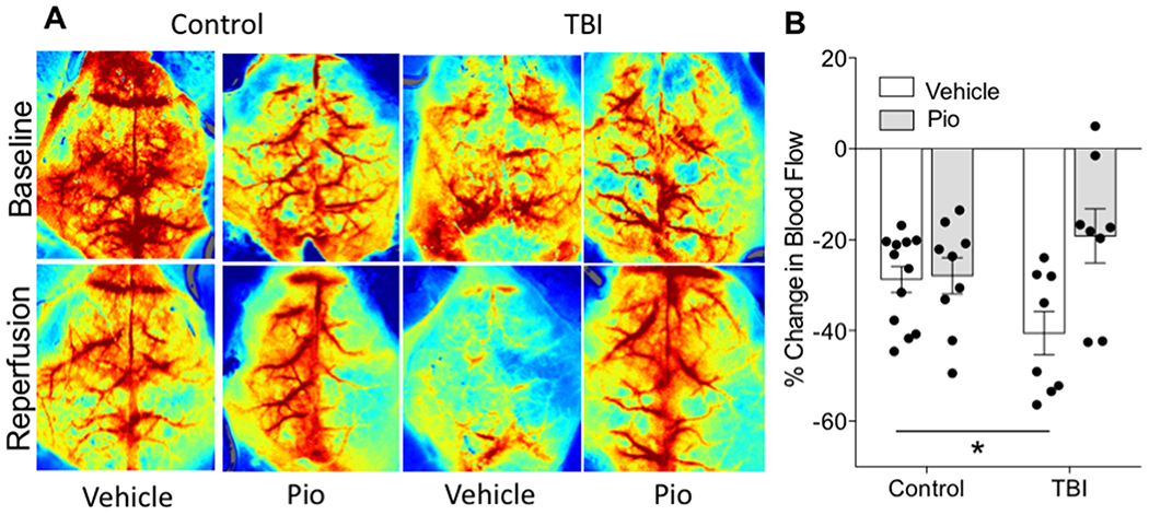
Recent studies have reported that TBI is an independent risk factor for subsequent stroke. Here, we tested the hypothesis that TBI would exacerbate experimental stroke outcomes via alternations in neuroimmune and neurometabolic function. We performed a mild closed-head TBI and then one week later induced an experimental stroke in adult male mice. Mice that had previously experienced TBI exhibited larger infarcts, greater functional deficits, and more pronounced neuroinflammatory responses to stroke. We hypothesized that impairments in central metabolic physiology mediated poorer outcomes after TBI. To test this, we treated mice with the insulin sensitizing drug pioglitazone (Pio) after TBI. Pio prevented the exacerbation of ischemic outcomes induced by TBI and also blocked the induction of insulin insensitivity by TBI. However, tissue respiratory function was not improved by Pio. Finally, TBI altered microvascular responses including promoting vascular accumulation of serum proteins and significantly impairing blood flow during the reperfusion period after stroke, both of which were reversed by treatment with Pio. Thus, TBI appears to exacerbate ischemic outcomes by impairing metabolic and microvascular physiology. These data have important implications because TBI patients experience strokes at greater rates than individuals without a history of head injury, but these data suggest that those strokes may also cause greater tissue damage and functional impairments in that population.

Keywords: Inflammation; Metabolism; Reperfusion; Stroke; Traumatic brain injury; Vasculature.


Conflict of interest statement

Declarations of interest

None

Figures

Figure 1.
Figure 1.. TBI prior to cerebral ischemia exacerbates infarct volume, edema, and functional deficits.
A) Mice that underwent a TBI 7 days prior to MCAO exhibited a significantly greater infarct volume, compared to those that had a control (CON) procedure prior to MCAO. B) Representative TTC stained tissue throughout the forebrain indicating healthy (red) and infarcted (white) tissue in the cortex and striatum. C) Mice that underwent a TBI prior to MCAO also exhibited significantly greater edema. D) Right-biased turning in the corner test revealed significant deficits in mice that underwent both a TBI and MCAO. Data are presented as mean +/− SEM. An asterisk (*) indicates significance at p < 0.05.
Figure 2.
Figure 2.. Axon degeneration and pro-inflammatory gene expression following TBI and cerebral ischemia.
A) Mice that underwent a TBI 7 days prior to MCAO exhibited more white matter degeneration than those that had either TBI or MCAO alone. Proinflammatory cytokine gene expression is shown relative to 18S for CD68, TNF, and IL1β in the ipsilateral cortex B) and striatum C). All data are considered significant at p < 0.05. An asterisk (*) indicates significance at p < 0.05.
Figure 3.
Figure 3.. Pioglitazone reduces brain vulnerability to ischemia after TBI.
A) Representative TTC staining after MCAO from mice that first underwent control or TBI and were treated with pioglitazone (Pio; 10 mg/kg) or vehicle (Veh). B) Mice that were treated with Pio after TBI has infarct volumes similar to mice that did not have a TBI prior to MCAO. C) Treatment with PIO prevented the TBI exacerbation of right-biased turning in the corner test. An asterisk (*) indicates significance at p < 0.05.
Figure 4.
Figure 4.. Treatment with pioglitazone improves central insulin signaling but not respiratory parameters after TBI.
Brain tissue from control (CON) and TBI mice was split into two wells and incubated ex vivo with aCSF alone or containing 10nm insulin. A) Representative western blot bands are shown for phosphorylated Akt (Ser473), total Akt, and GAPDH. B) Data are shown as percent change pAkt expression in response to insulin. Separate animals underwent TBI or control procedure and were treated with Pio or vehicle, and seven days later one mm tissue punches were collected from 250-micron sections through the forebrain and analyzed with Seahorse respirometry. TBI reduced C) non-mitochondrial O2 production, D) basal respiratory capacity, D) maximal respiratory capacity, and E) spare respiratory capacity. There were no TBI effects on either G) proton leak or H) ATP production. There were no effects of Pio on any parameter. Data are presented as mean +/− SEM. An asterisk (*) indicates significance at p < 0.05.
Figure 5.
Figure 5.. TBI induces vascular pathology that is attenuated by pioglitazone.
A-D) Representative sections indicate TBI induces accumulation of intravascular IgG (green) as well as mild extravasation that colocalizes with CD31 positive endothelial cells (blue) this is attenuated by pioglitazone. GFAP positive astrocytes (red) are also in close contact with IgG positive vessels. A’-D’) Corresponding insets of cortical staining. Size of insets indicated by white box in A). E) Among vehicle-treated mice TBI significantly increased IgG accumulation relative to CON, but Pio treatment reduced IgG accumulation to CON levels. An asterisk (*) indicates significance at p < 0.05.
Figure 6.
Figure 6.. TBI impairs reperfusion blood flow, reversal by pioglitazone.
Baseline and reperfusion (60 minutes after removal of the occluder) blood flow measurements assessed with laser speckle imaging. Warmer colors indicate greater blood flow. B) Assessment of relative change in blood flow from baseline to reperfusion in the ipsilateral hemisphere. Data are presented as mean +/− SEM. An asterisk (*) indicates significance at p < 0.05.

References

    1. Abcouwer SF, Shanmugam S, Gomez PF, Shushanov S, Barber AJ, Lanoue KF, Quinn PG, Kester M, Gardner TW, 2008. Effect of IL-1beta on survival and energy metabolism of R28 and RGC-5 retinal neurons. Invest Ophthalmol Vis Sci 49, 5581–5592. - PubMed
    1. Abdul-Muneer PM, Chandra N, Haorah J, 2015. Interactions of oxidative stress and neurovascular inflammation in the pathogenesis of traumatic brain injury. Mol Neurobiol 51, 966–979. - PMC - PubMed
    1. Albrecht JS, Liu X, Baumgarten M, Langenberg P, Rattinger GB, Smith GS, Gambert SR, Gottlieb SS, Zuckerman IH, 2014. Benefits and risks of anticoagulation resumption following traumatic brain injury. JAMA Intern Med 174, 1244–1251. - PMC - PubMed
    1. Albrecht JS, Liu X, Smith GS, Baumgarten M, Rattinger GB, Gambert SR, Langenberg P, Zuckerman IH, 2015. Stroke incidence following traumatic brain injury in older adults. J Head Trauma Rehabil 30, E62–67. - PMC - PubMed
    1. Arbel-Ornath M, Hudry E, Eikermann-Haerter K, Hou S, Gregory JL, Zhao L, Betensky RA, Frosch MP, Greenberg SM, Bacskai BJ, 2013. Interstitial fluid drainage is impaired in ischemic stroke and Alzheimer’s disease mouse models. Acta Neuropathol 126, 353–364. - PMC - PubMed

Publication types