. 2021 Jan-Jun:296:100777.
doi: 10.1016/j.jbc.2021.100777. Epub 2021 May 14.

Reduced virulence of the MARTX toxin increases the persistence of outbreak-associated Vibrio vulnificus in host reservoirs

Affiliations

Reduced virulence of the MARTX toxin increases the persistence of outbreak-associated Vibrio vulnificus in host reservoirs

Sanghyeon Choi et al. J Biol Chem. 2021 Jan-Jun.

Abstract

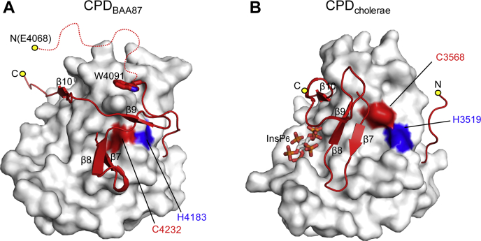
Opportunistic bacteria strategically dampen their virulence to allow them to survive and propagate in hosts. However, the molecular mechanisms underlying virulence control are not clearly understood. Here, we found that the opportunistic pathogen Vibrio vulnificus biotype 3, which caused an outbreak of severe wound and intestinal infections associated with farmed tilapia, secretes significantly less virulent multifunctional autoprocessing repeats-in-toxin (MARTX) toxin, which is the most critical virulence factor in other clinical Vibrio strains. The biotype 3 MARTX toxin contains a cysteine protease domain (CPD) evolutionarily retaining a unique autocleavage site and a distinct β-flap region. CPD autoproteolytic activity is attenuated following its autocleavage because of the β-flap region. This β-flap blocks the active site, disabling further autoproteolytic processing and release of the modularly structured effector domains within the toxin. Expression of this altered CPD consequently results in attenuated release of effectors by the toxin and significantly reduces the virulence of V. vulnificus biotype 3 in cells and in mice. Bioinformatic analysis revealed that this virulence mechanism is shared in all biotype 3 strains. Thus, these data provide new insights into the mechanisms by which opportunistic bacteria persist in an environmental reservoir, prolonging the potential to cause outbreaks.

Keywords: MARTX toxin; Vibrio vulnificus; cysteine protease domain; effector; infectious disease; virulence factor.


Conflict of interest statement

Conflicts of interest The authors declare that they have no conflicts of interest with the contents of this article.

Figures

Figure 1
Figure 1
The CPD of the V. vulnificus biotype 3 MARTX toxin is autocleaved atypically.A, multiple sequence alignment of CPDs of MARTX toxins from various Vibrio species. Completely conserved residues are highlighted in red, sequences for the β-flap region are indicated in dark yellow, X1-L-X2 motifs (autocleavage sites) are displayed in green, and autocleavage residues (VLE) of the V. vulnificus BAA87 CPD are indicated in blue. Within the β-flap region, secondary structure elements of the V. cholerae CPD (PDB ID 3EEB) are indicated. Multiple sequence alignment was carried out using MultiAlin, and the figure was generated with ESPript. The following Vibrio strains are shown: V. vulnificus BAA87 (WP_039507922.1), V. vulnificus MO6-24/O (WP_015728045.1), V. vulnificus FORC_009 (WP_060534095.1), V. cholerae N16961 (AAD21057.1), and V. anguillarum (RMZ64238.1). B, autocleavage site analysis of CPDBAA87. The N-terminal sequence of autocleaved CPDBAA87 is EVSGQ, as determined by Edman sequencing. The first amino acid (E) of the autocleaved CPD is highlighted in red. C and D, in vitro autocleavage assay. CPDBAA87, its mutants CPDAWT/ALA and CPDVLE/ALA, and CPDMO6-24/O were incubated with equivalent molar concentrations of InsP6, resolved by SDS-PAGE, and visualized by Coomassie staining. Autocleaved CPDs are indicated by arrows. The intensity of the cleaved protein on SDS-PAGE was quantified using NIH ImageJ and plotted. The plots are from three independent experiments. CPD, cysteine protease domain; MARTX, multifunctional autoprocessing repeats-in-toxin.
Figure 2
Figure 2
The structure of CPDBAA87suggests nonfunctionality for processing of effector domains.A, crystal structure of CPDBAA87, consisting of the core catalytic domain and β-flap. The helices, β-strands, and loop regions are shown in red, yellow, and green, respectively. B, superimposition of the catalytic domain of CPDBAA87 onto the corresponding domain of CPDcholerae (white). Residues corresponding to different secondary structures of CPDBAA87 are shown in color, and residues corresponding to CPDcholerae are shown in black. The catalytic residues of CPDBAA87 (H4183 and C4232) and CPDcholerae (H3519 and C3568) are indicated. C, superimposition of the β-flap of CPDBAA87 onto the corresponding region of CPDcholerae (white). Active residues are indicated for each CPD. D, the electrostatic surface potential of CPDcholerae shows a positively charged pocket for InsP6 binding (upper left panel). The residues involved in formation of the pocket are indicated. Residues involved in InsP6 binding are shown as a stick model within CPDcholerae displayed as a ribbon model (upper right panel). The β-flapcholerae residues critically involved in formation of the InsP6-binding pocket are indicated in red (upper panels). The electrostatic surface potential of CPDBAA87 reveals a highly basic pocket formed by the corresponding residues of CPDcholerae (lower left panel). The corresponding residues involved in the InsP6-binding pocket of CPDBAA87 are displayed, including the residues corresponding to the critical β-flapcholerae residues for InsP6 binding, which are shown in red (lower right panel). E, crater-like hydrophobic surface formed by structural movement of the β-hairpin in the β-flap region via interacting forces. Hydrogen bonds between residues are shown as dashed lines. CPD, cysteine protease domain; MARTX, multifunctional autoprocessing repeats-in-toxin.
Figure 3
Figure 3
Autocleaved CPDBAA87becomes nonfunctional for processing of MARTX toxin effectors.A, the steric configuration of the β-flap (ribbon model in red) blocking the active site of the autocleaved CPDBAA87. Catalytic residues are shown in red (Cys4232) and blue (His4183) on a surface model of the catalytic domain (in white). The N-terminal flexible region (Glu4068–Ala4090) is shown as a red dashed line, and Trp4091 is shown with a red stick model. B, the steric configuration of the β-flap, showing autocleaved CPDcholerae, which is functional for processing of MARTX toxin effectors. Catalytic residues are shown in red (Cys3568) and blue (His3519) on the surface model of the catalytic domain (in white). The two structures are displayed in the same orientation. CPD, cysteine protease domain; MARTX, multifunctional autoprocessing repeats-in-toxin.
Figure 4
Figure 4
Evolutionary insights into the disabled effector domain processing of CPDBAA87.A and B, in vitro autocleavage assay for CPD proteins. CPDs were incubated with InsP6 for the indicated durations. The intensities of the bands representing the cleaved protein on SDS-PAGE were quantified with NIH ImageJ and plotted. The plots are from three independent experiments. Autocleaved CPDs are indicated by arrows. C, in vitro MARTX effector domain processing assay. The purified effector region of MARTXBAA87 toxin was co-incubated with the indicated CPDs in the presence of 1 mM InsP6 for 1 h at 37 °C. Effector products processed by each CPD were analyzed by SDS-PAGE (left panel). The bar graph shows the proportion of the uncleaved effector region relative to the effector region in the absence of CPD (right panel). The intensity of each band representing the uncleaved effector region was quantified with NIH ImageJ, and the mean of individual values were statistically analyzed by a one-way ANOVA with multiple comparisons (∗∗p < 0.005; ∗∗∗∗p < 0.0001; n.s., not significant). CPD, cysteine protease domain; MARTX, multifunctional autoprocessing repeats-in-toxin.
Figure 5
Figure 5
The reduced virulence of the MARTXBAA87toxin is mediated by CPDBAA87.A, schematic representation of the V. vulnificus strains used for cell rounding assays and mouse survival experiments. Parental, V. vulnificus MO6-24/O strain; MO6-24/O/MARTXBAA87, V. vulnificus MO6-24/O expressing MARTXBAA87 (i.e., in which the MCF and CPD of MARTXMO6-24/O are substituted with the ExoY, DmX, and CPD of MARTXBAA87); MO6-24/O/MARTXBAA87/CPD/β-flapMO6-24/O, an engineered strain in which the β-flap of CPDBAA87 is substituted with that of MO6-24/O; MO6-24/O/MARTXBAA87/CPDMO6-24/O-mimic, an engineered strain in which CPDBAA87 in MO6-24/O/MARTXBAA87 is substituted with CPDMO6-24/O. Mutated residues and the substituted regions in CPDMO6-24/O-mimic are indicated. B, representative images of HeLa cells treated with PBS as a control or the indicated V. vulnificus strains at an multiplicity of infection of 0.1 for 120 min. C, quantification of rounded HeLa cells from images obtained after 60, 90, and 120 min of the treatments described in B. The proportion of rounded HeLa cells relative to the total number of HeLa cells was calculated. Data are shown as the mean ± standard deviation (SD) from three independent images (∗∗∗∗p < 0.0001). D, survival of mice challenged by subcutaneous injection of the indicated V. vulnificus strains (n = 10; pooled data from two independent experiments with five mice per group). Data from the MO6-24/O/MARTXBAA87 strain and the MO6-24/O/MARTXBAA87/CPDMO6-24/O-mimic strain were compared by the log-rank test. CPD, cysteine protease domain; MARTX, multifunctional autoprocessing repeats-in-toxin.
Figure 6
Figure 6
The functionality of CPDs correlates with the evolutionary classification of V. vulnificus biotypes.A, proposed model for MARTXBAA87 processing. Upon entry into host cells, the CPD of MARTXBAA87 is allosterically activated by binding to InsP6 and autocleaved (left panel). DmX released from the autocleaved CPD is then allosterically activated via the interaction with ARF and autocleaved (middle panel). The released DmX complexed with ARF disrupts the Golgi structure, and DmX may induce cell shrinking via modification of an unknown target(s) (16, 28). B, phylogenetic tree of CPDs from different V. vulnificus strains. Amino acid sequence analysis revealed that CPDs can be classified into three groups based on their sequences and predicted functional discrepancies, shown as arcs outside of the tree. The biotypes of V. vulnificus strains are indicated with different symbols: biotype 1, red circle; biotype 2, blue circle; biotype 3, green circle; unidentified, magenta diamond. ARF, ADP-ribosylation factor; CPD, cysteine protease domain; DmX, domain X effector; DUF1, domain of unknown function; ExoY, ExoY-like adenylate cyclase domain; MARTX, multifunctional autoprocessing repeats-in-toxin; RID, Rho GTPase-inactivation domain.

References

    1. Agarwal S., Agarwal S., Biancucci M., Satchell K.J. Induced autoprocessing of the cytopathic makes caterpillars floppy-like effector domain of the Vibrio vulnificus MARTX toxin. Cell. Microbiol. 2015;17:1494–1509. - PMC - PubMed
    1. Mikonranta L., Mappes J., Laakso J., Ketola T. Within-host evolution decreases virulence in an opportunistic bacterial pathogen. BMC Evol. Biol. 2015;15:165. - PMC - PubMed
    1. Hernandez-Cabanyero C., Amaro C. Phylogeny and life cycle of the zoonotic pathogen Vibrio vulnificus. Environ. Microbiol. 2020;22:4133–4148. - PubMed
    1. Baker-Austin C., Oliver J.D. Vibrio vulnificus: New insights into a deadly opportunistic pathogen. Environ. Microbiol. 2018;20:423–430. - PubMed
    1. Froelich B.A., Noble R.T. Factors affecting the uptake and retention of Vibrio vulnificus in oysters. Appl. Environ. Microbiol. 2014;80:7454–7459. - PMC - PubMed

Publication types

LinkOut - more resources