. 2021 May:67:103381.
doi: 10.1016/j.ebiom.2021.103381. Epub 2021 May 14.

Q493K and Q498H substitutions in Spike promote adaptation of SARS-CoV-2 in mice

Affiliations

Q493K and Q498H substitutions in Spike promote adaptation of SARS-CoV-2 in mice

Kun Huang et al. EBioMedicine. 2021 May.

Abstract

Background: An ideal animal model to study SARS-coronavirus 2 (SARS-CoV-2) pathogenesis and evaluate therapies and vaccines should reproduce SARS-CoV-2 infection and recapitulate lung disease like those seen in humans. The angiotensin-converting enzyme 2 (ACE2) is a functional receptor for SARS-CoV-2, but mice are resistant to the infection because their ACE2 is incompatible with the receptor-binding domain (RBD) of the SARS-CoV-2 spike protein .

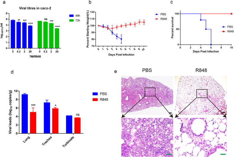
Methods: SARS-CoV-2 was passaged in BALB/c mice to obtain mouse-adapted virus strain. Complete genome deep sequencing of different generations of viruses was performed to characterize the dynamics of the adaptive mutations in SARS-CoV-2. Indirect immunofluorescence analysis and Biolayer interferometry experiments determined the binding affinity of mouse-adapted SARS-CoV-2 WBP-1 RBD to mouse ACE2 and human ACE2. Finally, we tested whether TLR7/8 agonist Resiquimod (R848) could also inhibit the replication of WBP-1 in the mouse model.

Findings: The mouse-adapted strain WBP-1 showed increased infectivity in BALB/c mice and led to severe interstitial pneumonia. We characterized the dynamics of the adaptive mutations in SARS-CoV-2 and demonstrated that Q493K and Q498H in RBD significantly increased its binding affinity towards mouse ACE2. Additionally, the study tentatively found that the TLR7/8 agonist Resiquimod was able to protect mice against WBP-1 challenge. Therefore, this mouse-adapted strain is a useful tool to investigate COVID-19 and develop new therapies.

Interpretation: We found for the first time that the Q493K and Q498H mutations in the RBD of WBP-1 enhanced its interactive affinities with mACE2. The mouse-adapted SARS-CoV-2 provides a valuable tool for the evaluation of novel antiviral and vaccine strategies. This study also tentatively verified the antiviral activity of TLR7/8 agonist Resiquimod against SARS-CoV-2 in vitro and in vivo.

Funding: This research was funded by the National Key Research and Development Program of China (2020YFC0845600) and Emergency Science and Technology Project of Hubei Province (2020FCA046) and Robert A. Welch Foundation (C-1565).

Keywords: Adaptive mutations; Angiotensin-converting enzyme 2; Mouse-adapted strain; SARS-CoV-2; TLR7/8 agonist.


Conflict of interest statement

Declaration of Competing Interest The authors declare no competing interests.

Figures

Fig 1
Fig. 1
Generation of mouse-adapted SARS-CoV-2 WBP-1. Wild type (WT) Wuhan-Hu-1 was passaged in old and young mice to obtain WBP-1 that was adapted to cause respiratory disease in mice. (a) Hematoxylin and eosin (H&E) staining of lung sections from mice infected with virus obtained through different passages (P2, P5, P8, P11) of SARS-CoV-2. Scale bars, 100 μm. (b) Viral copies were detected through qRT-PCR at three days post infection (dpi) in the lungs. (c) Five clonal isolates were obtained from the P11 virus pool by three rounds of plaque purification in Vero cells. The strain #2 that showed a high mortality rate in mice was defined as WBP-1. (d) Location of the mutations and deletion in the genome of WBP-1. WBP-1 obtained five synonymous (purple triangles), five nonsynonymous (red triangles), and a 740 bp deletion (black line). (e) Mutational frequency during SARS-CoV-2 experimental passage in pooled mouse groups. Single nucleotide variants are represented along the genome for the mice of passage 5, passage 8, passage 11 and WBP-1 virus.
Fig 2
Fig. 2
Characterization of mouse-adapted SARS-CoV-2 WBP-1 in mice. (a, b) Groups of mice (n = 5) were intranasally infected with 10-fold serial dilutions of WBP-1 virus. The infected mice were observed for weight loss and survival for 10 dpi. (c- f) Mice (n = 6) were intranasally infected with 2 LD50 of the WBP-1 virus and mice in the control group were mock-infected with DMEM. (c) Viral RNA copies were determined through qRT-PCR at 3 and 5 dpi in the heart, liver, lung, kidney, brain, intestine, trachea, turbinate, and spleen of WBP-1. (d) Viral titers in the lung were determined through plaque assays. (e) Cytokine (IL1β, TNFα, MCP1, and IL10) mRNA levels in the lung were evaluated at 3 and 5 dpi. Statistical significance was analyzed by unpaired Student's t tests. *p < 0.05; **p < 0.01; ***p < 0.001 ****p < 0.0001. (f) Hematoxylin and eosin (H&E) staining of lung sections from mice infected Mouse-adapted WBP-1 virus (Exfoliated necrotic cells, black arrow; Mononuclear lymphocytes, green arrow; Inflammatory exudate, red arrow; Vascular thrombosis, blue arrow). Scale bars (black), 100 μm. Scale bars (green), 50 μm. (g) Immunohistochemical detection of viral antigen using anti-Spike protein Rabbit monoclonal antibody in bronchi and alveoli. Scale bars (black), 100 μm. Scale bars (green), 50 μm.
Fig 3
Fig. 3
WBP-1 virus using mouse ACE2 to gain access to cells was determined by immunofluorescence. Hela cells with transfection of empty vector (Flag), hACE2, or mACE2 plasmids were infected with WT Wuhan-Hu-1 or mouse-adapted WBP-1. ACE2 expression was detcted using rabbit anti-ACE2 IgG followed by CoraLite594-conjugated goat anti-rabbit IgG (H+L). SARS-CoV-2 nucleocapsid expression was detected using mouse anti-NP IgG followed by CoraLite488-conjugated Affinipure goat anti-mouse IgG(H+L). Nuclei were stained with DAPI. Scale bars, 200 μm.
Fig 4
Fig. 4
Mouse-adapted WBP-1 virus RBD enhance interactive affinities with mACE2. (a-d) Modelling of SARS-CoV-2 RBD–ACE2 interface. (a) Wuhan-Hu-1 RBD interacts with of human ACE2. (b) WBP-1 RBD (Q493K/Q498H-RBD) interacts human ACE2. (c) Modelling of Wuhan-Hu-1 RBD and mouse ACE2. (d) Modelling of WBP-1 RBD (Q493K/Q498H-RBD) shows interaction with mouse ACE2. (e-l) The binding affinities of the RBD of WBP-1 and mACE2 or hACE2 were determined through BLI experiments. The sensors were dipped in mACE2-hFC and functionalized sensorgrams captured upon incubation of Q493K-RBD (e), Q498H-RBD (f), Q493K/Q498H-RBD (g), and WT-RBD (h) at 6.25 (black), 12.5 (blue), 25 (green), and 50 nM (red). The sensors were dipped in hACE2-hFC and functionalized sensorgrams captured upon incubation of Q493K-RBD (i), Q498H-RBD (j), Q493K/Q498H-RBD (k), and WT-RBD (l) at 50 (black), 100 (blue), 200 (green), and 400 nM (red).
Fig 5
Fig. 5
R848 inhibits SARS-CoV-2 in vitro and in vivo. (a) Caco2 cells were treated with R848 (0-20 μM) for 24 h before infection with viruses at a multiplicity of infection (MOI) of 1. Infected cells were further incubated in the R848. Samples were collected at 48 and 72 h post infection (hpi) and viral titers were determined using plaque assays. (b-e) (n = 10) Mice were intraperitoneally injected with R848 after immediately intranasal infection with the WBP-1 virus. Infected mice were further treated with the R848 or PBS once a day for four consecutive days. Mice were monitored for weight loss (b) and survival (c) for 10 days. (d) Viral loads in mice lung, trachea and turbinate were determined at 5 dpi. (e) Hematoxylin and eosin (H&E) staining of lung sections from PBS or R848 treated mice. Scale bars (black), 100 μm. Scale bars (green), 50 μm. (a) and (d), Log transformed data analyzed by Two-way ANOVA followed by Sidak's multiple comparisons. The line represents the mean and error bars represent standard deviation. *p < 0.05; **p < 0.01; ***p < 0.001 ****p < 0.0001.

Comment in

Similar articles

Cited by

References

    1. Zhu N., Zhang D., Wang W., Li X., Yang B., Song J. A novel coronavirus from patients with pneumonia in China, 2019. N Engl J Med. 2020;382(8):727–733. - PMC - PubMed
    1. Guan W.J., Ni Z.Y., Hu Y., Liang W.H., Ou C.Q., He J.X. Clinical characteristics of coronavirus disease 2019 in China. N Engl J Med. 2020;382(18):1708–1720. - PMC - PubMed
    1. Kim Y.I., Kim S.G., Kim S.M., Kim E.H., Park S.J., Yu K.M. Infection and rapid transmission of SARS-CoV-2 in Ferrets. Cell Host Microbe. 2020;27(5) 704-9 e2. - PMC - PubMed
    1. Sun S.H., Chen Q., Gu H.J., Yang G., Wang Y.X., Huang X.Y. A mouse model of SARS-CoV-2 infection and pathogenesis. Cell Host Microbe. 2020;28(1) 124-33 e4. - PMC - PubMed
    1. Shan C., Yao Y.F., Yang X.L., Zhou Y.W., Gao G., Peng Y. Infection with novel coronavirus (SARS-CoV-2) causes pneumonia in Rhesus macaques. Cell Res. 2020;30(8):670–677. - PMC - PubMed

MeSH terms

LinkOut - more resources